Advertisement

Main menu

  • Home
  • Content
    • Current Issue
    • Accepted Manuscripts
    • Article Preview
    • Past Issue Archive
    • Video Articles
    • AJNR Case Collection
    • Case of the Week Archive
    • Case of the Month Archive
    • Classic Case Archive
  • Special Collections
    • AJNR Awards
    • Low-Field MRI
    • Alzheimer Disease
    • ASNR Foundation Special Collection
    • Photon-Counting CT
    • View All
  • Multimedia
    • AJNR Podcasts
    • AJNR SCANtastic
    • Trainee Corner
    • MRI Safety Corner
    • Imaging Protocols
  • For Authors
    • Submit a Manuscript
    • Submit a Video Article
    • Submit an eLetter to the Editor/Response
    • Manuscript Submission Guidelines
    • Statistical Tips
    • Fast Publishing of Accepted Manuscripts
    • Graphical Abstract Preparation
    • Imaging Protocol Submission
    • Author Policies
  • About Us
    • About AJNR
    • Editorial Board
    • Editorial Board Alumni
  • More
    • Become a Reviewer/Academy of Reviewers
    • Subscribers
    • Permissions
    • Alerts
    • Feedback
    • Advertisers
    • ASNR Home

User menu

  • Alerts
  • Log in

Search

  • Advanced search
American Journal of Neuroradiology

American Journal of Neuroradiology

ASHNR American Society of Functional Neuroradiology ASHNR American Society of Pediatric Neuroradiology ASSR
  • Alerts
  • Log in

Advanced Search

  • Home
  • Content
    • Current Issue
    • Accepted Manuscripts
    • Article Preview
    • Past Issue Archive
    • Video Articles
    • AJNR Case Collection
    • Case of the Week Archive
    • Case of the Month Archive
    • Classic Case Archive
  • Special Collections
    • AJNR Awards
    • Low-Field MRI
    • Alzheimer Disease
    • ASNR Foundation Special Collection
    • Photon-Counting CT
    • View All
  • Multimedia
    • AJNR Podcasts
    • AJNR SCANtastic
    • Trainee Corner
    • MRI Safety Corner
    • Imaging Protocols
  • For Authors
    • Submit a Manuscript
    • Submit a Video Article
    • Submit an eLetter to the Editor/Response
    • Manuscript Submission Guidelines
    • Statistical Tips
    • Fast Publishing of Accepted Manuscripts
    • Graphical Abstract Preparation
    • Imaging Protocol Submission
    • Author Policies
  • About Us
    • About AJNR
    • Editorial Board
    • Editorial Board Alumni
  • More
    • Become a Reviewer/Academy of Reviewers
    • Subscribers
    • Permissions
    • Alerts
    • Feedback
    • Advertisers
    • ASNR Home
  • Follow AJNR on Twitter
  • Visit AJNR on Facebook
  • Follow AJNR on Instagram
  • Join AJNR on LinkedIn
  • RSS Feeds

AJNR Awards, New Junior Editors, and more. Read the latest AJNR updates

Research ArticleBRAIN

MR Imaging of Dural Arteriovenous Fistulas Draining into Cerebellar Cortical Veins

Seon-Kyu Lee, Robert A. Willinsky, Walter Montanera and Karel G. terBrugge
American Journal of Neuroradiology September 2003, 24 (8) 1602-1606;
Seon-Kyu Lee
  • Find this author on Google Scholar
  • Find this author on PubMed
  • Search for this author on this site
Robert A. Willinsky
  • Find this author on Google Scholar
  • Find this author on PubMed
  • Search for this author on this site
Walter Montanera
  • Find this author on Google Scholar
  • Find this author on PubMed
  • Search for this author on this site
Karel G. terBrugge
  • Find this author on Google Scholar
  • Find this author on PubMed
  • Search for this author on this site
  • Article
  • Figures & Data
  • Info & Metrics
  • Responses
  • References
  • PDF
Loading

Article Figures & Data

Figures

  • Tables
  • Fig 1.
    • Download figure
    • Open in new tab
    • Download powerpoint
    Fig 1.

    Patient 1, a 73-year-old man who presented with incoordination and ataxia.

    A, Parasagittal T1-weighted MR image shows a low-signal-intensity lesion involving the posterior inferior aspect of the cerebellar hemisphere.

    B, Axial T2-weighted MR image shows an ill-defined high-signal-intensity lesion (asterisk) in the left cerebellar hemisphere. Note multiple signal voids in the peripheral portion of the lesion.

    C, Axial gadolinium-enhanced T1-weighted image demonstrates intense peripheral enhancement of the left hemispheric lesion, with a central nonenhancing area.

    D, Left external carotid artery angiogram demonstrates DAVF (short thick black arrow) supplied by meningeal artery of cerebellar (Mening. a. of Cbll.) fossa coming from a neuromeningeal branch of the right ascending pharyngeal artery and left occipital artery (Occp. A.) draining through the inferior hemispheric vein (curved arrow).
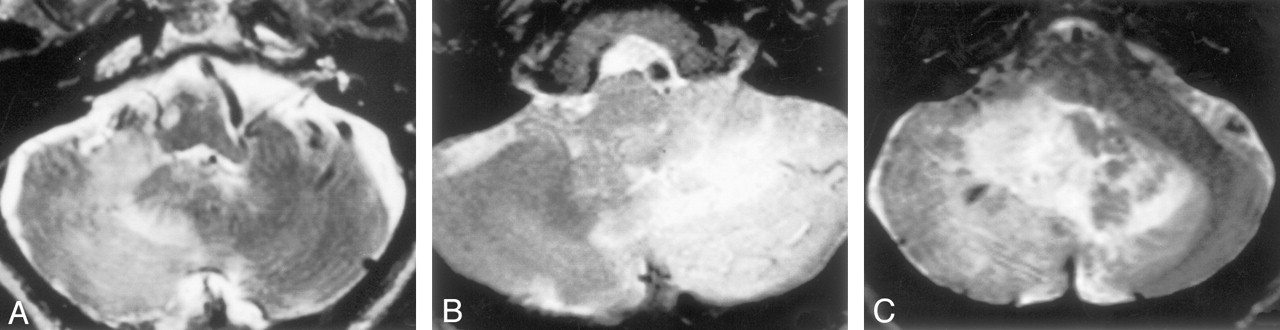
  • Fig 2.
    • Download figure
    • Open in new tab
    • Download powerpoint
    Fig 2.

    A–C, Axial T2-weighted MR images in patient 5 (A), patient 2 (B), and patient 4 (C) show high signal intensity of the lesion, with various degrees of signal voids.
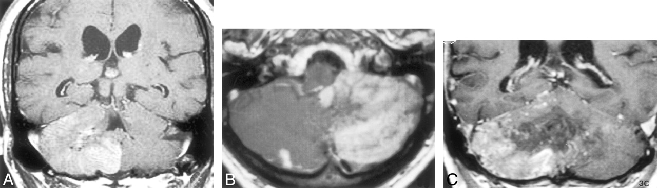
  • Fig 3.
    • Download figure
    • Open in new tab
    • Download powerpoint
    Fig 3.

    A–C, Gadolinium enhanced T1-weighted MR images in patient 3 (A, coronal), patient 2 (B, axial), and patient 4 (C, coronal) show diffuse peripheral enhancement.
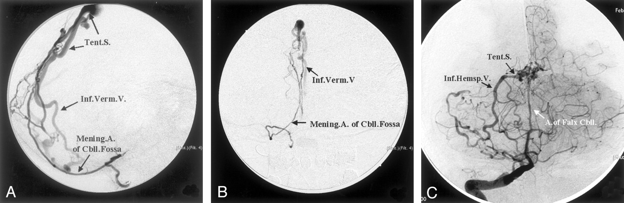
  • Fig 4.
    • Download figure
    • Open in new tab
    • Download powerpoint
    Fig 4.

    A and B, Lateral (A) and anterioposterior (B) selective angiograms of the meningeal artery of the cerebellar (Mening. A. of Cbll.) fossa in patient 3 show a DAVF draining into the inferior vermian vein (Inf. Verm. V.) via a short tentorial sinus (Tent.S.).

    C, Right vertebral angiogram in patient 4 demonstrates enlarged artery of the falx cerebelli (A.of Falx Cbll.) supplying a DAVF and draining into the right inferior hemispheric vein (Inf.Hemsp. V.) through the tentorial sinus (Tent.S.)

Tables

  • Figures
  • Clinical, MR, and angiographic findings of aggressive DAVF involving the cerebellar hemisphere

    CharacteristicPatient 1Patient 2Patient 3Patient 4Patient 5Patient 6
    Age (y)/sex73/M67/M72/M88/F79/M59/M
     Clinical findingsIncoordination, ataxiaVomitting, dizzinessImbalance, fatigueImbalance, ataxiaHemiplegia, ataxiaAtaxia
     HemisphereRightRightRightLeftLeftLeft
    MR findings
     TopographyInferior aspect of cerebellar hemisphereInferior aspect of cerebellar hemisphereInferior aspect of cerebellar hemisphereInferior aspect of cerebellar hemisphereInferior aspect of cerebellar hemisphereInferior aspect of cerebellar hemisphere
     Signal void lesions++++++++++
     T1 SILowLowLowLowLowLow
     T2 SIHighHighHighHighHighHigh
     Gd-enhancementPeripheralPeripheralPeripheralPeripheralPeripheralPeripheral
    Angiographic findings
     ICA feedersNoNoNoNoNoC5 branch
     ECA feedersOccipital a., ascending pharyngeal a.NoneOccipital a., ascending pharyngeal a.Occipital a.Occipital a.Occipital a., ascending pharyngeal a.
     VA feedersArtery of falx cerebelli, meningeal artery of cerebellar fossaArtery of falx cerebelliArtery of falx cerebelli, meningeal artery of cerebellar fossaArtery of falx cerebelliMeningeal artery of cerebellar fossaMeningeal artery of cerebellar fossa
     Venous drainageInferior hemispheric veinInferior hemispheric veinInferior vermian vein to inferior hemispheric veinInferior hemispheric veinInferior hemispheric veinInferior vermian vein to inferior hemispheric vein
    • Notes.—+ indicates few; ++, prominent; +++, significant; T1, T1-weighted images; T2, T2-weighted images; SI, signal intensity; Gd, gadolinium-enhanced T1-weighted images; ICA, internal carotid artery; ECA, external carotid artery; VA, vertebral artery; a, artery.

PreviousNext
Back to top

In this issue

American Journal of Neuroradiology: 24 (8)
American Journal of Neuroradiology
Vol. 24, Issue 8
1 Sep 2003
  • Table of Contents
  • Index by author
Advertisement
Print
Download PDF
Email Article

Thank you for your interest in spreading the word on American Journal of Neuroradiology.

NOTE: We only request your email address so that the person you are recommending the page to knows that you wanted them to see it, and that it is not junk mail. We do not capture any email address.

Enter multiple addresses on separate lines or separate them with commas.
MR Imaging of Dural Arteriovenous Fistulas Draining into Cerebellar Cortical Veins
(Your Name) has sent you a message from American Journal of Neuroradiology
(Your Name) thought you would like to see the American Journal of Neuroradiology web site.
CAPTCHA
This question is for testing whether or not you are a human visitor and to prevent automated spam submissions.
Cite this article
Seon-Kyu Lee, Robert A. Willinsky, Walter Montanera, Karel G. terBrugge
MR Imaging of Dural Arteriovenous Fistulas Draining into Cerebellar Cortical Veins
American Journal of Neuroradiology Sep 2003, 24 (8) 1602-1606;

Citation Manager Formats

  • BibTeX
  • Bookends
  • EasyBib
  • EndNote (tagged)
  • EndNote 8 (xml)
  • Medlars
  • Mendeley
  • Papers
  • RefWorks Tagged
  • Ref Manager
  • RIS
  • Zotero
0 Responses
Respond to this article
Share
Bookmark this article
MR Imaging of Dural Arteriovenous Fistulas Draining into Cerebellar Cortical Veins
Seon-Kyu Lee, Robert A. Willinsky, Walter Montanera, Karel G. terBrugge
American Journal of Neuroradiology Sep 2003, 24 (8) 1602-1606;
del.icio.us logo Twitter logo Facebook logo Mendeley logo
  • Tweet Widget
  • Facebook Like
  • Google Plus One

Jump to section

  • Article
    • Abstract
    • Methods
    • Results
    • Discussion
    • Conclusion
    • References
  • Figures & Data
  • Info & Metrics
  • Responses
  • References
  • PDF

Related Articles

  • No related articles found.
  • PubMed
  • Google Scholar

Cited By...

  • Standard and Guidelines: Intracranial Dural Arteriovenous Shunts
  • Susceptibility-Weighted MR Phase Imaging Can Demonstrate Retrograde Leptomeningeal Venous Drainage in Patients with Dural Arteriovenous Fistula
  • MR Angiography of Dural Arteriovenous Fistulas: Diagnosis and Follow-Up after Treatment Using a Time-Resolved 3D Contrast-Enhanced Technique
  • Assessment of Dural Arteriovenous Fistulae by Transcranial Color-Coded Duplex Sonography
  • Crossref
  • Google Scholar

This article has not yet been cited by articles in journals that are participating in Crossref Cited-by Linking.

More in this TOC Section

  • Statin Therapy Does Not Affect the Radiographic and Clinical Profile of Patients with TIA and Minor Stroke
  • Usefulness of Quantitative Susceptibility Mapping for the Diagnosis of Parkinson Disease
  • White Matter Alterations in the Brains of Patients with Active, Remitted, and Cured Cushing Syndrome: A DTI Study
Show more Brain

Similar Articles

Advertisement

Indexed Content

  • Current Issue
  • Accepted Manuscripts
  • Article Preview
  • Past Issues
  • Editorials
  • Editor's Choice
  • Fellows' Journal Club
  • Letters to the Editor
  • Video Articles

Cases

  • Case Collection
  • Archive - Case of the Week
  • Archive - Case of the Month
  • Archive - Classic Case

More from AJNR

  • Trainee Corner
  • Imaging Protocols
  • MRI Safety Corner
  • Book Reviews

Multimedia

  • AJNR Podcasts
  • AJNR Scantastics

Resources

  • Turnaround Time
  • Submit a Manuscript
  • Submit a Video Article
  • Submit an eLetter to the Editor/Response
  • Manuscript Submission Guidelines
  • Statistical Tips
  • Fast Publishing of Accepted Manuscripts
  • Graphical Abstract Preparation
  • Imaging Protocol Submission
  • Evidence-Based Medicine Level Guide
  • Publishing Checklists
  • Author Policies
  • Become a Reviewer/Academy of Reviewers
  • News and Updates

About Us

  • About AJNR
  • Editorial Board
  • Editorial Board Alumni
  • Alerts
  • Permissions
  • Not an AJNR Subscriber? Join Now
  • Advertise with Us
  • Librarian Resources
  • Feedback
  • Terms and Conditions
  • AJNR Editorial Board Alumni

American Society of Neuroradiology

  • Not an ASNR Member? Join Now

© 2025 by the American Society of Neuroradiology All rights, including for text and data mining, AI training, and similar technologies, are reserved.
Print ISSN: 0195-6108 Online ISSN: 1936-959X

Powered by HighWire