
Article Figures & Data
Figures
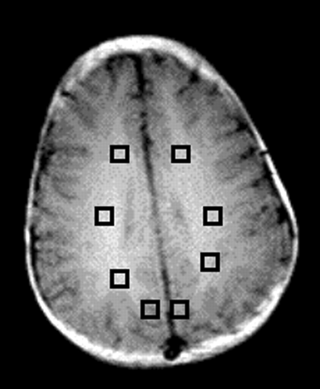
- Fig 1.
Regions of interest (squares) in the frontal-lobe white matter, centrum semiovale, and parietal white matter and gray matter were chosen for analysis.
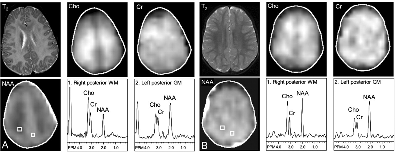
- Fig 2.
Severe PMD. T2-weighted MR images (T2); metabolic images of Cho, Cr and NAA; and selected spectra from the parietal white matter (WM) and parieto-occipital gray matter (GM). T2-weighted MR images show diffuse hyperintensity throughout the white matter, which also has markedly decreased NAA levels. Cho and Cr levels are also mildly elevated in the centrum semiovale. Gray matter spectra also show a lower ratio of NAA/Cho and NAA/Cr in the patient compared with that in the control subject.
A, Patient 1, aged 2 years 5 months.
B, Control subject 1, aged 1 year 9 months.
- Fig 3.
Mild PMD. T2-weighted MR imaging; metabolic images of Cho, Cr, and NAA; and selected spectra from the parietal white matter and parieto-occipital gray matter. T2-weighted MR images show patchy hyperintensity with greatest involvement in the centrum semiovale. NAA levels are bilaterally decreased in the parietal white matter. Gray-matter spectra are normal.
A, Patient 2, aged 3 years 8 months.
B, Control subject 2, aged 3 year 2 months.


Tables
Study No. of Patients Age, y Genetics Technique* Findings Grodd et al, 1991 4 0.7–5.7 Not given SV MRS, ratios 2 patterns: increased Cho and decreased NAA levels or decreased Cho and normal NAA levels Takanashi et al, 1997 2 5, 6 Point mutations: exon 5 (Pro210(4)→Leu[CTA]), exon 2 (Leu45[CTA]→Arg[CGA]) SV MRS, ratios Normal MRS results, trend for increased creatine levels Lam et al, 1998 1 19 Not given SV MRS, quantitation Slightly decreased NAA level in basal ganglia Spalice et al, 2000 2 1.5, 6 Connatal PMD, no mutation or duplication detected SV MRS, ratios Decreased Cho level Bonavita et al, 2001 9 6–43 Duplication (n=1), mutations G431A (n=6) and K150N (n=2) MRSI, ratios Decreased NAA level Garbern et al, 2002 2 11, 17 Deletion, mutation (G to A) MRS, MRSI, quantitation Decreased NAA level Hobson et al, 2002 1 11 Deletion 19 base pairs intron 3 PLP1/DM20 SV MRS, ratios Increased Cho, decreased NAA levels Takanashi et al, 2002 5 4–10 Duplication (classic PMD, mild form, n=4; severe connatal form, n = 1) SV MRS, quantitation Mildly increased NAA, creatine, and myo-inositol levels; normal Cho level * MRS indicates MR spectroscopy; SV, single voxel.
Individual Metabolite Levels Relative to Gray Matter Metabolite Ratios Cho Cr NAA NAA/Cho NAA/Cr Cho/Cr A. Frontal white matter Patient 1 1.17 1.19 0.84 1.16 1.63 1.41 Patient 2 1.28 1.01 1.02 1.62 2.63 1.63 Patient 3 1.68 0.95 0.87 1.27 2.32 1.83 Control subject 1 1.64 1.00 0.81 1.09 2.17 2.00 Control subject 2 1.51 0.92 0.94 1.26 2.57 2.03 Control subject 3 1.14 0.61 0.85 1.89 4.17 2.21 B. Centrum semiovale Patient 1 1.38 1.11 0.74 0.87 1.55 1.79 Patient 2 1.28 0.95 0.89 1.42 2.43 1.71 Patient 3 1.61 1.02 0.96 1.46 2.38 1.63 Control subject 1 1.35 0.81 0.82 1.34 2.71 2.03 Control subject 2 1.38 0.85 0.99 1.46 2.90 1.99 Control subject 3 1.55 0.85 0.99 1.61 3.46 2.15 C: Parietal white matter Patient 1 1.31 1.03 0.61 0.76 1.37 1.81 Patient 2 1.13 0.97 0.93 1.69 2.50 1.48 Patient 3 1.65 1.03 0.81 1.20 1.98 1.66 Control subject 1 1.42 0.92 0.84 1.29 2.42 1.87 Control subject 2 1.42 0.84 0.96 1.37 2.84 2.07 Control subject 3 1.45 0.66 0.84 1.46 3.80 2.60 D: Parieto-occipital gray matter* Patient 1 NA NA NA 1.61 2.31 1.43 Patient 2 NA NA NA 2.03 2.60 1.28 Patient 3 NA NA NA 2.44 2.52 1.04 Control subject 1 NA NA NA 2.19 2.67 1.22 Control subject 2 NA NA NA 2.03 2.50 1.23 Control subject 3 NA NA NA 2.52 2.99 1.18 Note.—Patient 1 was aged 2 years 5 months; patient 2, 3 years, 8 months; patient 3, 7 years, 6 months; control subject 1, 1 year, 9 months; control subject 2, 3 years, 2 months; and control subject 3, 7 years, 0 months.
* NA indicates not applicable.


