
Article Figures & Data
Figures
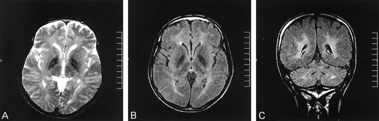
- Fig 1.
T2-weighted and fluid-attenuated inversion recovery images from the initial examination.
A, T2-weighted image reveals marked hypointensity in the globus pallidi with high signal intensity foci (eye-of-the-tiger appearance).
B, Fluid-attenuated inversion recovery image reveals marked hypointensity of the globus pallidi. Note that the eye-of-the-tiger appearance cannot be detected.
C, Fluid-attenuated inversion recovery image reveals high signal intensity lesions in the deep cerebral white matter and dentate nuclei.
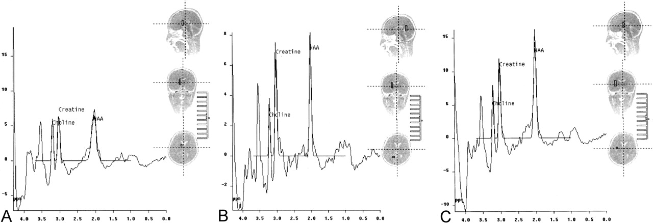
- Fig 2.
Spectra from the initial examination.
A, Proton MR spectroscopy (1500/40) reveals markedly decreased NAA in the globus pallidi.
B, Spectrum from deep cerebral white matter reveals decreased NAA and increased myoinositol peaks.
C, Spectrum from a normal region in the cerebral parenchyma.
- Fig 3.
Images obtained at the 10-month follow-up examination.
A, T2-weighted image reveals further hypointense appearance of the globus pallidi. Note loss of the eye-of-the-tiger appearance.
B, b=1000 s/mm2 image from trace diffusion MR imaging sequence (5700/139) reveals low signal intensity in the globus pallidi.
C, Corresponding ADC map (same section as that shown in B) reveals low ADC values (0.45–0.54 × 10⁻³ mm2/s) in the globus pallidi, compared with the unaffected thalamus (0.77 to 0.83 × 10⁻³ mm2/s).
D, b=1000 s/mm2 image is negative (normal appearing) for the deep cerebral white matter lesions.
E, Corresponding ADC map (same section as that shown in D) reveals slightly increased ADC values (1.08–1.12 × 10⁻³ mm2/s) compared with the ADC values from the normal white matter regions (0.78–0.87 × 10⁻³ mm2/s).


In this issue


Jump to section
Related Articles
Cited By...
- No citing articles found.