
Article Figures & Data
Figures
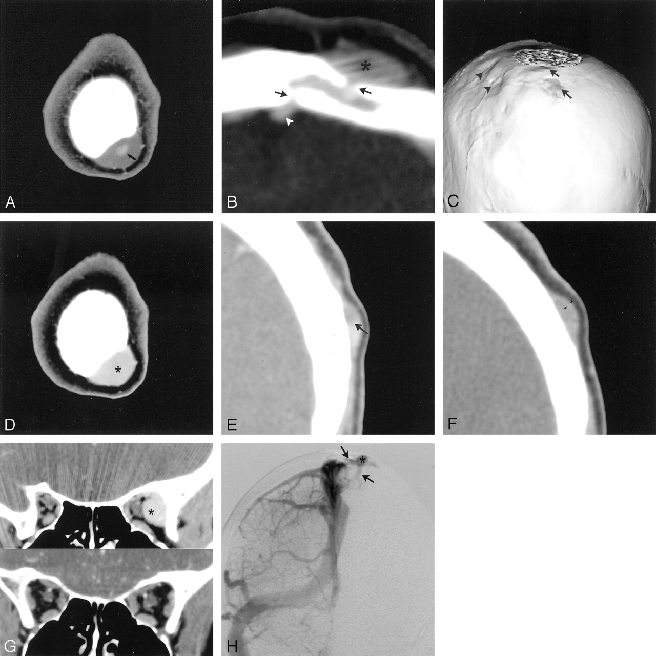
- Fig 1.
31-year-old woman with sinus pericranii and sudden onset of pain and swelling over the left scalp.
A, Source image from CTA (first pass) demonstrates only partial filling of a varicosity with contrast (arrow).
B, Reformatted oblique coronal CT through the earliest filling varicosity (asterisk) demonstrating defects within both the inner and outer tables (arrows) of the calvaria and filling of the diploe with venous blood. The connecting intracranial vein is denoted by the white arrowhead.
C, Volume-rendered CTA of the calvaria viewed from behind. Multiple calvarial depressions are seen along the left parietal and temporal bones (arrowheads). These depressions underlie slow-filling varicosities. The varicosities with the more direct connections to the dural sinuses already show enhancement (arrows).
D, Source image from the axial CTV through the same level as in A. The varicosity (asterisk) now demonstrates homogeneous enhancement.
E, Delayed coronal CT image demonstrates a sharply demarcated filling defect within a varicosity (arrow). The higher signal intensity represents flowing venous blood.
F, Delayed coronal CT through the region of greatest tenderness demonstrates only peripheral enhancement (arrowheads). This was only minimally compressible.
G, Coronal CT image (above) acquired with the head extended and dependent demonstrates an orbital varix (asterisk). A coronally reformatted CT image (below) shows the same region, but without the venous anomaly.
H, Angiographic anteroposterior view (venous phase) of a selective right internal carotid artery injection demonstrates the venous connections (arrow) to a pericranial varicosity (asterisk). Flow is both into and out of this pericranial varicosity.
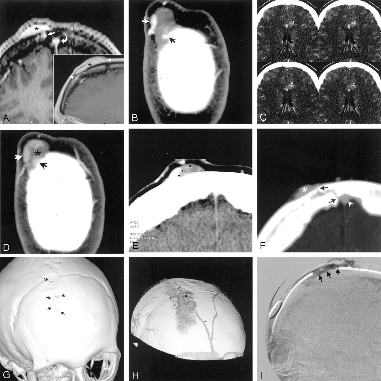
- Fig 2.
53-year-old woman with sinus pericranii and extreme pain involving the right forehead and scalp.
A, Coronal postcontrast gradient T1-weighted (full image) and sagittal postcontrast spin-echo T1-weighted (inset) MR images demonstrate the homogeneous appearance of the pericranial varicosity (asterisks). The coronal image clearly demonstrates one of the trans-osseous connections (arrow) as well as the adjacent superior sagittal sinus (curved arrow). The arrowheads in the inset demarcate the extent of the varicosity.
B, Source image from a CTA through the right frontal mass shows enhancement of preferential channels of flow (arrows) with most the varicosity remaining unenhanced.
C, Axial source images from the CTA demonstrating a small developmental venous anomaly just adjacent to a cavernous angioma. The arrows point to the single draining venous trunk while the cavernous angioma is of increased attenuation just posterior to the vein.
D, Source image from the axial CTV through the same region as Fig 2B. The varicosity is largely thrombosed (asterisk) in this portion. The patent channels of flow have become less distinct (arrows), and the anterior aspect now demonstrates enhancement.
E, Coronal postcontrast CT clearly depicts (arrowheads) the flowing venous blood from the adjacent thrombus (asterisk).
F, Reformatted oblique axial image from the CTV data through the inferior aspect of the pericranial varicosity. One of the transosseous pathways from the superior sagittal sinus (arrowhead) to the pericranial varicosity (asterisk) is clearly demonstrated. The defects within the inner and outer table of the frontal bones are denoted by arrows.
G, Volume-rendered image of the skull viewed obliquely from the front. Multiple small defects (arrows) are seen within the outer table of the right frontal bone. Compare this image to Fig H.
H, Volume-rendered image from CTV data shows the pericranial varicosity and adjacent scalp veins (arrowheads). The region of the clot is demarcated by an asterisk. The view is similar to that depicted in Fig 2G with the calvaria tilted more to the left.
I, Late venous phase image (lateral projection) from a selective injection of the left internal carotid artery depicting the pericranial varicosity (arrows).


