Advertisement

Main menu

  • Home
  • Content
    • Current Issue
    • Accepted Manuscripts
    • Article Preview
    • Past Issue Archive
    • AJNR Case Collection
    • Case of the Week Archive
    • Classic Case Archive
    • Case of the Month Archive
  • Special Collections
    • Spinal CSF Leak Articles (Jan 2020-June 2024)
    • 2024 AJNR Journal Awards
    • Most Impactful AJNR Articles
  • Multimedia
    • AJNR Podcast
    • AJNR Scantastics
    • Video Articles
  • For Authors
    • Submit a Manuscript
    • Author Policies
    • Fast publishing of Accepted Manuscripts
    • Graphical Abstract Preparation
    • Manuscript Submission Guidelines
    • Imaging Protocol Submission
    • Submit a Case for the Case Collection
  • About Us
    • About AJNR
    • Editorial Board
  • More
    • Become a Reviewer/Academy of Reviewers
    • Subscribers
    • Permissions
    • Alerts
    • Feedback
    • Advertisers
    • ASNR Home
  • Other Publications
    • ajnr

User menu

  • Alerts
  • Log in

Search

  • Advanced search
American Journal of Neuroradiology

American Journal of Neuroradiology

ASHNR American Society of Functional Neuroradiology ASHNR American Society of Pediatric Neuroradiology ASSR
  • Alerts
  • Log in

Advanced Search

  • Home
  • Content
    • Current Issue
    • Accepted Manuscripts
    • Article Preview
    • Past Issue Archive
    • AJNR Case Collection
    • Case of the Week Archive
    • Classic Case Archive
    • Case of the Month Archive
  • Special Collections
    • Spinal CSF Leak Articles (Jan 2020-June 2024)
    • 2024 AJNR Journal Awards
    • Most Impactful AJNR Articles
  • Multimedia
    • AJNR Podcast
    • AJNR Scantastics
    • Video Articles
  • For Authors
    • Submit a Manuscript
    • Author Policies
    • Fast publishing of Accepted Manuscripts
    • Graphical Abstract Preparation
    • Manuscript Submission Guidelines
    • Imaging Protocol Submission
    • Submit a Case for the Case Collection
  • About Us
    • About AJNR
    • Editorial Board
  • More
    • Become a Reviewer/Academy of Reviewers
    • Subscribers
    • Permissions
    • Alerts
    • Feedback
    • Advertisers
    • ASNR Home
  • Follow AJNR on Twitter
  • Visit AJNR on Facebook
  • Follow AJNR on Instagram
  • Join AJNR on LinkedIn
  • RSS Feeds

Welcome to the new AJNR, Updated Hall of Fame, and more. Read the full announcements.


AJNR is seeking candidates for the position of Associate Section Editor, AJNR Case Collection. Read the full announcement.


Getting new auth cookie, if you see this message a lot, tell someone!
Research ArticleINTERVENTIONAL

Treatment of Internal Carotid Artery Aneurysms with a Covered Stent: Experience in 24 Patients with Mid-Term Follow-up Results

Isil Saatci, H. Saruhan Cekirge, M. Halil Ozturk, Anil Arat, Fikret Ergungor, Zeki Sekerci, Engin Senveli, Uygur Er, Sami Turkoglu, Osman E. Ozcan and Tuncalp Ozgen
American Journal of Neuroradiology November 2004, 25 (10) 1742-1749;
Isil Saatci
  • Find this author on Google Scholar
  • Find this author on PubMed
  • Search for this author on this site
H. Saruhan Cekirge
  • Find this author on Google Scholar
  • Find this author on PubMed
  • Search for this author on this site
M. Halil Ozturk
  • Find this author on Google Scholar
  • Find this author on PubMed
  • Search for this author on this site
Anil Arat
  • Find this author on Google Scholar
  • Find this author on PubMed
  • Search for this author on this site
Fikret Ergungor
  • Find this author on Google Scholar
  • Find this author on PubMed
  • Search for this author on this site
Zeki Sekerci
  • Find this author on Google Scholar
  • Find this author on PubMed
  • Search for this author on this site
Engin Senveli
  • Find this author on Google Scholar
  • Find this author on PubMed
  • Search for this author on this site
Uygur Er
  • Find this author on Google Scholar
  • Find this author on PubMed
  • Search for this author on this site
Sami Turkoglu
  • Find this author on Google Scholar
  • Find this author on PubMed
  • Search for this author on this site
Osman E. Ozcan
  • Find this author on Google Scholar
  • Find this author on PubMed
  • Search for this author on this site
Tuncalp Ozgen
  • Find this author on Google Scholar
  • Find this author on PubMed
  • Search for this author on this site
  • Article
  • Figures & Data
  • Info & Metrics
  • Responses
  • References
  • PDF
Loading

Article Figures & Data

Figures

  • Tables
  • Fig 1.
    • Download figure
    • Open in new tab
    • Download powerpoint
    Fig 1.

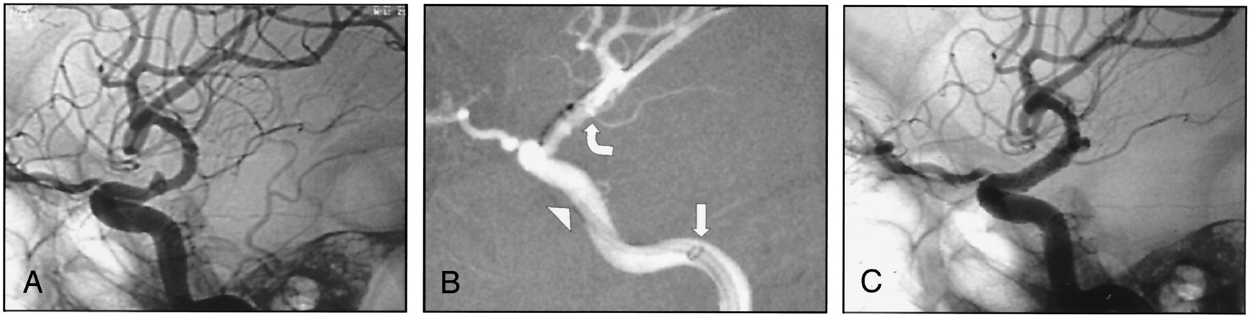
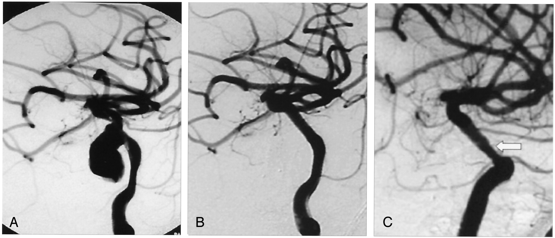
    A, Lateral angiogram obtained before treatment shows a right ruptured dissecting ICA aneurysm.

    B, 4 × 9 mm Jomed covered stent placed across the aneurysm neck with the support of a long reinforced Arrow sheath in the proximal petrous (straight arrow) and a 6-French Envoy guiding catheter in the distal petrous ICA (arrowhead). Extreme care was taken not to cover the anterior choroidal artery origin with the graft (curved arrow).

    C, Post-treatment lateral view shows exclusion of the aneurysm and the reconstructed internal carotid artery.

  • Fig 2.
    • Download figure
    • Open in new tab
    • Download powerpoint
    Fig 2.

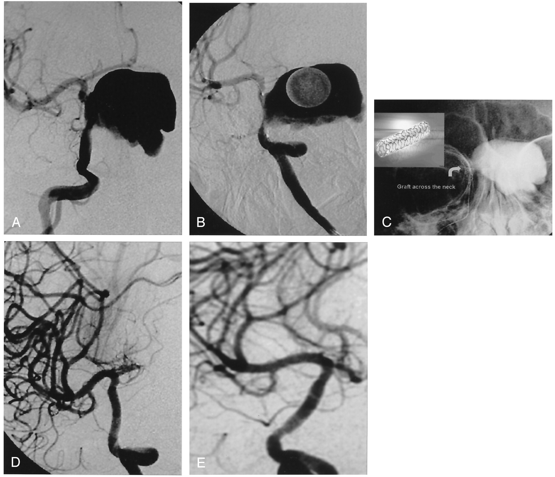
    A, Giant right carotico-ophthalmic aneurysm.

    B, Oblique image of stent graft placed across the aneurysm neck before its deployment. Note that the ophthalmic artery is covered with the graft but not the anterior choroidal artery.

    C, Non-subtracted view of the deployed stent graft (curved arrow). Note the contrast material trapped in the aneurysm sac because of the immediate exclusion.

    D, Oblique angiogram obtained after treatment reveals the reconstruction of the ICA. Note that the ophthalmic artery is not filling but the anterior choroidal artery origin is preserved.

    E, Oblique control angiogram obtained 18 months later shows no stenosis.

  • Fig 3.
    • Download figure
    • Open in new tab
    • Download powerpoint
    Fig 3.

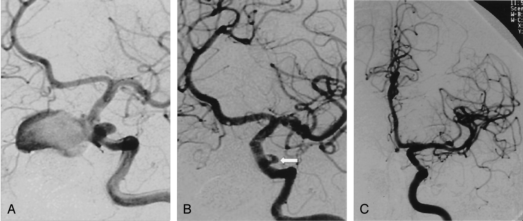
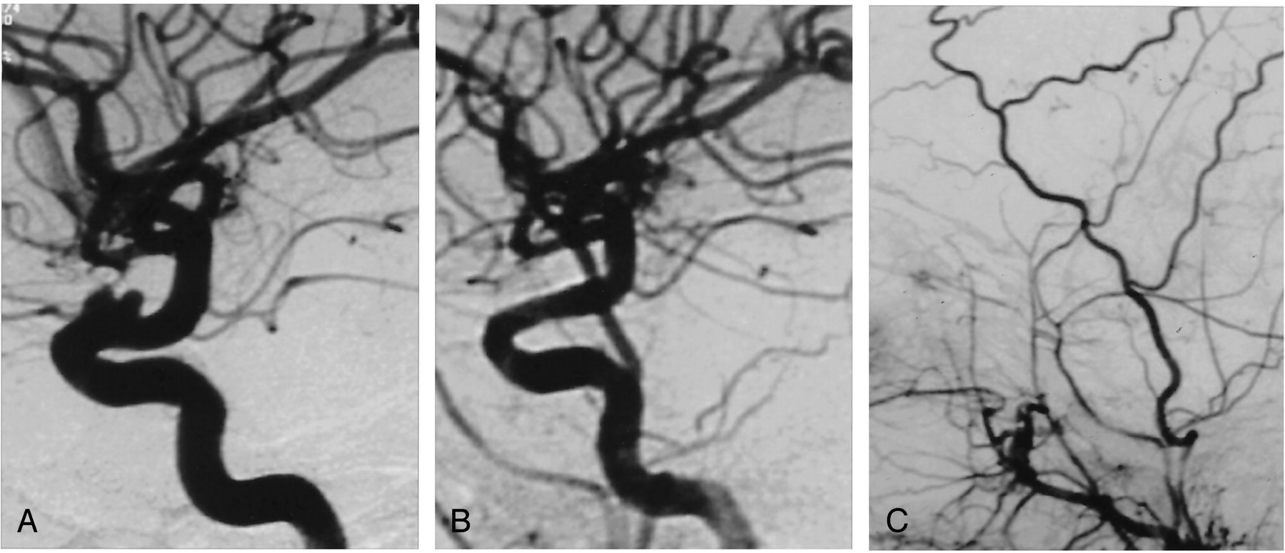
    A, Oblique angiogram of the left carotid artery reveals two posttraumatic aneurysms. One is giant, extending medially, and the other is small, extending laterally. Both originate from the cavernous portion of the ICA.

    B, Oblique post-treatment angiogram obtained after deployment of the stent graft reveals exclusion of the giant aneurysm but persistent endoleak into the small aneurysm (arrow).

    C, Control angiogram obtained 2 years later reveals spontaneous occlusion of the endoleak and excellent reconstruction of the ICA, with no stenosis.

  • Fig 4.
    • Download figure
    • Open in new tab
    • Download powerpoint
    Fig 4.

    A, Oblique posttraumatic angiogram of a left cavernous ICA aneurysm.

    B, Oblique angiogram obtained after stent graft deployment shows reconstruction of the ICA with no residual aneurysm filling.

    C, Oblique control angiogram obtained 6 months later reveals intimal hyperplasia (arrow) with no hemodynamic significance.

  • Fig 5.
    • Download figure
    • Open in new tab
    • Download powerpoint
    Fig 5.

    A, Lateral angiogram shows a broad based, bilobulated carotico-ophthalmic ICA aneurysm for which a previous seal test for Onyx treatment failed.

    B, Lateral angiogram obtained after treatment shows reconstruction of the ICA with the stent graft deployed across the aneurysm neck. Note the occlusion of the ophthalmic artery origin covered by the graft.

    C, Retrograde filling of the ophthalmic artery via external carotid artery.

Tables

  • Figures
  • Patient demographics, aneurysm location and size, clinical data, post-procedural clinical and angiographic follow-up

    Patient No.Age (yr)SexAneurysm LocationSize (mm)Mass EffectTraumaControl AngiographyMass Effect Early Outcome*Mass Effect Late Outcome*
    155MR cav33 × 286n Palsy+6 monthFull recoveryStable
    222MR cav34 × 29Headache+1 yearImprovedFull recovery
    348FR petrocav27 × 31Headache+NAImprovedNA
    449ML cav†15 × 206n and 3n Palsy−NAFull recoveryNA
    5α18ML cav16 × 24None+6 monthNANA
    6α23ML cav†39 × 332n Palsy+(β)2 yearFull recoveryStable
    L cav6 × 6NANA
    754MR cav20 × 17Headache−6 monthImprovedFull recovery
    8α43FR cav†22 × 156n Palsy+6 monthNo changeFull recovery 6 months
    932ML cav14 × 21None+(β)6 monthNANA
    1025ML petrocav†5 × 7None+(β)1 yearNANA
    1135MR cav21 × 17Headache+1 yearImprovedFull recovery
    12α34MR caroticooph37 × 25Total ophthalmoplegia+18 monthFull recoveryStable
    13α59FL caroticooph14 × 17None (SAH)−6 monthNANA
    14α43ML cav†27 × 20Total ophthalmoplegia+18 monthPartial recoveryFull recovery 6 months
    1518ML petrocav12 × 14None+(β)6 monthNANA
    1622MR petrocav9 × 6None+(β)6 monthNANA
    17α34ML cav28 × 343n Palsy−1 yearPartial recoveryFull recovery 6 months
    1817FL cav18 × 146n Palsy+1 yearFull recoveryStable
    1923ML cav16 × 11None−NANANA
    20α27FL caroticooph9 × 7None−6 monthNANA
    2129ML cav4 × 3None+(β)6 monthNANA
    2236FL petrous24 × 28Headache+6 monthFull recoveryStable
    2324FR caroticooph3 × 4None (SAH)−1 yearNANA
    2434FR cavernous22 × 273n Palsy+6 monthPartial recoveryFull recovery
    • Note.—α indicates patients in whom ophthalmic artery was covered with the stent graft; M, male; F, female; R, right; L, left; cav, cavernous internal carotid artery; petrocav, petrous and cavernous portion of the internal carotid artery; caroticooph, carotico-ophthalmic segment of the internal carotid artery; 6n, sixth nerve; 3n, third nerve; 2n, second nerve; SAH, subarachnoid hemorrhage; β, patients who had nasal bleeding; NA, not applicable because the time interval since treatment was <6 months and no control angiography had yet been performed.

    • * Control Angiography, latest angiography performed; Mass Effect Early Outcome, evaluation of mass effect symptom at end of second week; Mass Effect Late Outcome, evaluation during clinical follow-up (during third month unless otherwise indicated).

    • † Aneurysms were partially thrombosed.

PreviousNext
Back to top

In this issue

American Journal of Neuroradiology: 25 (10)
American Journal of Neuroradiology
Vol. 25, Issue 10
1 Nov 2004
  • Table of Contents
  • Index by author
Advertisement
Print
Download PDF
Email Article

Thank you for your interest in spreading the word on American Journal of Neuroradiology.

NOTE: We only request your email address so that the person you are recommending the page to knows that you wanted them to see it, and that it is not junk mail. We do not capture any email address.

Enter multiple addresses on separate lines or separate them with commas.
Treatment of Internal Carotid Artery Aneurysms with a Covered Stent: Experience in 24 Patients with Mid-Term Follow-up Results
(Your Name) has sent you a message from American Journal of Neuroradiology
(Your Name) thought you would like to see the American Journal of Neuroradiology web site.
CAPTCHA
This question is for testing whether or not you are a human visitor and to prevent automated spam submissions.
Cite this article
Isil Saatci, H. Saruhan Cekirge, M. Halil Ozturk, Anil Arat, Fikret Ergungor, Zeki Sekerci, Engin Senveli, Uygur Er, Sami Turkoglu, Osman E. Ozcan, Tuncalp Ozgen
Treatment of Internal Carotid Artery Aneurysms with a Covered Stent: Experience in 24 Patients with Mid-Term Follow-up Results
American Journal of Neuroradiology Nov 2004, 25 (10) 1742-1749;

Citation Manager Formats

  • BibTeX
  • Bookends
  • EasyBib
  • EndNote (tagged)
  • EndNote 8 (xml)
  • Medlars
  • Mendeley
  • Papers
  • RefWorks Tagged
  • Ref Manager
  • RIS
  • Zotero
0 Responses
Respond to this article
Share
Bookmark this article
Treatment of Internal Carotid Artery Aneurysms with a Covered Stent: Experience in 24 Patients with Mid-Term Follow-up Results
Isil Saatci, H. Saruhan Cekirge, M. Halil Ozturk, Anil Arat, Fikret Ergungor, Zeki Sekerci, Engin Senveli, Uygur Er, Sami Turkoglu, Osman E. Ozcan, Tuncalp Ozgen
American Journal of Neuroradiology Nov 2004, 25 (10) 1742-1749;
del.icio.us logo Twitter logo Facebook logo Mendeley logo
  • Tweet Widget
  • Facebook Like
  • Google Plus One

Jump to section

  • Article
    • Abstract
    • Patients and Techniques
    • Results
    • Discussion
    • References
  • Figures & Data
  • Info & Metrics
  • Responses
  • References
  • PDF

Related Articles

  • No related articles found.
  • PubMed
  • Google Scholar

Cited By...

  • Treatment of 14 intracranial aneurysms with the FRED system
  • The sea anchor technique: a novel method to aid in stent-assisted embolization of giant cerebral aneurysms
  • Geometric, Hemodynamic, and Pathological Study of a Distal Internal Carotid Artery Aneurysm Model in Dogs
  • Carotid Vessel Evaluation Via a 3-D Workstation
  • Stent usage in the treatment of intracranial aneurysms: past, present and future
  • Pipeline Embolization Device in Aneurysmal Subarachnoid Hemorrhage
  • Treatment of Intracranial Aneurysms Using the Pipeline Flow-Diverter Embolization Device: A Single-Center Experience with Long-Term Follow-Up Results
  • The Woven EndoBridge: A New Aneurysm Occlusion Device
  • The balloon anchor technique: a novel technique for distal access through a giant aneurysm
  • Partially Thrombosed Intracranial Aneurysms Presenting with Mass Effect: Long-Term Clinical and Imaging Follow-Up after Endovascular Treatment
  • Endovascular treatment of recurrent intracranial aneurysms with re-coiling or covered stents
  • Placement of Covered Stents for the Treatment of Direct Carotid Cavernous Fistulas
  • A Second-Generation, Endoluminal, Flow-Disrupting Device for Treatment of Saccular Aneurysms
  • A New Endoluminal, Flow-Disrupting Device for Treatment of Saccular Aneurysms
  • Crossref
  • Google Scholar

This article has not yet been cited by articles in journals that are participating in Crossref Cited-by Linking.

More in this TOC Section

  • SAVE vs. Solumbra Techniques for Thrombectomy
  • CT Perfusion&Reperfusion in Acute Ischemic Stroke
  • Delayed Reperfusion Post-Thrombectomy&Thrombolysis
Show more Interventional

Similar Articles

Advertisement

Indexed Content

  • Current Issue
  • Accepted Manuscripts
  • Article Preview
  • Past Issues
  • Editorials
  • Editors Choice
  • Fellow Journal Club
  • Letters to the Editor

Cases

  • Case Collection
  • Archive - Case of the Week
  • Archive - Case of the Month
  • Archive - Classic Case

Special Collections

  • Special Collections

Resources

  • News and Updates
  • Turn around Times
  • Submit a Manuscript
  • Author Policies
  • Manuscript Submission Guidelines
  • Evidence-Based Medicine Level Guide
  • Publishing Checklists
  • Graphical Abstract Preparation
  • Imaging Protocol Submission
  • Submit a Case
  • Become a Reviewer/Academy of Reviewers
  • Get Peer Review Credit from Publons

Multimedia

  • AJNR Podcast
  • AJNR SCANtastic
  • Video Articles

About Us

  • About AJNR
  • Editorial Board
  • Not an AJNR Subscriber? Join Now
  • Alerts
  • Feedback
  • Advertise with us
  • Librarian Resources
  • Permissions
  • Terms and Conditions

American Society of Neuroradiology

  • Not an ASNR Member? Join Now

© 2025 by the American Society of Neuroradiology All rights, including for text and data mining, AI training, and similar technologies, are reserved.
Print ISSN: 0195-6108 Online ISSN: 1936-959X

Powered by HighWire