
Data supplements
Figure Correction
In the article Blood Brain-Barrier Disruption of Nonionic Iodinated Contrast Medium Following Coil Embolization of a Ruptured Intracerebral Aneurysm, AJNR 25:1783-1786, November/December 2004, the figures were incorrectly labeled. The correct figures and legends are as follows:
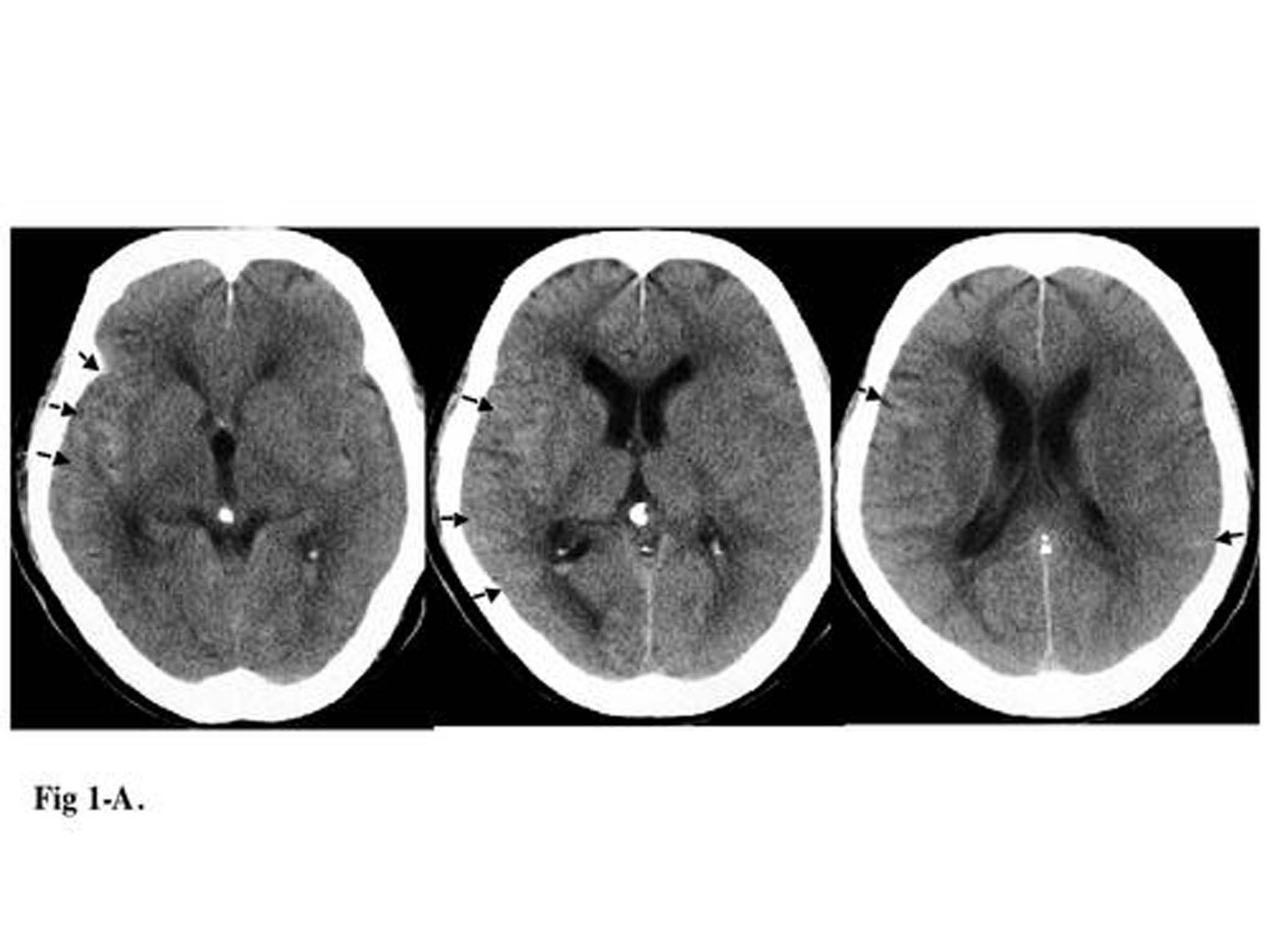
FIG 1. A 72-year-old man presented with subarachnoid hemorrhage secondary to a ruptured anterior communicating artery complex aneurysm. The patient underwent primary endovascular coil treatment of the aneurysm to occlusion.
Files in this Data Supplement:
- Figure 1A (128KB) - Pretreatment CT scan showing hyperattenuated area mainly in the right Sylvian fissure (arrow).
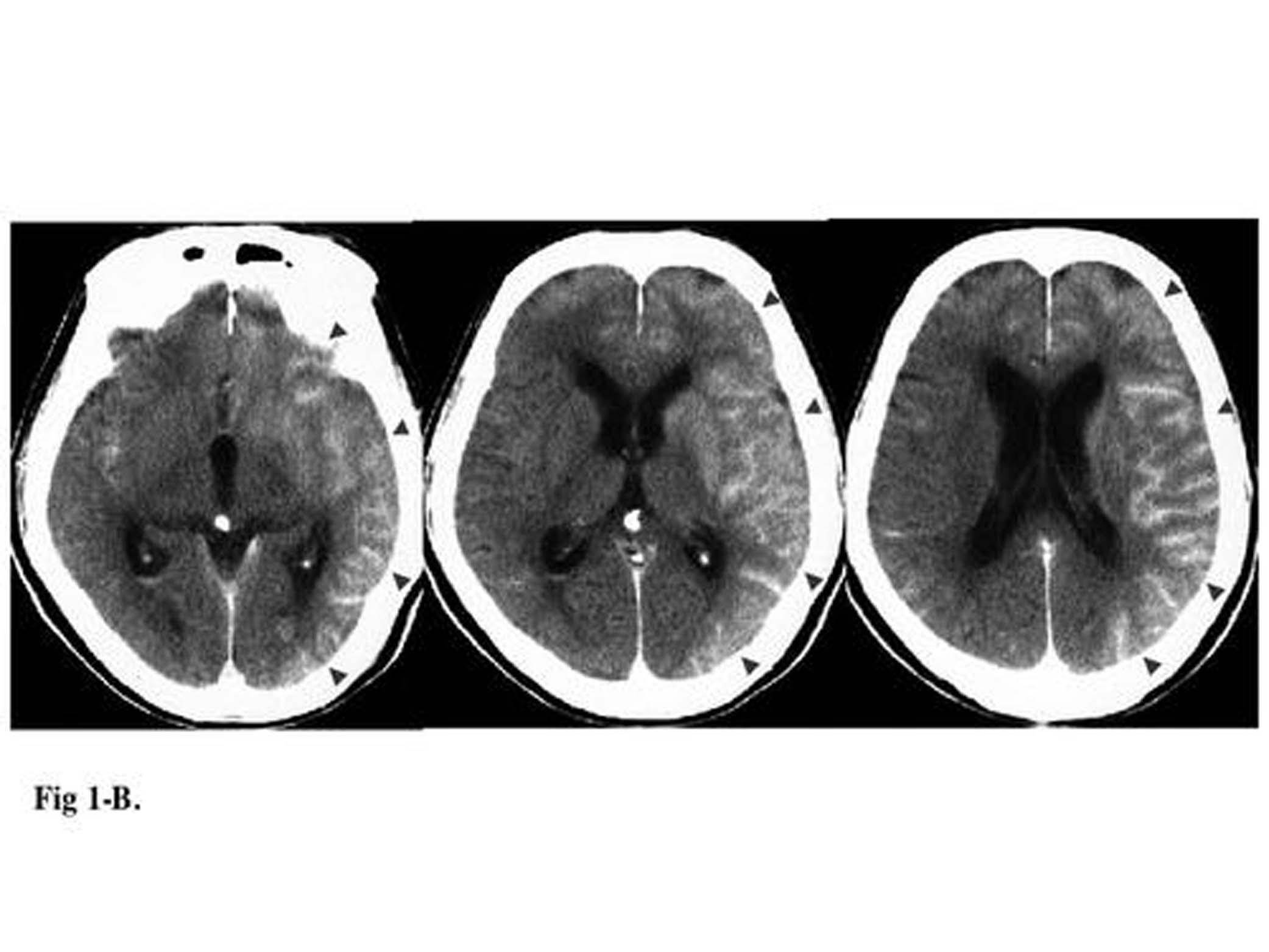
- Figure 1B (130KB) - Postprocedural CT scan, obtained 1 hour after the procedure, demonstrating extensive gyriform enhancement of the cerebral cortex, significantly greater on the left (arrowhead).
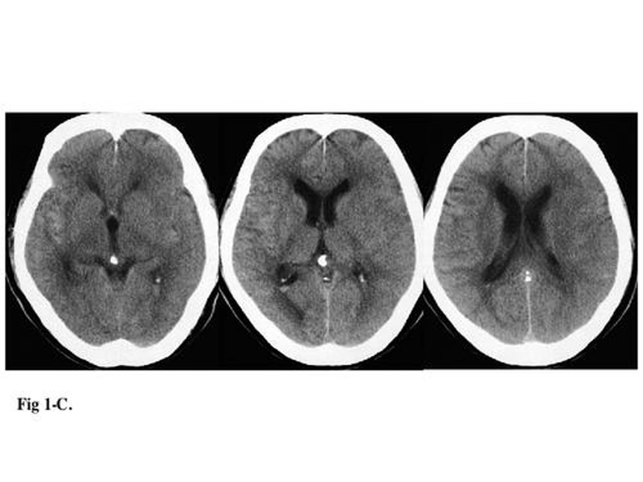
- Figure 1C (127KB) - Follow-up CT scan, obtained 11 hours after the procedure, demonstrates resolution of the previously demonstrated gyriform cortical enhancement, with only slight diffuse vasogenic edema shown in the left cortical mantle.
- Figure 1A (3.10MB) - Pretreatment CT scan showing hyperattenuated area mainly in the right Sylvian fissure (arrow).
- Figure 1B (3.10MB) - Postprocedural CT scan, obtained 1 hour after the procedure, demonstrating extensive gyriform enhancement of the cerebral cortex, significantly greater on the left (arrowhead).
- Figure 1C (3.10MB) - Follow-up CT scan, obtained 11 hours after the procedure, demonstrates resolution of the previously demonstrated gyriform cortical enhancement, with only slight diffuse vasogenic edema shown in the left cortical mantle.
In this issue


Jump to section
Related Articles
Cited By...
- Contrast Injection from an Intermediate Catheter Placed in an Intradural Artery is Associated with Contrast-Induced Encephalopathy following Neurointervention
- Loss of NLRX1 Exacerbates Neural Tissue Damage and NF-{kappa}B Signaling following Brain Injury
- Contrast encephalopathy after coiling in the setting of obstructive sleep apnoea
- Subarachnoid Hyperattenuation on Flat Panel Detector-Based Conebeam CT Immediately after Uneventful Coil Embolization of Unruptured Intracranial Aneurysms
- Does the Application of X-Ray Contrast Agents Impair the Clinical Effect of Intravenous Recombinant Tissue-Type Plasminogen Activator in Acute Ischemic Stroke Patients?
- Safety of performing CT angiography in stroke patients treated with intravenous thrombolysis
- Intra-Arterial Iodinated Radiographic Contrast Material Injection Administration in a Rat Middle Cerebral Artery Occlusion and Reperfusion Model: Possible Effects on Intracerebral Hemorrhage