Advertisement

Main menu

  • Home
  • Content
    • Current Issue
    • Accepted Manuscripts
    • Article Preview
    • Past Issue Archive
    • AJNR Case Collection
    • Case of the Week Archive
    • Classic Case Archive
    • Case of the Month Archive
  • Special Collections
    • Spinal CSF Leak Articles (Jan 2020-June 2024)
    • 2024 AJNR Journal Awards
    • Most Impactful AJNR Articles
  • Multimedia
    • AJNR Podcast
    • AJNR Scantastics
    • Video Articles
  • For Authors
    • Submit a Manuscript
    • Author Policies
    • Fast publishing of Accepted Manuscripts
    • Graphical Abstract Preparation
    • Manuscript Submission Guidelines
    • Imaging Protocol Submission
    • Submit a Case for the Case Collection
  • About Us
    • About AJNR
    • Editorial Board
  • More
    • Become a Reviewer/Academy of Reviewers
    • Subscribers
    • Permissions
    • Alerts
    • Feedback
    • Advertisers
    • ASNR Home
  • Other Publications
    • ajnr

User menu

  • Alerts
  • Log in

Search

  • Advanced search
American Journal of Neuroradiology

American Journal of Neuroradiology

ASHNR American Society of Functional Neuroradiology ASHNR American Society of Pediatric Neuroradiology ASSR
  • Alerts
  • Log in

Advanced Search

  • Home
  • Content
    • Current Issue
    • Accepted Manuscripts
    • Article Preview
    • Past Issue Archive
    • AJNR Case Collection
    • Case of the Week Archive
    • Classic Case Archive
    • Case of the Month Archive
  • Special Collections
    • Spinal CSF Leak Articles (Jan 2020-June 2024)
    • 2024 AJNR Journal Awards
    • Most Impactful AJNR Articles
  • Multimedia
    • AJNR Podcast
    • AJNR Scantastics
    • Video Articles
  • For Authors
    • Submit a Manuscript
    • Author Policies
    • Fast publishing of Accepted Manuscripts
    • Graphical Abstract Preparation
    • Manuscript Submission Guidelines
    • Imaging Protocol Submission
    • Submit a Case for the Case Collection
  • About Us
    • About AJNR
    • Editorial Board
  • More
    • Become a Reviewer/Academy of Reviewers
    • Subscribers
    • Permissions
    • Alerts
    • Feedback
    • Advertisers
    • ASNR Home
  • Follow AJNR on Twitter
  • Visit AJNR on Facebook
  • Follow AJNR on Instagram
  • Join AJNR on LinkedIn
  • RSS Feeds

Welcome to the new AJNR, Updated Hall of Fame, and more. Read the full announcements.


AJNR is seeking candidates for the position of Associate Section Editor, AJNR Case Collection. Read the full announcement.


OtherINTERVENTIONAL

Simultaneous Bilateral Internal Carotid Artery 3D Rotational Angiography

Joon K. Song, Yasunari Niimi, Jonathan L. Brisman and Alejandro Berenstein
American Journal of Neuroradiology November 2004, 25 (10) 1787-1789;
Joon K. Song
  • Find this author on Google Scholar
  • Find this author on PubMed
  • Search for this author on this site
Yasunari Niimi
  • Find this author on Google Scholar
  • Find this author on PubMed
  • Search for this author on this site
Jonathan L. Brisman
  • Find this author on Google Scholar
  • Find this author on PubMed
  • Search for this author on this site
Alejandro Berenstein
  • Find this author on Google Scholar
  • Find this author on PubMed
  • Search for this author on this site
  • Article
  • Figures & Data
  • Info & Metrics
  • Responses
  • References
  • PDF
Loading

Abstract

Summary: We simultaneously injected both internal carotid arteries (ICAs) during 3D rotational digital subtraction angiography in a patient with a ruptured anterior communicating artery (Acom) aneurysm and vasospasm. Shaded surface display (SSD) images from this acquisition provided more anatomic detail than did SSD images created from individual ICA 3D rotational digital subtraction angriography. In the evaluation of complex Acom aneurysms, this technique can facilitate the choice of optimal working projections for safer aneurysm coiling.

With increasing clinical application, 3D rotational cerebral angiography helps overcome the limitations of 2D angiography by more efficiently and more definitively visualizing aneurysm anatomy (1–6). We present a case of an extended application of 3D rotational angiography. In a patient with a ruptured anterior communicating artery (Acom) aneurysm and vasospasm, both internal carotid arteries (ICAs) were simultaneously injected during 3D rotational angiogram acquisition to more clearly define complex aneurysm and adjacent vessel anatomy before endovascular treatment.

Case Report

A 48-year-old right-handed man collapsed at home from a subarachnoid hemorrhage. On examination, the patient was lethargic with left hemiparesis. Cerebral angiography showed a complex Acom aneurysm and vasospasm (Fig 1A and B). The aneurysm appeared to originate at the junction of the right A1 segment and a prominent callosomarginal artery originating from the base of the aneurysm. Left internal carotid injection showed the left A2 segment seeming to originate from its proximal neck.

Fig 1.
  • Download figure
  • Open in new tab
  • Download powerpoint
Fig 1.

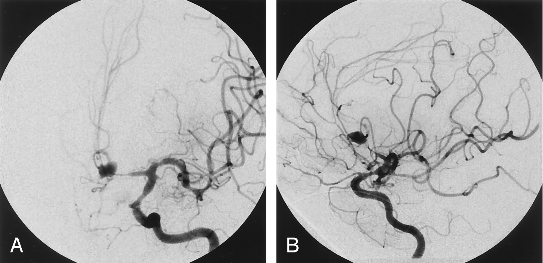
Left ICA angiogram, frontal (A) and lateral (B) oblique projections, show Acom aneurysm associated with severe distal A1 and A2 segment vasospasm.

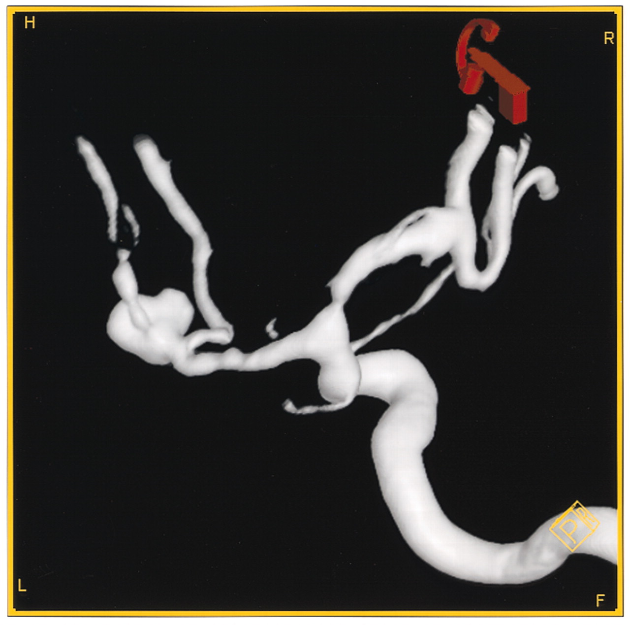
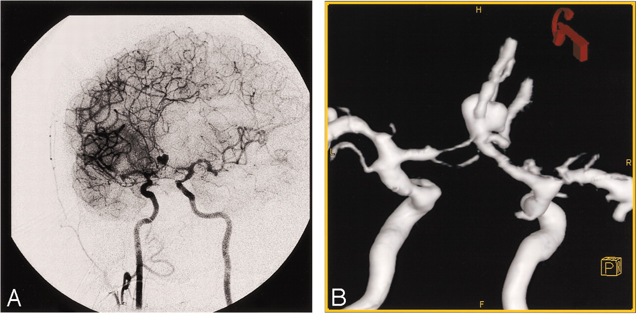
We performed 3D rotational angiography by injection of contrast medium into the right and left ICA separately (Siemens Biplane, Axiom Artis BA; Leonardo InSpace workstation, Erlangen, Germany). The resultant 3D shaded surface display (SSD) reconstruction (Fig 2), however, showed incomplete visualization of the complete aneurysm and adjacent arteries. Because of the midline location of this aneurysm, the frequent competing flows at the Acom level, superimposed vasospasm, and the complex anatomic relationships of the different vessels near the aneurysm, we performed simultaneous bilateral ICA 3D rotational angiography (Fig 3A). By using bilateral groin percutaneous access, separate catheters were placed into each ICA. One catheter was connected to a power injector with injection of 3 mL/s for 7 seconds while the other ICA catheter was simultaneously hand-injected with 20 mL of contrast medium. The resultant 3D SSD better showed the relationship of the base of the aneurysm to both A1 segments of the anterior cerebral arteries, left A2 anterior cerebral artery (ACA) segment, and branches of a prominent right callosomarginal artery arising from the base of the aneurysm (Fig 3B).

Fig 2.
  • Download figure
  • Open in new tab
  • Download powerpoint
Fig 2.

Left ICA oblique angiogram during 3D rotational angiography shows attenuation of the distal A1 segment and the complex Acom aneurysm. The SSD reconstructions did not convincingly reveal the relationship of the aneurysm and the adjacent anterior cerebral artery vessels.

Fig 3.
  • Download figure
  • Open in new tab
  • Download powerpoint
Fig 3.

Simultaneous bilateral ICA injections during 3D rotational angiogram acquisition reveal heart-shaped Acom aneurysm and both A1 segments (A). 3D SSD reconstructions from the simultaneous bilateral ICA injection 3D rotational angiogram shows clearly the relation of the aneurysm and both A1 segments as well as the right callosomarginal artery branches arising from the base of the aneurysm (B).

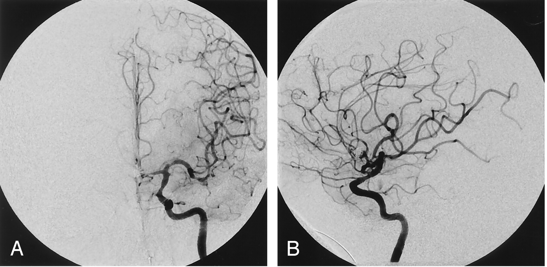
With this additional knowledge, we were able to occlude the aneurysm successfully with coils, while preserving both the anterior cerebral arteries (Fig 4A and B). The patient made a significant recovery and was discharged to a rehabilitation facility, independently caring for himself and ambulating with a stable mild left hemiparesis.

Fig 4.
  • Download figure
  • Open in new tab
  • Download powerpoint
Fig 4.

Postcoiling left ICA angiograms, frontal (A) and lateral (B) projections, show aneurysm occlusion and preservation of the left ACA branches, including opacification of both pericallosal arteries.

Discussion

3D rotational digital subtraction angiography with SSD display provides significantly more detailed information for evaluating cerebral aneurysms than that provided by the conventional 2D technique (1–6). 3D rotational angiography is performed with the injection of a target vessel such as an ICA or vertebral artery, usually with a power injector (1–6). In almost all cases with patients who are cooperative or who are under general anesthesia, where patient motion does not degrade the reconstructions, standard protocols are sufficient to produce adequate anatomic images for endovascular decision-making. In our case, however, because of vasospasm and anatomic variation in pericallosal and callosomarginal origins, standard 3D rotational angiography of each ICA did not cross-fill the other A1 segment.

Although the technical possibility of cross-compression of the contralateral carotid artery during 3D rotational angiography has been mentioned, cross-compression for Acom aneurysms has not been performed in any series (1). Cross-compression was considered in our case; however, considerations of operator exposure to radiation and creating misregistration artifact during rotation of the image intensifier dissuaded us from cross-compression. In patients with a complex unruptured Acom aneurysm, good-quality CTA can provide similar information and can be obtained before treatment. In patients with an acutely ruptured aneurysm, however, obtaining a CTA immediately before angiography is rarely performed.

Conclusion

Simultaneous bilateral ICA 3D rotational angiography can be a useful technique in the pretreatment evaluation of complex Acom aneurysms. Simultaneous different-vessel-injection 3D rotational angiography may be useful in the real-time treatment evaluation of selected brain arteriovenous malformations, dural fistula, and facial malformations.

References

  1. ↵
    Hirai T, Korogi Y, Suginohara K, et al. Clinical usefulness of unsubtracted 3D digital angiography compared with rotational digital angiography in the pretreatment evaluation of intracranial aneurysms. AJNR Am J Neuroradiol 2003;24:1067–1074
    Abstract/FREE Full Text
  2. Ernemann UU, Gronewaller E, Duffner FB, et al. Influence of geometric and hemodynamic parameters on aneurysm visualization during three-dimensional rotational angiography: an in vitro study. AJNR Am J Neuroradiol 2003;24:597–603
    Abstract/FREE Full Text
  3. Sugahara T, Y Korogi, K Nakashima, et al. Comparison of 2D and 3D digital subtraction angiography in evaluation of intracranial aneurysms. AJNR Am J Neuroradiol 2002;23:1545–1552
    Abstract/FREE Full Text
  4. Hochmuth A, Spetzger U, Schumacher M. Comparison of three-dimensional rotational angiography with digital subtraction angiography in the assessment of ruptured cerebral aneurysms. AJNR Am J Neuroradiol 2002;23:1199–1205
    Abstract/FREE Full Text
  5. Abe T, Hirohata M, Tanaka N, et al. Clinical benefits of rotational 3D angiography in endovascular treatment of ruptured cerebral aneurysm. AJNR Am J Neuroradiol 2002;23:686–688
    Abstract/FREE Full Text
  6. ↵
    Anxionnat R, Bracard S, Ducrocq X, et al. Intracranial aneurysms: clinical value of 3D digital subtraction angiography in the therapeutic decision and endovascular treatment. Radiology 2001;218:799–808
    CrossRefPubMed
  • Received April 19, 2004.
  • Accepted after revision May 19, 2004.
  • Copyright © American Society of Neuroradiology
View Abstract
PreviousNext
Back to top

In this issue

American Journal of Neuroradiology: 25 (10)
American Journal of Neuroradiology
Vol. 25, Issue 10
1 Nov 2004
  • Table of Contents
  • Index by author
Advertisement
Print
Download PDF
Email Article

Thank you for your interest in spreading the word on American Journal of Neuroradiology.

NOTE: We only request your email address so that the person you are recommending the page to knows that you wanted them to see it, and that it is not junk mail. We do not capture any email address.

Enter multiple addresses on separate lines or separate them with commas.
Simultaneous Bilateral Internal Carotid Artery 3D Rotational Angiography
(Your Name) has sent you a message from American Journal of Neuroradiology
(Your Name) thought you would like to see the American Journal of Neuroradiology web site.
CAPTCHA
This question is for testing whether or not you are a human visitor and to prevent automated spam submissions.
Cite this article
Joon K. Song, Yasunari Niimi, Jonathan L. Brisman, Alejandro Berenstein
Simultaneous Bilateral Internal Carotid Artery 3D Rotational Angiography
American Journal of Neuroradiology Nov 2004, 25 (10) 1787-1789;

Citation Manager Formats

  • BibTeX
  • Bookends
  • EasyBib
  • EndNote (tagged)
  • EndNote 8 (xml)
  • Medlars
  • Mendeley
  • Papers
  • RefWorks Tagged
  • Ref Manager
  • RIS
  • Zotero
0 Responses
Respond to this article
Share
Bookmark this article
Simultaneous Bilateral Internal Carotid Artery 3D Rotational Angiography
Joon K. Song, Yasunari Niimi, Jonathan L. Brisman, Alejandro Berenstein
American Journal of Neuroradiology Nov 2004, 25 (10) 1787-1789;
del.icio.us logo Twitter logo Facebook logo Mendeley logo
  • Tweet Widget
  • Facebook Like
  • Google Plus One

Jump to section

  • Article
    • Abstract
    • Case Report
    • Discussion
    • Conclusion
    • References
  • Figures & Data
  • Info & Metrics
  • Responses
  • References
  • PDF

Related Articles

  • No related articles found.
  • PubMed
  • Google Scholar

Cited By...

  • No citing articles found.
  • Crossref
  • Google Scholar

This article has not yet been cited by articles in journals that are participating in Crossref Cited-by Linking.

More in this TOC Section

  • SAVE vs. Solumbra Techniques for Thrombectomy
  • CT Perfusion&Reperfusion in Acute Ischemic Stroke
  • Delayed Reperfusion Post-Thrombectomy&Thrombolysis
Show more Interventional

Similar Articles

Advertisement

Indexed Content

  • Current Issue
  • Accepted Manuscripts
  • Article Preview
  • Past Issues
  • Editorials
  • Editors Choice
  • Fellow Journal Club
  • Letters to the Editor

Cases

  • Case Collection
  • Archive - Case of the Week
  • Archive - Case of the Month
  • Archive - Classic Case

Special Collections

  • Special Collections

Resources

  • News and Updates
  • Turn around Times
  • Submit a Manuscript
  • Author Policies
  • Manuscript Submission Guidelines
  • Evidence-Based Medicine Level Guide
  • Publishing Checklists
  • Graphical Abstract Preparation
  • Imaging Protocol Submission
  • Submit a Case
  • Become a Reviewer/Academy of Reviewers
  • Get Peer Review Credit from Publons

Multimedia

  • AJNR Podcast
  • AJNR SCANtastic
  • Video Articles

About Us

  • About AJNR
  • Editorial Board
  • Not an AJNR Subscriber? Join Now
  • Alerts
  • Feedback
  • Advertise with us
  • Librarian Resources
  • Permissions
  • Terms and Conditions

American Society of Neuroradiology

  • Not an ASNR Member? Join Now

© 2025 by the American Society of Neuroradiology All rights, including for text and data mining, AI training, and similar technologies, are reserved.
Print ISSN: 0195-6108 Online ISSN: 1936-959X

Powered by HighWire