Advertisement

Main menu

  • Home
  • Content
    • Current Issue
    • Accepted Manuscripts
    • Article Preview
    • Past Issue Archive
    • AJNR Case Collection
    • Case of the Week Archive
    • Classic Case Archive
    • Case of the Month Archive
  • Special Collections
    • Spinal CSF Leak Articles (Jan 2020-June 2024)
    • 2024 AJNR Journal Awards
    • Most Impactful AJNR Articles
  • Multimedia
    • AJNR Podcast
    • AJNR Scantastics
    • Video Articles
  • For Authors
    • Submit a Manuscript
    • Author Policies
    • Fast publishing of Accepted Manuscripts
    • Graphical Abstract Preparation
    • Manuscript Submission Guidelines
    • Imaging Protocol Submission
    • Submit a Case for the Case Collection
  • About Us
    • About AJNR
    • Editorial Board
  • More
    • Become a Reviewer/Academy of Reviewers
    • Subscribers
    • Permissions
    • Alerts
    • Feedback
    • Advertisers
    • ASNR Home
  • Other Publications
    • ajnr

User menu

  • Alerts
  • Log in

Search

  • Advanced search
American Journal of Neuroradiology

American Journal of Neuroradiology

ASHNR American Society of Functional Neuroradiology ASHNR American Society of Pediatric Neuroradiology ASSR
  • Alerts
  • Log in

Advanced Search

  • Home
  • Content
    • Current Issue
    • Accepted Manuscripts
    • Article Preview
    • Past Issue Archive
    • AJNR Case Collection
    • Case of the Week Archive
    • Classic Case Archive
    • Case of the Month Archive
  • Special Collections
    • Spinal CSF Leak Articles (Jan 2020-June 2024)
    • 2024 AJNR Journal Awards
    • Most Impactful AJNR Articles
  • Multimedia
    • AJNR Podcast
    • AJNR Scantastics
    • Video Articles
  • For Authors
    • Submit a Manuscript
    • Author Policies
    • Fast publishing of Accepted Manuscripts
    • Graphical Abstract Preparation
    • Manuscript Submission Guidelines
    • Imaging Protocol Submission
    • Submit a Case for the Case Collection
  • About Us
    • About AJNR
    • Editorial Board
  • More
    • Become a Reviewer/Academy of Reviewers
    • Subscribers
    • Permissions
    • Alerts
    • Feedback
    • Advertisers
    • ASNR Home
  • Follow AJNR on Twitter
  • Visit AJNR on Facebook
  • Follow AJNR on Instagram
  • Join AJNR on LinkedIn
  • RSS Feeds

Welcome to the new AJNR, Updated Hall of Fame, and more. Read the full announcements.


AJNR is seeking candidates for the position of Associate Section Editor, AJNR Case Collection. Read the full announcement.


OtherINTERVENTIONAL

Endovascular Management of Dolichoectasia of the Posterior Cerebral Artery Report

Kuo H. Chao, Howard A. Riina, Linda Heier, Philip E. Steig and Y. Pierre Gobin
American Journal of Neuroradiology November 2004, 25 (10) 1790-1791;
Kuo H. Chao
  • Find this author on Google Scholar
  • Find this author on PubMed
  • Search for this author on this site
Howard A. Riina
  • Find this author on Google Scholar
  • Find this author on PubMed
  • Search for this author on this site
Linda Heier
  • Find this author on Google Scholar
  • Find this author on PubMed
  • Search for this author on this site
Philip E. Steig
  • Find this author on Google Scholar
  • Find this author on PubMed
  • Search for this author on this site
Y. Pierre Gobin
  • Find this author on Google Scholar
  • Find this author on PubMed
  • Search for this author on this site
  • Article
  • Figures & Data
  • Info & Metrics
  • Responses
  • References
  • PDF
Loading

Abstract

Summary: Congenital or nonatherosclerotic dolichoectasia is a rare condition; its etiology, natural history, and indications and technique of treatment are not yet clarified. During a workup for recent headaches and left-sided paresthesia in a 48 year-old female patient, brain MR imaging and cerebral angiography showed a large dolichoectasia of the P2 segment of the right posterior cerebral artery (PCA). The patient passed endovascular testing for occlusion of P2 with both balloon test occlusion and selective amytal testing. Endovascular coil occlusion of the right PCA dolichoectasia was successfully performed with hydrogel coils.

Dolichoectasia, which is elongation and fusiform dilatation of the intracranial vessels, has an incidence of 0.06–5.8% (1–4). Several types of dolichoectasia are distinguished—atherosclerotic and congenital types—the latter seen in younger patients without atherosclerosis or other known causes of cerebral aneurysms (5–7). A third type, resulting from dissection, can present with a dolichoectactic appearance (8–9). Atherosclerotic dolichoectasia is the more common type and is seen predominantly in the proximal vertebrobasilar arteries followed by internal carotid arteries. The age at onset is usually greater than 40 years with a male predominance (1, 3, 6, 10, 11). The congenital type is more rare and generally found in people younger than 40 years, with a female predominance; it more frequently involves distal branches of the cerebral arteries and is possibly more common in the posterior cerebral arteries (PCAs; 5, 6, 7, 12).

Atherosclerotic dolichoectasia presents with symptoms resulting from mass effect on adjacent brain stem and cranial nerves, with stroke from thromboembolism and thrombosis of perforating branches, and rarely with subarachnoid hemorrhage from rupture. Others present with nonlocalizing symptoms of headaches, seizures, and dementia (1, 3, 7). Congenital and dissecting dolichoectasia presents with similar symptoms but possibly with a greater proportion with subarachnoid hemorrhage, 17–27% versus 10% (1, 7–9, 12). Management is controversial, with options reported that include observation with follow-up, proximal occlusion, proximal and distal occlusion (trapping), resection with reanastomosis, transposition, and wrapping (7, 8, 12, 13).

Case Report

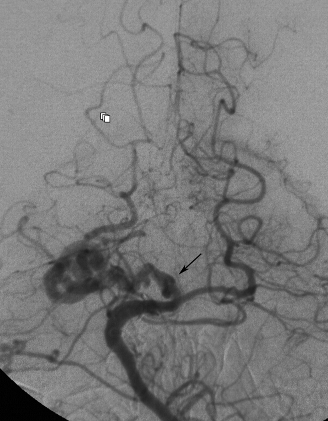
A 48-year-old right-handed female patient with a right aortic arch and history of tetralogy of Fallot repair as a child presented with recent episodes of severe headaches that resolved and episodes of left-sided numbness. On examination, the patient had no neurologic deficit. MR imaging of the brain was performed, which demonstrated tortuous dilated vessels in the right perimesencephalic region and mass effect on the brain stem. Cerebral angiography demonstrated a right proximal PCA dolichoectasia of the P2 segment supplied by a patent P1 segment and posterior communicating artery (Fig 1). Other areas of milder dolichoectasia were present with mild enlargement and tortuosity involving the right intracranial internal carotid artery, basilar artery, and distal right anterior inferior cerebellar artery.

Fig 1.
  • Download figure
  • Open in new tab
  • Download powerpoint
Fig 1.

Anteroposterior left vertebral angiogram demonstrates the right PCA dolichoectasia (arrow) just distal to the P1 segment. Mild dolichoectasia of the basilar artery and distal right anterior inferior cerebellar artery is also present.

After consultation with neuroscience specialists with backgrounds in neurology, neurosurgery, and interventional neuroradiology, a diverse range of management options were considered. Those who felt observational follow-up to be appropriate felt from their experience with a limited number of patients with this disorder, that the risk of subarachnoid hemorrhage was low and that the main risk of the disease was progressive increase of mass effect. Others saw this entity to be like a saccular aneurysm, with a propensity for catastrophic rupture. These options were presented to the patient with the uncertainty of the natural history based on current medical knowledge. The patient preferred endovascular occlusion after a series of endovascular testing to confirm a low therapeutic risk.

A balloon test occlusion was performed (Hyperform balloon 4 mm × 7 mm; Micro Therapeutics, Irvine, CA) in the proximal aspect of the P2 segment followed by selective Wada testing performed by injection of 30 mg of sodium amytal in the same location. The patient developed only a left homonymous hemianopsia during the selective Wada testing. Therefore, coil occlusion was performed of the PCA dolichoectasia with three hydrogel coils (Microvention, Aliso Viejo, CA). The patient developed no complication and had no recurrence of her presenting symptoms.

Discussion

Dolichoectasia defined as dilatation and elongation, and thus tortuosity of the vessel, may be the end product of different pathologies that weaken the vessel wall. Histologic findings suggest distinct etiologic subtypes of dolichoectasia, an acquired type from atherosclerosis and a congenital type with additional deficiencies in the muscular media layer (2, 5, 10, 14, 15). A third type resulting from dissection demonstrates intramural hemorrhage (8, 9).

Although thought to be rare, the risk of subarachnoid hemorrhage with dolichoectasia specifically with the congenital type, may be higher than previously thought (7, 12, 15). In a series of 31 patients with dolichoectasia, two presented with subarachnoid hemorrhage. The type of dolichoectasia was not identified (1). In a more recent series of 11 patients with congenital dolichoectasia, three presented with subarachnoid hemorrhage (7). Furthermore, in a large series of 120 fusiform aneurysms, of which 111 were nonatherosclerotic, 20% present with subarachnoid hemorrhage. Although, some of the cases may in fact have been giant saccular aneurysms rather than dolichoectasia, some evidence suggests giant saccular aneurysms belong to the spectrum of the same disease (10, 12, 13). A dissecting aneurysm would be another possible diagnosis and may account for the high rate of subarachnoid hemorrhage in some series with relatively low rates of subarachnoid hemorrhage in actual congenital dolichoectasia (7–9). Our case did not have the appearance of dissection with intramural hemorrhage, and similar but less extensive morphological analysis in other vessels in this patient suggests a congenital origin.

The patient opted for endovascular treatment rather than follow-up. Proximal occlusion of the PCA has been performed surgically and is usually well tolerated (7, 12). Complications from occlusion of the P2 segment may develop secondary to occlusion of a perforator or from distal hemodynamic failure. Thus, double testing was performed; a balloon test occlusion of P2 at the origin of the dolichoectasia demonstrated excellent collateral from the middle cerebral artery pial anastomosis. Also, the amytal test, performed by injection of amytal at the origin of the dolichoectasia, resulted in hemianopsia but no other motor, sensory or memory deficit, which suggests that no perforator arose from the dolichoectasia.

Conclusion

Congenital dolichoectasia, a rare cerebrovascular disorder, has an uncertain natural history and experience is limited. Parent artery occlusion may offer effective low risk treatment if test occlusion is carefully performed.

References

  1. ↵
    Yu YL, Moseley IF, Pullicino P, McDonald WI. The clinical picture of ectasia of the intracerebral arteries. J Neurol Neurosurg Psychiatry 1982;45:29–36
    Abstract/FREE Full Text
  2. ↵
    Nijensohn DE, Saez RJ, Reagan TJ. Clinical significance of basilar artery aneurysms. Neurology 1974;24:301–305
    Abstract/FREE Full Text
  3. ↵
    Dziewas R, Freund M, Ludemann P, et al. Treatment options in vertebrobasilar dolichoectasia: case report and review of the literature. Eur Neurol 2003;49:245–247
    CrossRefPubMed
  4. ↵
    Resta M, Gentile A, Cuonzo FD, et al. Clinical-angiographic correlations in 132 patients with megadolichovertebrobasilar anomaly. Neuroradiology 1984;26:213–216
    CrossRefPubMed
  5. ↵
    Araki Y, Mori S, Kanoh M, et al. Congenital hemicerebral arterial ectasia complicating unilateral megalencephaly. Br J Radiol 1987;60:395–400
    Abstract/FREE Full Text
  6. ↵
    Doran SE, Deveikis JP, Chandler WF. Dolichoectasia of the anterior cerebral arteries in an adolescent. AJNR Am J Neuroradiol 1995;16:1548–1550
    Abstract
  7. ↵
    Findlay JM, Hao C, Emery D. Non-atherosclerotic fusiform cerebral aneurysms. Can J Neurol Sci 2002;29:41–48
    PubMed
  8. ↵
    Mizutani T, Yoshimasa M, Kojima H, Suzuki H. Proposed classification of nonatherosclerotic cerebral fusiform and dissecting aneurysms. Neurosurgery 1999;45:253–260
    CrossRefPubMed
  9. ↵
    Nakatomi H, Segawa H, Kurata A, et al. Clinicopathological study of intracranial fusiform and dolichoectatic aneurysms. Stroke 2000;31:896–900
    Abstract/FREE Full Text
  10. ↵
    Hegedus K. Ectasia of the basilar artery with special reference to possible pathogenesis. Surg Neurol 1985;24:463–469
    CrossRefPubMed
  11. ↵
    Moseley IF, Holland IM. Ectasia of the basilar artery: the breadth of the clinical spectrum and the diagnostic value of computed tomography. Neuroradiology 1979;18:83–91
    CrossRefPubMed
  12. ↵
    Drake CG, Peerless SJ. Giant fusiform intracranial aneurysms: review of 120 patients treated surgically from 1965 to 1992. J Neurosurg 1997;87:141–162
    CrossRefPubMed
  13. ↵
    Anson JA, Lawton MT, Spetzler RF. Characteristics and surgical treatment of dolichoectatic and fusiform aneurysms. J Neurosurg 1996;84:185–191
    PubMed
  14. ↵
    Sacks JG, Lindenburg L. Dolicho-ectatic intracranial arteries: symptomatology and pathogenesis of arterial elongation and distention. Johns Hopkins Med J 1969;125:95–106
    PubMed
  15. ↵
    Ferry PC, Kerber C, Peterson D, Gallo AA. Arteriectasis, subarachnoid hemorrhage in a three-month-old infant. Neurology 1974;24:494–500
    Abstract/FREE Full Text
  • Received January 30, 2004.
  • Accepted after revision March 7, 2004.
  • Copyright © American Society of Neuroradiology
View Abstract
PreviousNext
Back to top

In this issue

American Journal of Neuroradiology: 25 (10)
American Journal of Neuroradiology
Vol. 25, Issue 10
1 Nov 2004
  • Table of Contents
  • Index by author
Advertisement
Print
Download PDF
Email Article

Thank you for your interest in spreading the word on American Journal of Neuroradiology.

NOTE: We only request your email address so that the person you are recommending the page to knows that you wanted them to see it, and that it is not junk mail. We do not capture any email address.

Enter multiple addresses on separate lines or separate them with commas.
Endovascular Management of Dolichoectasia of the Posterior Cerebral Artery Report
(Your Name) has sent you a message from American Journal of Neuroradiology
(Your Name) thought you would like to see the American Journal of Neuroradiology web site.
CAPTCHA
This question is for testing whether or not you are a human visitor and to prevent automated spam submissions.
Cite this article
Kuo H. Chao, Howard A. Riina, Linda Heier, Philip E. Steig, Y. Pierre Gobin
Endovascular Management of Dolichoectasia of the Posterior Cerebral Artery Report
American Journal of Neuroradiology Nov 2004, 25 (10) 1790-1791;

Citation Manager Formats

  • BibTeX
  • Bookends
  • EasyBib
  • EndNote (tagged)
  • EndNote 8 (xml)
  • Medlars
  • Mendeley
  • Papers
  • RefWorks Tagged
  • Ref Manager
  • RIS
  • Zotero
0 Responses
Respond to this article
Share
Bookmark this article
Endovascular Management of Dolichoectasia of the Posterior Cerebral Artery Report
Kuo H. Chao, Howard A. Riina, Linda Heier, Philip E. Steig, Y. Pierre Gobin
American Journal of Neuroradiology Nov 2004, 25 (10) 1790-1791;
del.icio.us logo Twitter logo Facebook logo Mendeley logo
  • Tweet Widget
  • Facebook Like
  • Google Plus One

Jump to section

  • Article
    • Abstract
    • Case Report
    • Discussion
    • Conclusion
    • References
  • Figures & Data
  • Info & Metrics
  • Responses
  • References
  • PDF

Related Articles

  • No related articles found.
  • PubMed
  • Google Scholar

Cited By...

  • Endoluminal Reconstruction for Nonsaccular Aneurysms of the Proximal Posterior Cerebral Artery with the Pipeline Embolization Device
  • Endovascular Reconstruction for Treatment of Vertebrobasilar Dolichoectasia: Long-Term Outcomes
  • Crossref
  • Google Scholar

This article has not yet been cited by articles in journals that are participating in Crossref Cited-by Linking.

More in this TOC Section

  • SAVE vs. Solumbra Techniques for Thrombectomy
  • CT Perfusion&Reperfusion in Acute Ischemic Stroke
  • Delayed Reperfusion Post-Thrombectomy&Thrombolysis
Show more Interventional

Similar Articles

Advertisement

Indexed Content

  • Current Issue
  • Accepted Manuscripts
  • Article Preview
  • Past Issues
  • Editorials
  • Editors Choice
  • Fellow Journal Club
  • Letters to the Editor

Cases

  • Case Collection
  • Archive - Case of the Week
  • Archive - Case of the Month
  • Archive - Classic Case

Special Collections

  • Special Collections

Resources

  • News and Updates
  • Turn around Times
  • Submit a Manuscript
  • Author Policies
  • Manuscript Submission Guidelines
  • Evidence-Based Medicine Level Guide
  • Publishing Checklists
  • Graphical Abstract Preparation
  • Imaging Protocol Submission
  • Submit a Case
  • Become a Reviewer/Academy of Reviewers
  • Get Peer Review Credit from Publons

Multimedia

  • AJNR Podcast
  • AJNR SCANtastic
  • Video Articles

About Us

  • About AJNR
  • Editorial Board
  • Not an AJNR Subscriber? Join Now
  • Alerts
  • Feedback
  • Advertise with us
  • Librarian Resources
  • Permissions
  • Terms and Conditions

American Society of Neuroradiology

  • Not an ASNR Member? Join Now

© 2025 by the American Society of Neuroradiology All rights, including for text and data mining, AI training, and similar technologies, are reserved.
Print ISSN: 0195-6108 Online ISSN: 1936-959X

Powered by HighWire