Advertisement

Main menu

  • Home
  • Content
    • Current Issue
    • Accepted Manuscripts
    • Article Preview
    • Past Issue Archive
    • AJNR Case Collection
    • Case of the Week Archive
    • Classic Case Archive
    • Case of the Month Archive
  • Special Collections
    • Spinal CSF Leak Articles (Jan 2020-June 2024)
    • 2024 AJNR Journal Awards
    • Most Impactful AJNR Articles
  • Multimedia
    • AJNR Podcast
    • AJNR Scantastics
    • Video Articles
  • For Authors
    • Submit a Manuscript
    • Author Policies
    • Fast publishing of Accepted Manuscripts
    • Graphical Abstract Preparation
    • Manuscript Submission Guidelines
    • Imaging Protocol Submission
    • Submit a Case for the Case Collection
  • About Us
    • About AJNR
    • Editorial Board
  • More
    • Become a Reviewer/Academy of Reviewers
    • Subscribers
    • Permissions
    • Alerts
    • Feedback
    • Advertisers
    • ASNR Home
  • Other Publications
    • ajnr

User menu

  • Alerts
  • Log in

Search

  • Advanced search
American Journal of Neuroradiology

American Journal of Neuroradiology

ASHNR American Society of Functional Neuroradiology ASHNR American Society of Pediatric Neuroradiology ASSR
  • Alerts
  • Log in

Advanced Search

  • Home
  • Content
    • Current Issue
    • Accepted Manuscripts
    • Article Preview
    • Past Issue Archive
    • AJNR Case Collection
    • Case of the Week Archive
    • Classic Case Archive
    • Case of the Month Archive
  • Special Collections
    • Spinal CSF Leak Articles (Jan 2020-June 2024)
    • 2024 AJNR Journal Awards
    • Most Impactful AJNR Articles
  • Multimedia
    • AJNR Podcast
    • AJNR Scantastics
    • Video Articles
  • For Authors
    • Submit a Manuscript
    • Author Policies
    • Fast publishing of Accepted Manuscripts
    • Graphical Abstract Preparation
    • Manuscript Submission Guidelines
    • Imaging Protocol Submission
    • Submit a Case for the Case Collection
  • About Us
    • About AJNR
    • Editorial Board
  • More
    • Become a Reviewer/Academy of Reviewers
    • Subscribers
    • Permissions
    • Alerts
    • Feedback
    • Advertisers
    • ASNR Home
  • Follow AJNR on Twitter
  • Visit AJNR on Facebook
  • Follow AJNR on Instagram
  • Join AJNR on LinkedIn
  • RSS Feeds

Welcome to the new AJNR, Updated Hall of Fame, and more. Read the full announcements.


AJNR is seeking candidates for the position of Associate Section Editor, AJNR Case Collection. Read the full announcement.


OtherBRAIN

Distinguishing between Anterior Cerebral Artery and Middle Cerebral Artery Perfusion by Color-Coded Perfusion Direction Mapping with Arterial Spin Labeling

Toshiaki Taoka, Satoru Iwasaki, Hiroyuki Nakagawa, Akio Fukusumi, Shinji Hirohashi, Masahiko Sakamoto, Kimihiko Kichikawa and Katsutoshi Murata
American Journal of Neuroradiology February 2004, 25 (2) 248-251;
Toshiaki Taoka
  • Find this author on Google Scholar
  • Find this author on PubMed
  • Search for this author on this site
Satoru Iwasaki
  • Find this author on Google Scholar
  • Find this author on PubMed
  • Search for this author on this site
Hiroyuki Nakagawa
  • Find this author on Google Scholar
  • Find this author on PubMed
  • Search for this author on this site
Akio Fukusumi
  • Find this author on Google Scholar
  • Find this author on PubMed
  • Search for this author on this site
Shinji Hirohashi
  • Find this author on Google Scholar
  • Find this author on PubMed
  • Search for this author on this site
Masahiko Sakamoto
  • Find this author on Google Scholar
  • Find this author on PubMed
  • Search for this author on this site
Kimihiko Kichikawa
  • Find this author on Google Scholar
  • Find this author on PubMed
  • Search for this author on this site
Katsutoshi Murata
  • Find this author on Google Scholar
  • Find this author on PubMed
  • Search for this author on this site
  • Article
  • Figures & Data
  • Info & Metrics
  • Responses
  • References
  • PDF
Loading

Article Figures & Data

Figures

  • Fig 1.
    • Download figure
    • Open in new tab
    • Download powerpoint
    Fig 1.

    Imaging plane for arterial spin labeling method. We used three imaging planes: plane A, sagittal plane; plane B, 10-degree laterally tilted sagittal plane; plane C, 20-degree laterally tilted sagittal plane. All three imaging planes included the internal carotid artery top. ACA is located on the medial side of the imaging plane, and MCA is located on the lateral side. In the frontal lobe, plane A includes the superior frontal gyrus and plane C includes the middle frontal gyrus. Plane B is located around the superior frontal sulcus in most cases.

  • Fig 2.
    • Download figure
    • Open in new tab
    • Download powerpoint
    Fig 2.

    Images of a 50-year-old man with right internal carotid artery-posterior communicating artery aneurysm. Area with perfusion from the medial side is displayed by orange gradation, and area with perfusion from the lateral side is displayed by blue gradation. Three imaging planes were set up.

    A, Plane A, sagittal plane, including internal carotid artery top. On plane A, the orange area (indicating perfusion from the ACA) is dominant (ratio of ACA area, 82%).

    B, Plane B, 10-degree laterally tilted sagittal plane, including internal carotid artery top. On plane B, the blue area (indicating perfusion from the MCA) is becoming dominant (ratio of ACA area, 54%).

    C, Plane C, 20-degree laterally tilted sagittal plane, including internal carotid artery top. On plane C, which is located near the superior frontal sulcus, the blue area is dominant (ratio of ACA area, 44%). Color scale shows P values.

    D−F, Corresponding half Fourier single shot turbo spin-echo images are shown.

    G, Digital subtration angiogram shows watershed (arrow) located in the middle of the middle frontal gyrus.
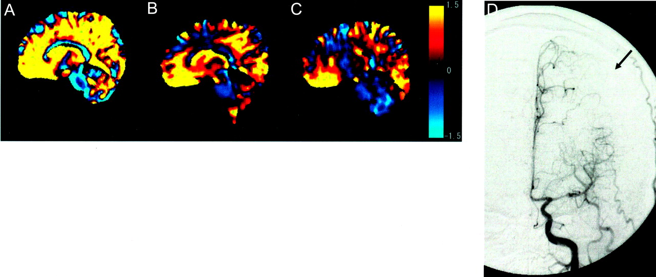
  • Fig 3.
    • Download figure
    • Open in new tab
    • Download powerpoint
    Fig 3.

    Images of a 45-year-old man with MCA stenosis at the horizontal part.

    A, On plane A, area with ACA perfusion is 87%, which is similar to the ratio of the patient whose images are shown in Figure 2.

    B and C, Tilted plane, however, shows that area with ACA perfusion did not diminish as in participants without MCA stenosis (70% on plane B and 69% on plane C). Color scale shows P values.

    D, Digital subtraction angiogram shows that territory of ACA has spread into the lateral part of middle frontal gyrus, indicating lateral shift of watershed area (arrow).
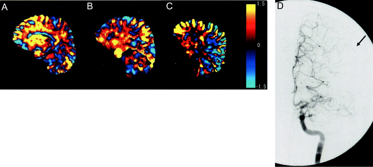
  • Fig 4.
    • Download figure
    • Open in new tab
    • Download powerpoint
    Fig 4.

    Images of a 70-year-old man with MCA occlusion at horizontal part. Digital subtraction angiogram (D) shows that territory of ACA has spread wide into the lateral area (arrow). Area with ACA perfusion is larger in the tilted plane than in the images shown in Figures 2 and 3 (86% on plane A, 78% on plane B, and 79% on plane C). Color scale shows t values.

  • Fig 5.
    • Download figure
    • Open in new tab
    • Download powerpoint
    Fig 5.

    Percentage of area with ACA perfusion in participants with intact MCAs and with MCA stenosis or occlusion. In participants with intact MCAs, as the imaging plane shifts to the lateral side, the areas with ACA perfusion become smaller. In participants with MCA stenosis or occlusion, the area with ACA perfusion is larger than in participants with intact MCAs.

PreviousNext
Back to top

In this issue

American Journal of Neuroradiology: 25 (2)
American Journal of Neuroradiology
Vol. 25, Issue 2
1 Feb 2004
  • Table of Contents
  • Index by author
Advertisement
Print
Download PDF
Email Article

Thank you for your interest in spreading the word on American Journal of Neuroradiology.

NOTE: We only request your email address so that the person you are recommending the page to knows that you wanted them to see it, and that it is not junk mail. We do not capture any email address.

Enter multiple addresses on separate lines or separate them with commas.
Distinguishing between Anterior Cerebral Artery and Middle Cerebral Artery Perfusion by Color-Coded Perfusion Direction Mapping with Arterial Spin Labeling
(Your Name) has sent you a message from American Journal of Neuroradiology
(Your Name) thought you would like to see the American Journal of Neuroradiology web site.
CAPTCHA
This question is for testing whether or not you are a human visitor and to prevent automated spam submissions.
Cite this article
Toshiaki Taoka, Satoru Iwasaki, Hiroyuki Nakagawa, Akio Fukusumi, Shinji Hirohashi, Masahiko Sakamoto, Kimihiko Kichikawa, Katsutoshi Murata
Distinguishing between Anterior Cerebral Artery and Middle Cerebral Artery Perfusion by Color-Coded Perfusion Direction Mapping with Arterial Spin Labeling
American Journal of Neuroradiology Feb 2004, 25 (2) 248-251;

Citation Manager Formats

  • BibTeX
  • Bookends
  • EasyBib
  • EndNote (tagged)
  • EndNote 8 (xml)
  • Medlars
  • Mendeley
  • Papers
  • RefWorks Tagged
  • Ref Manager
  • RIS
  • Zotero
0 Responses
Respond to this article
Share
Bookmark this article
Distinguishing between Anterior Cerebral Artery and Middle Cerebral Artery Perfusion by Color-Coded Perfusion Direction Mapping with Arterial Spin Labeling
Toshiaki Taoka, Satoru Iwasaki, Hiroyuki Nakagawa, Akio Fukusumi, Shinji Hirohashi, Masahiko Sakamoto, Kimihiko Kichikawa, Katsutoshi Murata
American Journal of Neuroradiology Feb 2004, 25 (2) 248-251;
del.icio.us logo Twitter logo Facebook logo Mendeley logo
  • Tweet Widget
  • Facebook Like
  • Google Plus One

Jump to section

  • Article
    • Abstract
    • Methods
    • Results
    • Discussion
    • Conclusion
    • References
  • Figures & Data
  • Info & Metrics
  • Responses
  • References
  • PDF

Related Articles

  • No related articles found.
  • PubMed
  • Google Scholar

Cited By...

  • Assessment of the Contribution of the External Carotid Artery to Brain Perfusion in Patients With Internal Carotid Artery Occlusion
  • Crossref
  • Google Scholar

This article has not yet been cited by articles in journals that are participating in Crossref Cited-by Linking.

More in this TOC Section

  • Multimodal CT Provides Improved Performance for Lacunar Infarct Detection
  • Statin Therapy Does Not Affect the Radiographic and Clinical Profile of Patients with TIA and Minor Stroke
  • Optimal MRI Sequence for Identifying Occlusion Location in Acute Stroke: Which Value of Time-Resolved Contrast-Enhanced MRA?
Show more Brain

Similar Articles

Advertisement

Indexed Content

  • Current Issue
  • Accepted Manuscripts
  • Article Preview
  • Past Issues
  • Editorials
  • Editors Choice
  • Fellow Journal Club
  • Letters to the Editor

Cases

  • Case Collection
  • Archive - Case of the Week
  • Archive - Case of the Month
  • Archive - Classic Case

Special Collections

  • Special Collections

Resources

  • News and Updates
  • Turn around Times
  • Submit a Manuscript
  • Author Policies
  • Manuscript Submission Guidelines
  • Evidence-Based Medicine Level Guide
  • Publishing Checklists
  • Graphical Abstract Preparation
  • Imaging Protocol Submission
  • Submit a Case
  • Become a Reviewer/Academy of Reviewers
  • Get Peer Review Credit from Publons

Multimedia

  • AJNR Podcast
  • AJNR SCANtastic
  • Video Articles

About Us

  • About AJNR
  • Editorial Board
  • Not an AJNR Subscriber? Join Now
  • Alerts
  • Feedback
  • Advertise with us
  • Librarian Resources
  • Permissions
  • Terms and Conditions

American Society of Neuroradiology

  • Not an ASNR Member? Join Now

© 2025 by the American Society of Neuroradiology All rights, including for text and data mining, AI training, and similar technologies, are reserved.
Print ISSN: 0195-6108 Online ISSN: 1936-959X

Powered by HighWire