Advertisement

Main menu

  • Home
  • Content
    • Current Issue
    • Accepted Manuscripts
    • Article Preview
    • Past Issue Archive
    • AJNR Case Collection
    • Case of the Week Archive
    • Classic Case Archive
    • Case of the Month Archive
  • Special Collections
    • Spinal CSF Leak Articles (Jan 2020-June 2024)
    • 2024 AJNR Journal Awards
    • Most Impactful AJNR Articles
  • Multimedia
    • AJNR Podcast
    • AJNR Scantastics
    • Video Articles
  • For Authors
    • Submit a Manuscript
    • Author Policies
    • Fast publishing of Accepted Manuscripts
    • Graphical Abstract Preparation
    • Manuscript Submission Guidelines
    • Imaging Protocol Submission
    • Submit a Case for the Case Collection
  • About Us
    • About AJNR
    • Editorial Board
  • More
    • Become a Reviewer/Academy of Reviewers
    • Subscribers
    • Permissions
    • Alerts
    • Feedback
    • Advertisers
    • ASNR Home
  • Other Publications
    • ajnr

User menu

  • Alerts
  • Log in

Search

  • Advanced search
American Journal of Neuroradiology

American Journal of Neuroradiology

ASHNR American Society of Functional Neuroradiology ASHNR American Society of Pediatric Neuroradiology ASSR
  • Alerts
  • Log in

Advanced Search

  • Home
  • Content
    • Current Issue
    • Accepted Manuscripts
    • Article Preview
    • Past Issue Archive
    • AJNR Case Collection
    • Case of the Week Archive
    • Classic Case Archive
    • Case of the Month Archive
  • Special Collections
    • Spinal CSF Leak Articles (Jan 2020-June 2024)
    • 2024 AJNR Journal Awards
    • Most Impactful AJNR Articles
  • Multimedia
    • AJNR Podcast
    • AJNR Scantastics
    • Video Articles
  • For Authors
    • Submit a Manuscript
    • Author Policies
    • Fast publishing of Accepted Manuscripts
    • Graphical Abstract Preparation
    • Manuscript Submission Guidelines
    • Imaging Protocol Submission
    • Submit a Case for the Case Collection
  • About Us
    • About AJNR
    • Editorial Board
  • More
    • Become a Reviewer/Academy of Reviewers
    • Subscribers
    • Permissions
    • Alerts
    • Feedback
    • Advertisers
    • ASNR Home
  • Follow AJNR on Twitter
  • Visit AJNR on Facebook
  • Follow AJNR on Instagram
  • Join AJNR on LinkedIn
  • RSS Feeds

Welcome to the new AJNR, Updated Hall of Fame, and more. Read the full announcements.


AJNR is seeking candidates for the position of Associate Section Editor, AJNR Case Collection. Read the full announcement.


OtherBRAIN

Double Origin of the Posterior Inferior Cerebellar Artery

Walter S. Lesley and Henry J. Dalsania
American Journal of Neuroradiology March 2004, 25 (3) 425-427;
Walter S. Lesley
  • Find this author on Google Scholar
  • Find this author on PubMed
  • Search for this author on this site
Henry J. Dalsania
  • Find this author on Google Scholar
  • Find this author on PubMed
  • Search for this author on this site
  • Article
  • Figures & Data
  • Info & Metrics
  • Responses
  • References
  • PDF
Loading

Abstract

Summary: Double origin of the posterior inferior cerebellar artery (PICA) has rarely been demonstrated by angiography in the peer-reviewed literature. Of the three previous reports, this PICA variant arose from the left vertebral artery. We report a right-sided, double-origin PICA.

Developmental anomalies of the intracranial arteries are not infrequently encountered during angiography. Fenestrated and duplicated arteries comprise a large component of such variations, and of these, most occur in the vertebrobasilar circulation. Although the posterior inferior cerebellar artery (PICA) commonly has a variable course, caliber, length, and target territory, fenestrations and bifid origins are exceedingly rare. In the peer-reviewed literature, three double origin PICA cases have been documented by angiography and all arose from the left vertebral artery (VA) (1, 2). This report describes a right-sided double origin PICA.

Case Report

A 66-year-old right-handed Caucasian man with emphysema and a medical history of paraplegia secondary to remote motor vehicle accident presented with new onset seizures. Physical examination revealed an awake but intermittently attentive elderly gentleman. Neurologic evaluation revealed left blepharospasm, upper extremity rigidity greater on the right side, bilateral lower extremity weakness, and generalized absent deep tendon reflexes. Left-sided pulmonary crackles were heard on auscultation. Blood cultures and lumbar puncture CSF evaluation were negative. Diffuse periventricular leukoencephalopathy was shown by cranial CT. Mediastinal lymphadenopathy and a 2-cm right lung mass were also discovered with the use of CT. Cerebral MR angiography showed major vessel patency, but the peripheral cerebral vasculature detail was limited. As a result, cerebral conventional angiography was performed to exclude vasculitis as a cause of seizures.

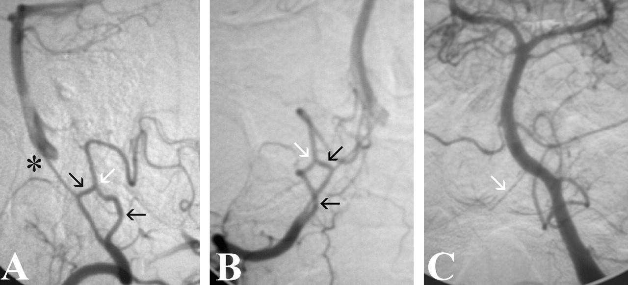
Standard biplane digital subtraction angiography of the bilateral VA and carotid arteries showed a dominant left VA with faint reflux into the distal aspect of a hypoplastic right VA (Fig 1C). The right PICA was clearly identified as a double origin variant in that both the distal and proximal PICA component vessels originated from the right V4 segment and fused at the midportion of the anterior medullary PICA segment (Fig 1A and B). With the exception of cervical carotid atherosclerosis, the remaining angiographic findings were normal. Before a complete workup could be performed, the patient left the hospital against medical advice and was subsequently lost to follow-up.

Fig 1.
  • Download figure
  • Open in new tab
  • Download powerpoint
Fig 1.

Double origin of the right PICA. Angiograms of the right VA show two separate and distinct PICA vessels (black arrows) arising from the V4 segment. They unite at the midportion of the anterior medullary segment of the PICA (white arrows). The vertebrobasilar junction (asterisk) is opacified at the convergence of the hypoplastic right and the dominant left V5 segments.

A, Lateral projection.

B, Frontal projection

C, For comparison, the right V5 segment (arrow) is seen filling retrograde on the frontal projection angiogram of the left VA. The left PICA, basilar, and bilateral anterior inferior cerebellar arteries are normal.

Discussion

Developmental variations of the cerebral vasculature are familiar findings in the angiography suite. The most recognizable oddities include the fenestrated, duplicated, and anomalous origin arteries. The double origin PICA might appear to represent, at least initially, a hybrid of all three.

Fenestration and duplication are terms that are frequently but mistakenly used interchangeably in the literature (3). Fenestration or “partial duplication” is defined as a division of a vessel lumen, resulting in two distinct endothelium-lined channels that may or may not share an adventitial layer (4, 5). Duplication, on the other hand, requires two distinct arteries with separate origins and no distal arterial convergence. The incidence rate of fenestration is, in general, greater at postmortem examination than at angiographic examination (4, 5). Nevertheless, based on autopsy and angiography series, cerebral arterial fenestration occurs more commonly in the vertebral and basilar arteries than in the middle cerebral, anterior cerebral, and internal carotid arteries (5, 6). The incidence of basilar artery fenestration ranges from 0.04% to 6% (5–7), whereas VA fenestrations are found in 0.3% to 2% of the population (6, 8). Of interest, only one instance of PICA fenestration has been reported (9). In this unique case, the fenestration was located on the anterior medullary segment of the PICA in a patient with nonaneurysmal subarachnoid hemorrhage.

Anomalous and duplicated intracranial arteries are frequently revealed by angiography. These variants, including the duplicated or anomalous posterior cerebral arterial origin of the superior cerebellar artery, present daily in the catheter laboratory. On the contrary, other vascular curiosities such as the duplicated basilar artery are exceedingly rare (10).

The double origin PICA represents a persistent anastomosis of the normal lateral spinal artery (1). Originating lateral to the medulla, the lateral spinal artery, which forms the caudal component of the double origin PICA, typically arises from either the PICA or the intradural VA. The rostral component of the double origin is the PICA proper, which derives embryologically from a hypertrophied radiculopial artery. The double origin PICA should not be confused with the common anterior inferior cerebellar artery-PICA configuration that Icardo et al (11) refer to as “duplication in the origin” of the PICA.

Of the four known cases, only this one illustrates the double origin PICA’s potential for right-sidedness. Taken together, an overall left-sided inclination may exist for this anomaly, which is intriguing in light of the findings of Hasegawa et al (12) who found a nearly double (9:5 ratio) left-sided preponderance in VA duplication. No explanation for such side-preference has been proposed.

Aneurysms infrequently arise from the PICA, accounting for 0.5% to 3% of all intracranial aneurysms (13, 14). However, certain congenital anomalies may predispose to aneurysmal formation. For example, Campos et al (15) reported a 35.5% prevalence of aneurysm within a fenestrated vertebrobasilar junction. Of the angiographically proved double origin PICA cases, one (25% incidence) harbored a 5-mm aneurysm near the convergence of the two proximal PICA segments (2). The preliminary evidence derived from this small cohort suggests a possible association of the double origin PICA with subsequent aneurysmal development.

Conclusion

This case report describes the angiographic documentation of an incidental but rare double origin PICA. This congenital anomaly can arise from either VA. An increased risk of associated PICA aneurysm may exist for patients with double origin PICA.

Footnotes

  • This work was presented at the 41st Annual Meeting of the American Society of Neuroradiology, Washington, DC, 2003.

References

  1. ↵
    Lasjaunias P, Vallee B, Person H, Ter Brugge K, Chiu M. The lateral spinal artery of the upper cervical spinal cord: anatomy, normal variations, and angiographic aspects. J Neurosurg 1985;63:235–241
    PubMed
  2. ↵
    Pasco A, Thouveny F, Papon X, et al. Ruptured aneurysm on a double origin of the posterior inferior cerebellar artery: a pathological entity in an anatomical variation: report of two cases and review of the literature. J Neurosurg 2002;96:127–131
  3. ↵
    Sim E, Vaccaro AR, Berzlanovich A, Thaler H, Ullrich CG. Fenestration of the extracranial vertebral artery: review of the literature. Spine 2001;26:E139–E142
    PubMed
  4. ↵
    Hacein-Bey L, Muszynski CA, Varelas PN. Saccular aneurysm associated with posterior cerebral artery fenestration manifesting as a subarachnoid hemorrhage in a child. AJNR Am J Neuroradiol 2002;23:1291–1294
    Abstract/FREE Full Text
  5. ↵
    Sanders WP, Sorek PA, Mehta BA. Fenestration of intracranial arteries with special attention to associated aneurysms and other anomalies. AJNR Am J Neuroradiol 1993;14:675–680
    Abstract/FREE Full Text
  6. ↵
    San-Galli F, Leman C, Kien P, Khazaal J, Phillips SD, Guerin J. Cerebral arterial fenestrations associated with intracranial saccular aneurysms. Neurosurgery 1992;30:279–283
    PubMed
  7. ↵
    Graves VB, Strother CM, Weir B, Duff TA. Vertebrobasilar junction aneurysms associated with fenestration: treatment with Guglielmi detachable coils. AJNR Am J Neuroradiol 1996;17:35–40
    Abstract
  8. ↵
    Yoshimoto H, Maeda H, Aoyama H, Kanazawa J, Kitaoka T, Uozumi T. Enlargement of cerebellar arteriovenous malformation associated with fenestration of the vertebral artery: case report. Neurol Med Chir (Tokyo) 1992;32(8):585–588
  9. ↵
    Theodosopoulos PV, Lawton MT. Fenestration of the posteroinferior cerebellar artery: case report. Neurosurgery 2000;47:463–465
    CrossRefPubMed
  10. ↵
    Goldstein JH, Woodcock R, Do HM, Phillips CD, Dion JE. Complete duplication or extreme fenestration of the basilar artery. AJNR Am J Neuroradiol 1999;20:149–150
    Abstract/FREE Full Text
  11. ↵
    Icardo JM, Ojeda JL, Garcia-Porrero JA, Hurle JM. The cerebellar arteries: cortical patterns and vascularization of the cerebellar nuclei. Acta Anat (Basel) 1982;113:108–116
    PubMed
  12. ↵
    Hasegawa T, Ito H, Hwang WZ, Yamamoto S. Single extracranial-intracranial duplication of the vertebral artery. Surg Neurol 1986;25:369–372
    CrossRefPubMed
  13. ↵
    Arai K, Endo S, Hirashima Y, Takaku A. Posterior inferior cerebellar artery aneurysm associated with fenestration of the vertebral artery: case report. Neurol Med Chir (Tokyo) 1989;29:29–31
    PubMed
  14. ↵
    Nishizaki T, Tamaki N, Nishida Y, Fujita K, Matsumoto S. Aneurysms of the distal posterior inferior cerebellar artery: experience with three cases and review of the literature. Neurosurgery 1985;16:829–832
    PubMed
  15. ↵
    Campos J, Fox AJ, Viñuela F, et al. Saccular aneurysms in basilar artery fenestration. AJNR Am J Neuroradiol 1987;8:233–236
    Abstract/FREE Full Text
  • Received May 28, 2003.
  • Accepted after revision June 8, 2003.
  • Copyright © American Society of Neuroradiology
View Abstract
PreviousNext
Back to top

In this issue

American Journal of Neuroradiology: 25 (3)
American Journal of Neuroradiology
Vol. 25, Issue 3
1 Mar 2004
  • Table of Contents
  • Index by author
Advertisement
Print
Download PDF
Email Article

Thank you for your interest in spreading the word on American Journal of Neuroradiology.

NOTE: We only request your email address so that the person you are recommending the page to knows that you wanted them to see it, and that it is not junk mail. We do not capture any email address.

Enter multiple addresses on separate lines or separate them with commas.
Double Origin of the Posterior Inferior Cerebellar Artery
(Your Name) has sent you a message from American Journal of Neuroradiology
(Your Name) thought you would like to see the American Journal of Neuroradiology web site.
CAPTCHA
This question is for testing whether or not you are a human visitor and to prevent automated spam submissions.
Cite this article
Walter S. Lesley, Henry J. Dalsania
Double Origin of the Posterior Inferior Cerebellar Artery
American Journal of Neuroradiology Mar 2004, 25 (3) 425-427;

Citation Manager Formats

  • BibTeX
  • Bookends
  • EasyBib
  • EndNote (tagged)
  • EndNote 8 (xml)
  • Medlars
  • Mendeley
  • Papers
  • RefWorks Tagged
  • Ref Manager
  • RIS
  • Zotero
0 Responses
Respond to this article
Share
Bookmark this article
Double Origin of the Posterior Inferior Cerebellar Artery
Walter S. Lesley, Henry J. Dalsania
American Journal of Neuroradiology Mar 2004, 25 (3) 425-427;
del.icio.us logo Twitter logo Facebook logo Mendeley logo
  • Tweet Widget
  • Facebook Like
  • Google Plus One

Jump to section

  • Article
    • Abstract
    • Case Report
    • Discussion
    • Conclusion
    • Footnotes
    • References
  • Figures & Data
  • Info & Metrics
  • Responses
  • References
  • PDF

Related Articles

  • No related articles found.
  • PubMed
  • Google Scholar

Cited By...

  • No citing articles found.
  • Crossref
  • Google Scholar

This article has not yet been cited by articles in journals that are participating in Crossref Cited-by Linking.

More in this TOC Section

  • Fast Contrast-Enhanced 4D MRA and 4D Flow MRI Using Constrained Reconstruction (HYPRFlow): Potential Applications for Brain Arteriovenous Malformations
  • Quiet PROPELLER MRI Techniques Match the Quality of Conventional PROPELLER Brain Imaging Techniques
  • Predictors of Reperfusion in Patients with Acute Ischemic Stroke
Show more Brain

Similar Articles

Advertisement

Indexed Content

  • Current Issue
  • Accepted Manuscripts
  • Article Preview
  • Past Issues
  • Editorials
  • Editors Choice
  • Fellow Journal Club
  • Letters to the Editor

Cases

  • Case Collection
  • Archive - Case of the Week
  • Archive - Case of the Month
  • Archive - Classic Case

Special Collections

  • Special Collections

Resources

  • News and Updates
  • Turn around Times
  • Submit a Manuscript
  • Author Policies
  • Manuscript Submission Guidelines
  • Evidence-Based Medicine Level Guide
  • Publishing Checklists
  • Graphical Abstract Preparation
  • Imaging Protocol Submission
  • Submit a Case
  • Become a Reviewer/Academy of Reviewers
  • Get Peer Review Credit from Publons

Multimedia

  • AJNR Podcast
  • AJNR SCANtastic
  • Video Articles

About Us

  • About AJNR
  • Editorial Board
  • Not an AJNR Subscriber? Join Now
  • Alerts
  • Feedback
  • Advertise with us
  • Librarian Resources
  • Permissions
  • Terms and Conditions

American Society of Neuroradiology

  • Not an ASNR Member? Join Now

© 2025 by the American Society of Neuroradiology All rights, including for text and data mining, AI training, and similar technologies, are reserved.
Print ISSN: 0195-6108 Online ISSN: 1936-959X

Powered by HighWire