Advertisement

Main menu

  • Home
  • Content
    • Current Issue
    • Accepted Manuscripts
    • Article Preview
    • Past Issue Archive
    • AJNR Case Collection
    • Case of the Week Archive
    • Classic Case Archive
    • Case of the Month Archive
  • Special Collections
    • Spinal CSF Leak Articles (Jan 2020-June 2024)
    • 2024 AJNR Journal Awards
    • Most Impactful AJNR Articles
  • Multimedia
    • AJNR Podcast
    • AJNR Scantastics
    • Video Articles
  • For Authors
    • Submit a Manuscript
    • Author Policies
    • Fast publishing of Accepted Manuscripts
    • Graphical Abstract Preparation
    • Manuscript Submission Guidelines
    • Imaging Protocol Submission
    • Submit a Case for the Case Collection
  • About Us
    • About AJNR
    • Editorial Board
  • More
    • Become a Reviewer/Academy of Reviewers
    • Subscribers
    • Permissions
    • Alerts
    • Feedback
    • Advertisers
    • ASNR Home
  • Other Publications
    • ajnr

User menu

  • Alerts
  • Log in

Search

  • Advanced search
American Journal of Neuroradiology

American Journal of Neuroradiology

ASHNR American Society of Functional Neuroradiology ASHNR American Society of Pediatric Neuroradiology ASSR
  • Alerts
  • Log in

Advanced Search

  • Home
  • Content
    • Current Issue
    • Accepted Manuscripts
    • Article Preview
    • Past Issue Archive
    • AJNR Case Collection
    • Case of the Week Archive
    • Classic Case Archive
    • Case of the Month Archive
  • Special Collections
    • Spinal CSF Leak Articles (Jan 2020-June 2024)
    • 2024 AJNR Journal Awards
    • Most Impactful AJNR Articles
  • Multimedia
    • AJNR Podcast
    • AJNR Scantastics
    • Video Articles
  • For Authors
    • Submit a Manuscript
    • Author Policies
    • Fast publishing of Accepted Manuscripts
    • Graphical Abstract Preparation
    • Manuscript Submission Guidelines
    • Imaging Protocol Submission
    • Submit a Case for the Case Collection
  • About Us
    • About AJNR
    • Editorial Board
  • More
    • Become a Reviewer/Academy of Reviewers
    • Subscribers
    • Permissions
    • Alerts
    • Feedback
    • Advertisers
    • ASNR Home
  • Follow AJNR on Twitter
  • Visit AJNR on Facebook
  • Follow AJNR on Instagram
  • Join AJNR on LinkedIn
  • RSS Feeds

Welcome to the new AJNR, Updated Hall of Fame, and more. Read the full announcements.


AJNR is seeking candidates for the position of Associate Section Editor, AJNR Case Collection. Read the full announcement.


OtherBRAIN

A Small Pulmonary Arteriovenous Malformation as a Cause of Recurrent Brain Embolism

Kenichi Todo, Hiroshi Moriwaki, Masahiro Higashi, Kohji Kimura and Hiroaki Naritomi
American Journal of Neuroradiology March 2004, 25 (3) 428-430;
Kenichi Todo
  • Find this author on Google Scholar
  • Find this author on PubMed
  • Search for this author on this site
Hiroshi Moriwaki
  • Find this author on Google Scholar
  • Find this author on PubMed
  • Search for this author on this site
Masahiro Higashi
  • Find this author on Google Scholar
  • Find this author on PubMed
  • Search for this author on this site
Kohji Kimura
  • Find this author on Google Scholar
  • Find this author on PubMed
  • Search for this author on this site
Hiroaki Naritomi
  • Find this author on Google Scholar
  • Find this author on PubMed
  • Search for this author on this site
  • Article
  • Figures & Data
  • Info & Metrics
  • Responses
  • References
  • PDF
Loading

Abstract

Summary: We report a case of recurrent paradoxical brain embolism mediated through a small pulmonary arteriovenous malformation (PAVM) with a 1.8-mm-diameter feeding artery. In this case, the further recurrent stroke was prevented successfully by PAVM embolization. Although embolization therapy is currently recommended only for PAVMs with feeding arteries greater than 3 mm in diameter, the therapy may be needed also in the smaller PAVMs.

Pulmonary arteriovenous malformation (PAVM) is well recognized as a cause of paradoxical brain embolism. Brain infarction associated with PAVMs most likely occurs in patients with feeding arteries of more than 3-mm diameter and not in those of smaller size (1–5). We report the case of a PAVM patient with a 1.8-mm-diameter feeding artery who had recurrent paradoxical brain embolism and was successfully treated with embolotherapy.

Case Report

A 67-year-old right-handed woman was admitted in February 2002 after suddenly developing dizziness, nausea, and speech disturbance. She was admitted to our hospital 7 hours after the onset of symptoms. At the time of admission, her blood pressure was 140/80 mm Hg. She had a regular heart rate of 60 beats per minute and a respiratory rate of 16 breaths per minute. Neither pathologic breath sounds nor heart murmurs were auscultated. No edema was present in her extremities. No cutaneous vascular malformations were observed. She had no episodes of hemoptysis or dyspnea. In neurologic examinations, she had dysarthria and left limb ataxia. Laboratory findings showed normal blood cell counts, normal liver and renal functions, and normal serum cholesterol and blood glucose levels. Plasma protein C, protein S, and antithrombin III levels were within normal ranges. Arterial blood gas analysis showed mild hypoxemia, with PaO2 84.6 mm Hg and PaCO2 43.2 mm Hg in room air. Chest radiographic and electrocardiographic findings were normal. Carotid ultrasonography failed to detect atherosclerotic changes and showed normal blood flow velocities in the common carotid arteries and vertebral arteries. Transthoracic echocardiography failed to find any embolic sources, including valvular pathology, dilated cardiomyopathy, akinetic left ventricular segment, and intracardiac thrombus. Cranial CT demonstrated an equivocal hypoattenuated area in the left cerebellar hemisphere but no abnormality in other areas. Diffusion-weighted MR images disclosed a high-signal-intensity area in the left cerebellar hemisphere (Fig 1A). Twelve hours after the onset of stroke, intraarterial digital subtraction cerebral angiography revealed no stenotic lesion in the extra- or intracranial vertebral arteries or basilar artery. With suspicion of embolic brain infarction, anticoagulation therapy with 10,000 U/day heparin was started 18 hours after the onset of symptoms, although the embolic source could not be identified.

Fig 1.
  • Download figure
  • Open in new tab
  • Download powerpoint
Fig 1.

Diffusion-weighted MR images. A, Diffusion-weighted image on day 1 shows a high-signal-intensity area in the left cerebellar hemisphere (arrow). B, Diffusion-weighted image on day 5 shows a new ischemic lesion in the right cerebellar hemisphere (arrow).

On day 3, the patient also developed right limb ataxia. Activated partial thromboplastin time was 28.0 seconds on admission and was prolonged to 43.1 second on day 3. The second CT scan showed only a hypoattenuated area in the left cerebellar hemisphere. Diffusion-weighted imaging on day 5 confirmed the development of new ischemic lesion in the right cerebellar hemisphere (Fig 1B). On day 6, transcranial Doppler sonography with saline contrast material injection (C-TCD) was performed to examine the presence of the right-to-left shunt. The C-TCD study was performed by using a mixture of saline solution (9 mL) and air (1 mL) agitated between two 10-mL syringes connected by a three-way stopcock. The solution was injected into the left antecubital vein within 2–3 seconds. C-TCD depicted multiple high-intensity transient signals (HITS) at the basilar artery during normal respiration (Fig 2A), which suggested the presence of continuous right-to-left shunt (6). Transesophageal echocardiography with saline contrast material injection into the antecubital vein confirmed a significant entry of microbubbles into the left atrium. Transesophageal echocardiography failed to show any embolic sources, including valvular pathology, intracardiac thrombus, atrial myxoma, spontaneous echo contrast of the left atrium, and complex atheroma of the aortic arch. Holter electrocardiographic monitoring failed to find atrial fibrillation or flutter. Radioisotope venography of the lower extremities displayed a filling defect with collateral pathways in the left popliteal vein, indicating the association of deep vein thrombosis. A contrast-enhanced CT scan of the patient’s chest revealed a small nodular lesion connected with the pulmonary artery and vein branches in the left pulmonary lower lobe. Selective pulmonary angiography on day 13 demonstrated a single PAVM with a 1.8-mm-diamter feeding artery at the corresponding area (Fig 3). The diagnosis of paradoxical brain embolism mediated through the PAVM was, thus, established. Subsequently, the PAVM was obstructed by using an embolization coil (Cook, Bloomington, IN). Following the embolization therapy, C-TCD detected no more HITS (Fig 2B), and the hypoxemia was slightly improved (PaO2 92.4 mm Hg in room air). On day 22, she was discharged without neurologic deficits. C-TCD never detected HITS on day 105. MR imaging on day 126 showed only old brain infarction equivalent to that on day 5. The patient had no recurrent attacks for the following 12 months.

Fig 2.
  • Download figure
  • Open in new tab
  • Download powerpoint
Fig 2.

C-TCD findings before and after the embolization therapy. A, C-TCD shows many HITS (arrows) from the basilar artery before the embolization therapy. B, C-TCD failed to depict any HITS after the embolization therapy.

Fig 3.
  • Download figure
  • Open in new tab
  • Download powerpoint
Fig 3.

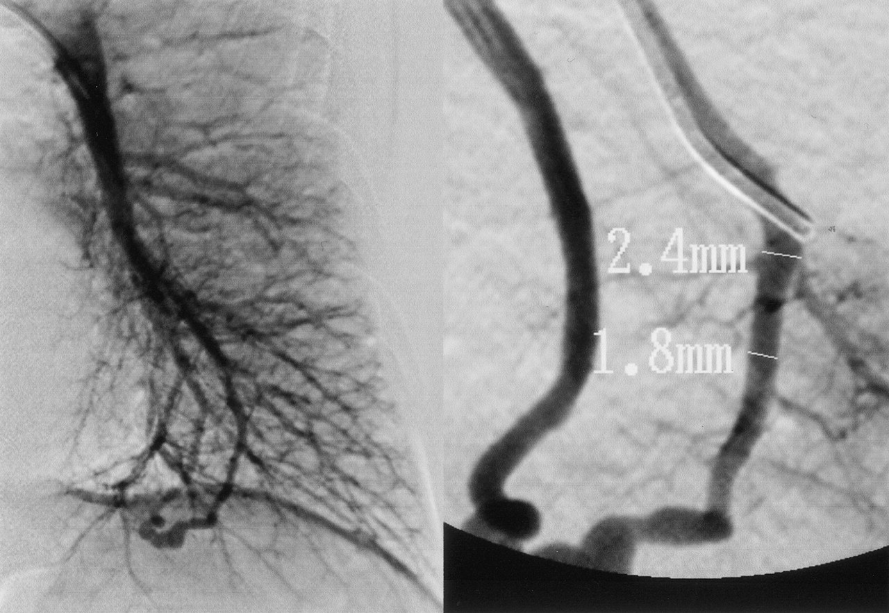
Selective pulmonary angiogram shows a single PAVM with a feeding artery diameter of 1.8 mm.

Discussion

PAVMs are commonly associated in hereditary hemorrhagic telangiectasia (HHT): approximately 15–35% of HHT patients have PAVMs, and 50–85% of PAVMs patients have HHT (1). The present patient, however, had no clinical signs suggestive of HHT. PAVMs increase the risk of paradoxical embolism because of continuous right-to-left shunt. Brain infarction is one of the most important complications that may occur in PAVM patients (1, 2). Stroke is often the first manifestation of PAVM and even of HHT (7).

Moussouttas et al (1) documented that brain infarction most likely occurs in PAVM patients with feeding arteries more than 3 mm in diameter and not in those with smaller size. Accordingly, they recommended embolization therapy for all PAVMs with feeding artery diameters exceeding 3 mm. To the best of our knowledge, the occurrence of brain infarction in PAVMs with less than 3 mm in feeding artery diameter has not been previously reported. The present patient had a small-sized PAVM, the diameter of which was 1.8 mm. In the present patient, the other embolic sources—including atrial fibrillation or flutter, bacterial endocarditis, valvular pathology, myocardial infarction, dilated cardiomyopathy, akinetic left ventricular segment, intracardiac thrombus, atrial myxoma, spontaneous echo contrast of the left atrium, or complex atheroma of the aortic arch—were ruled out, and other causes of stroke—such as atherosclerotic arterial changes, nonatherosclerotic arteriopathies, coagulopathies, hematologic or systemic disorders, or migranous infarction—were found to be negative (8, 9). The patient had deep vein thrombosis, which may cause arterial embolism under the presence of right-to-left shunt. Thus, paradoxical embolism is only one possible mechanism that can explain recurrent strokes in this patient. After the embolization therapy, this patient had no more recurrent stroke, and C-TCD never detected HITS, indicating successful obstruction of right-to-left shunt (10).

A recent study reported that the stroke recurrence rate in patients with patent foramen ovale (PFO) was unaltered regardless of whether the diameter was more than 2 mm or less (11). Paradoxically, PAVMs and PFO share the similar mechanisms in the development of brain infarction through the right-to-left shunt. The shunt flow goes through PAVMs continuously, whereas it can pass though PFO only intermittently at the time of right atrial pressure increase. In this context, PAVM is regarded to be more risky than PFO as a route of paradoxical embolism. The size of right-to-left shunt may not be a critical factor for stroke occurrence. Embolization therapy appears to be useful for preventing recurrent attacks in stroke patients with a small PAVM in whom other causes of cerebral infarction are definitely ruled out. A randomized controlled study may be necessary to elucidate the requirement of embolization therapy in small-sized PAVMs.

Footnotes

  • Supported in part by the Research Grants for Cardiovascular Diseases (14–1) from the Ministry of the Health and Welfare of Japan.

References

  1. ↵
    Moussouttas M, Fayad P, Rosenblatt M, et al. Pulmonary arteriovenous malformations: cerebral ischemia and neurologic manifestations. Neurology 2000;55:959–964
    Abstract/FREE Full Text
  2. ↵
    Shovlin CL, Letarte M. Hereditary hemorrhagic telangiectasia and pulmonary arteriovenous malformations: issues in clinical management and review of pathogenic mechanisms. Thorax 1999;54:714–729
    FREE Full Text
  3. Hughes JM. Intrapulmonary shunts: coils to transplantation. J R Coll Phys 1994;28:247–253
  4. Haitjema TJ, Overtoom TT, Westermann CJ, Lammers JW. Embolisation of pulmonary arteriovenous malformations: results and follow-up in 32 patients. Thorax 1995;50:719–723
    Abstract/FREE Full Text
  5. ↵
    White RI, Pollack JS. Pulmonary arteriovenous malformations: diagnosis by three-dimensional helical CT, a breakthrough without contrast media. Radiology 1994;191:613–614
    PubMed
  6. ↵
    Chimowitz MI, Nemec JJ, Marwick TH, et al. Transcranial Doppler ultrasound identifies patients with right-to-left cardiac or pulmonary shunts. Neurology 1991;41:1902–1904
    Abstract/FREE Full Text
  7. ↵
    Hewes RC, Auster M, White RI Jr. Cerebral embolism: first manifestation of pulmonary arteriovenous malformation in patients with hereditary hemorrhagic telangiectasia. Cardiovasc Intervent Radiol 1985;8:151–155
    PubMed
  8. ↵
    Mas JL, Arquizan C, Lamy C, et al. Recurrent cerebrovascular events associated with patent foramen ovale, atrial septal aneurysm, or both. N Engl J Med 2001;345:1740–1746
    CrossRefPubMed
  9. ↵
    Adams HP Jr, Bendixen BH, Kappelle LJ, et al. Classification of subtype of acute ischemic stroke: definitions for use in a multicenter clinical trial: TOAST: Trial of Org 10172 in Acute Stroke Treatment. Stroke 1993;24:35–41
    Abstract/FREE Full Text
  10. ↵
    Kimura K, Minematsu K, Wada K, et al. Transcranial Doppler of a paradoxical brain embolism associated with a pulmonary arteriovenous fistula. AJNR Am J Neuroradiol 1999;20:1881–1884
    Abstract/FREE Full Text
  11. ↵
    Homma S, Sacco RL, Di Tullio MR, et al. Effect of medical treatment in stroke patients with patent foramen ovale: Patent Foramen Ovale in Cryptogenic Study. Circulation 2002;105:2625–2631
    Abstract/FREE Full Text
  • Received June 5, 2003.
  • Accepted after revision June 11, 2003.
  • Copyright © American Society of Neuroradiology
View Abstract
PreviousNext
Back to top

In this issue

American Journal of Neuroradiology: 25 (3)
American Journal of Neuroradiology
Vol. 25, Issue 3
1 Mar 2004
  • Table of Contents
  • Index by author
Advertisement
Print
Download PDF
Email Article

Thank you for your interest in spreading the word on American Journal of Neuroradiology.

NOTE: We only request your email address so that the person you are recommending the page to knows that you wanted them to see it, and that it is not junk mail. We do not capture any email address.

Enter multiple addresses on separate lines or separate them with commas.
A Small Pulmonary Arteriovenous Malformation as a Cause of Recurrent Brain Embolism
(Your Name) has sent you a message from American Journal of Neuroradiology
(Your Name) thought you would like to see the American Journal of Neuroradiology web site.
CAPTCHA
This question is for testing whether or not you are a human visitor and to prevent automated spam submissions.
Cite this article
Kenichi Todo, Hiroshi Moriwaki, Masahiro Higashi, Kohji Kimura, Hiroaki Naritomi
A Small Pulmonary Arteriovenous Malformation as a Cause of Recurrent Brain Embolism
American Journal of Neuroradiology Mar 2004, 25 (3) 428-430;

Citation Manager Formats

  • BibTeX
  • Bookends
  • EasyBib
  • EndNote (tagged)
  • EndNote 8 (xml)
  • Medlars
  • Mendeley
  • Papers
  • RefWorks Tagged
  • Ref Manager
  • RIS
  • Zotero
0 Responses
Respond to this article
Share
Bookmark this article
A Small Pulmonary Arteriovenous Malformation as a Cause of Recurrent Brain Embolism
Kenichi Todo, Hiroshi Moriwaki, Masahiro Higashi, Kohji Kimura, Hiroaki Naritomi
American Journal of Neuroradiology Mar 2004, 25 (3) 428-430;
del.icio.us logo Twitter logo Facebook logo Mendeley logo
  • Tweet Widget
  • Facebook Like
  • Google Plus One

Jump to section

  • Article
    • Abstract
    • Case Report
    • Discussion
    • Footnotes
    • References
  • Figures & Data
  • Info & Metrics
  • Responses
  • References
  • PDF

Related Articles

  • No related articles found.
  • PubMed
  • Google Scholar

Cited By...

  • Personalised stroke evaluation and management: tailoring individualised patient care for hereditary haemorrhagic telangiectasia
  • Pulmonary arteriovenous malformation as a cause of ischaemic stroke in the elderly
  • Stroke resulting from an isolated pulmonary arteriovenous malformation
  • Diagnosis of Secondary Source of Right-to-Left Shunt With Balloon Occlusion of Patent Foramen Ovale and Power M-Mode Transcranial Doppler
  • Crossref
  • Google Scholar

This article has not yet been cited by articles in journals that are participating in Crossref Cited-by Linking.

More in this TOC Section

  • Fast Contrast-Enhanced 4D MRA and 4D Flow MRI Using Constrained Reconstruction (HYPRFlow): Potential Applications for Brain Arteriovenous Malformations
  • Quiet PROPELLER MRI Techniques Match the Quality of Conventional PROPELLER Brain Imaging Techniques
  • Predictors of Reperfusion in Patients with Acute Ischemic Stroke
Show more Brain

Similar Articles

Advertisement

Indexed Content

  • Current Issue
  • Accepted Manuscripts
  • Article Preview
  • Past Issues
  • Editorials
  • Editors Choice
  • Fellow Journal Club
  • Letters to the Editor

Cases

  • Case Collection
  • Archive - Case of the Week
  • Archive - Case of the Month
  • Archive - Classic Case

Special Collections

  • Special Collections

Resources

  • News and Updates
  • Turn around Times
  • Submit a Manuscript
  • Author Policies
  • Manuscript Submission Guidelines
  • Evidence-Based Medicine Level Guide
  • Publishing Checklists
  • Graphical Abstract Preparation
  • Imaging Protocol Submission
  • Submit a Case
  • Become a Reviewer/Academy of Reviewers
  • Get Peer Review Credit from Publons

Multimedia

  • AJNR Podcast
  • AJNR SCANtastic
  • Video Articles

About Us

  • About AJNR
  • Editorial Board
  • Not an AJNR Subscriber? Join Now
  • Alerts
  • Feedback
  • Advertise with us
  • Librarian Resources
  • Permissions
  • Terms and Conditions

American Society of Neuroradiology

  • Not an ASNR Member? Join Now

© 2025 by the American Society of Neuroradiology All rights, including for text and data mining, AI training, and similar technologies, are reserved.
Print ISSN: 0195-6108 Online ISSN: 1936-959X

Powered by HighWire