
Article Figures & Data
Figures
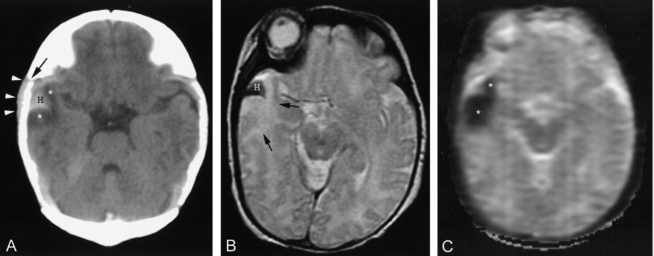
- Fig 1.
Images of a neonate (patient 4) with perinatal depression and episodes of apnea beginning immediately after birth.
A, Axial view unenhanced CT scan of the head shows parenchymal hemorrhage (H) extending to the brain surface and asymmetric soft-tissue swelling (arrowheads). A minor amount of edema is present next to the hemorrhage (*). Note proximity to the pterion (arrow).
B, Axial view T2-weighted MR image (3200/85/1; echo train length, 8) shows a low intensity hemorrhage (H) with adjacent edema (arrows).
C, ADC map (2014/103/1; b max, 750 s/mm2) shows decreased diffusion in area of edema next to the hemorrhage (*).
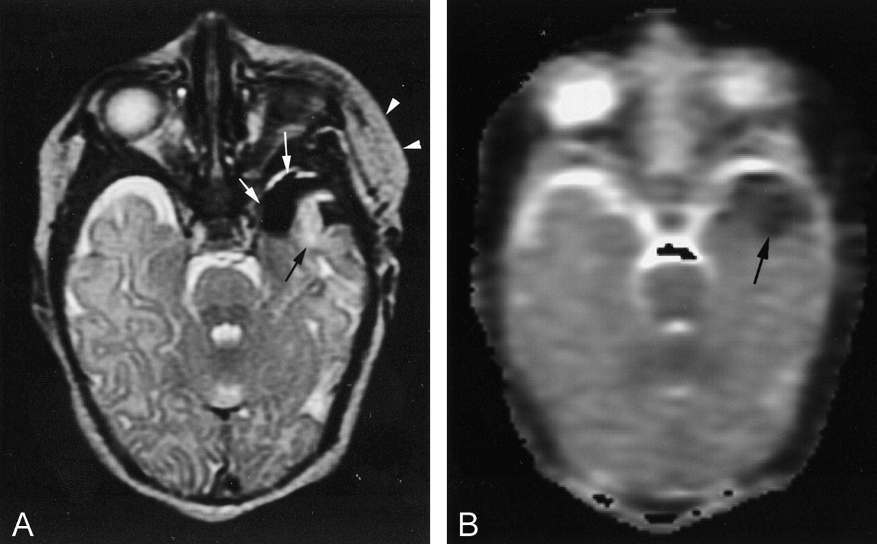
- Fig 2.
Images of a neonate (patient 3) with apnea and seizures at 24 hr of life.
A, Axial view T2-weighted MR image (3200/85/1; echo train length, 8), obtained above the level of parenchymal hemorrhage, shows soft-tissue swelling (arrowheads), subpial extension of hemorrhage (white arrows), and parenchymal edema (black arrow).
B, ADC map indicates decreased diffusion in the edematous area (arrow).
- Fig 3.
Images of a neonate (patient 5) with apnea at 8 hr of life.
A, Axial view unenhanced CT scan of the head shows leptomeningeal hemorrhage (H) under an expanded squamosal suture (arrowheads). Adjacent low attenuation edema is present (arrows).
B, Axial view T2-weighted MR image (3200/85/1; echo train length, 8) shows low intensity hemorrhage (H) and high intensity adjacent edema (arrows).
C, Decreased diffusion (arrows) is indicated on the ADC map (2014/103/1; b max, 750 s/mm2).
D, Axial view T2-weighted MR image (3200/85/1; echo train length, 8), obtained at 16 months of age, shows encephalomalacia (arrows). No underlying vascular abnormality is apparent.
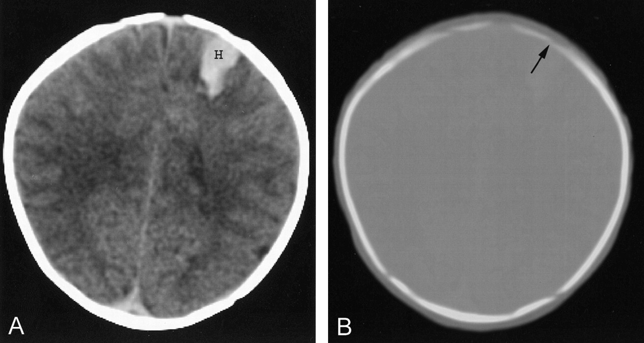
- Fig 4.
Unenhanced CT scan of a neonate (patient 7) with seizures at 36 hr of life.
A, Brain window shows a parenchymal hemorrhage (H) extending to the brain surface in close proximity to the coronal suture (arrow). Leptomeningeal hemorrhage was evident at other levels (not shown).
B, Bone window also shows a parenchymal hemorrhage (H) extending to the brain surface in close proximity to the coronal suture (arrow).
- Fig 5.
Unenhanced CT scans of a neonate (patient 6) with focal seizures at 18 hr of life.
A, CT scan shows a right parietal parenchymal hematoma (H).
B, CT scan obtained at a slightly higher anatomic section reveals additional hemorrhage (H) with extension into the sulci, indicating leptomeningeal bleeding (arrows). A large overlying cephalohematoma (arrowheads) is also present.


Tables
Patient No. Maternal Age (yr) Gestational Age (wk) Prenatal History Delivery Apgar Score at 1,5 min Weight (kg) Presentation 1 35 41 Unremarkable Vaginal, uncomplicated 9,9 4.2 Apnea and seizures at 12 hr 2 36 40 Unremarkable Vaginal, uncomplicated 8,9 4.1 Apnea at 6 hr 3 37 39 Unremarkable Vaginal, uncomplicated 9,9 3.2 Apnea and seizures at 24 hr 4 38 40 Maternal hypertension Vaginal, low forceps 2,8 3.5 Apnea at birth; perinatal depression 5 32 38 Maternal thalassemia minor Vaginal, uncomplicated 7,8 3.2 Apnea at 8 hr 6 31 40 Unremarkable Vaginal, vacuum assisted 2,7 3.0 Focal seizures at 18 hr 7 34 39 Unremarkable Vaginal, uncomplicated 6,9 3.6 Focal seizures at 36 hr Patient No. Location Adjacent Suture STS ↓ ADC* MRA MRV SDH LH 1 Inferior/anterior/lateral lt temporal lobe Pterion Yes Yes Normal NA No Yes 2 Inferior/anterior/lateral rt temporal lobe Pterion No Yes NA NA No Yes 3 Inferior/anterior/lateral lt temporal lobe Pterion Yes Yes Normal NA No Yes 4 Inferior/anterior/lateral rt temporal lobe Pterion Yes Yes NA Normal No Yes 5 Lateral lt temporal lobe Squamosal Yes Yes NA NA No Yes 6 Rt parietal lobe None Yes No NA Normal Yes Yes 7 Lt frontal lobe Coronal No No NA NA No Yes Note.—STS indicates soft tissue swelling; ADC, apparent diffusion coefficient; MRA, MR angiography; MRV, MR venography; SDH, subdural hematoma; LH, leptomeningeal hemorrhage; lt, left; rt, right; NA, not available.
* Apparent diffusion coefficient decreased in parenchyma adjacent to hemorrhage.
In this issue


Jump to section
Related Articles
Cited By...
- Imaging Patterns of Neonatal Subpial Hemorrhage: Provisional Statements on Neurologic Outcomes
- Intracranial Hemorrhage in Term and Late-Preterm Neonates: An Institutional Perspective
- An In-Depth Analysis of Brain and Spine Neuroimaging in Children with Abusive Head Trauma: Beyond the Classic Imaging Findings
- Apnea Spells in a Term Neonate
- Idiopathic Neonatal Subpial Hemorrhage with Underlying Cerebral Infarct: Imaging Features and Clinical Outcome
- Retrospective Analysis of Delayed Intraparenchymal Hemorrhage after Flow-Diverter Treatment: Presentation of a Retrospective Multicenter Trial
- Superficial Echogenic Lesions Detected on Neonatal Cranial Sonography: Possible Indicators of Severe Birth Injury
- Neonatal Seizures
- Early versus late MRI in asphyxiated newborns treated with hypothermia
- Isolated cerebral cortical tears in children: aetiology, characterisation and differentiation from non-accidental head injury
- Neonatal Intracranial Hemorrhage May Be More Frequent than Previously Suspected