
Article Figures & Data
Figures
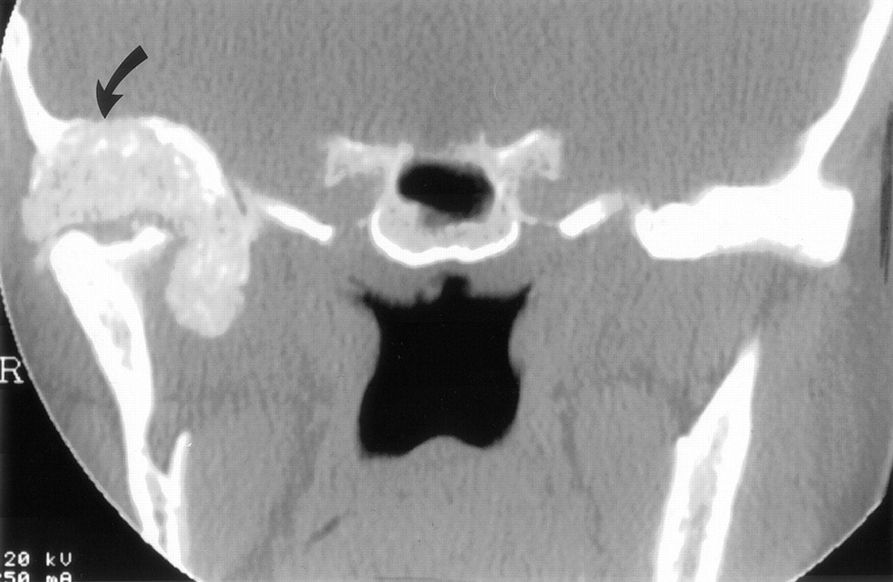
- Fig 1.
Coronal CT demonstrates a large calcified mass centered within the glenoid fossa bulging into the right middle cranial fossa (arrows). Note mass effect and remodeling of the right mandibular condyle.
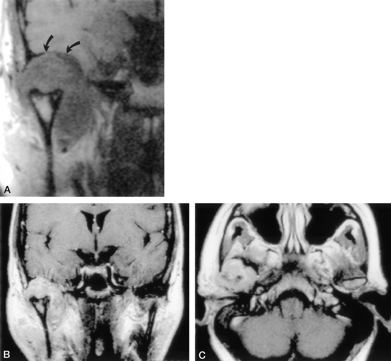
- Fig 2.
Coronal T1-weighted image (A) demonstrates a large TMJ mass of intermediate signal intensity bulging into the middle cranial fossa (arrows). Note marked widening of the joint space (TR/TE/NEX = 358/25/4). Postcontrast T1-weighted coronal (B) and axial (C) images reveal inhomogeneous enhancement of the mass (TR/TE/NEX = 550/25/1).
- Fig 3.
Needle biopsy under CT guidance. Axial section shows the central position of the needle within the lesion, which encroaches on the middle ear cavity and abuts the ossicles (arrow).
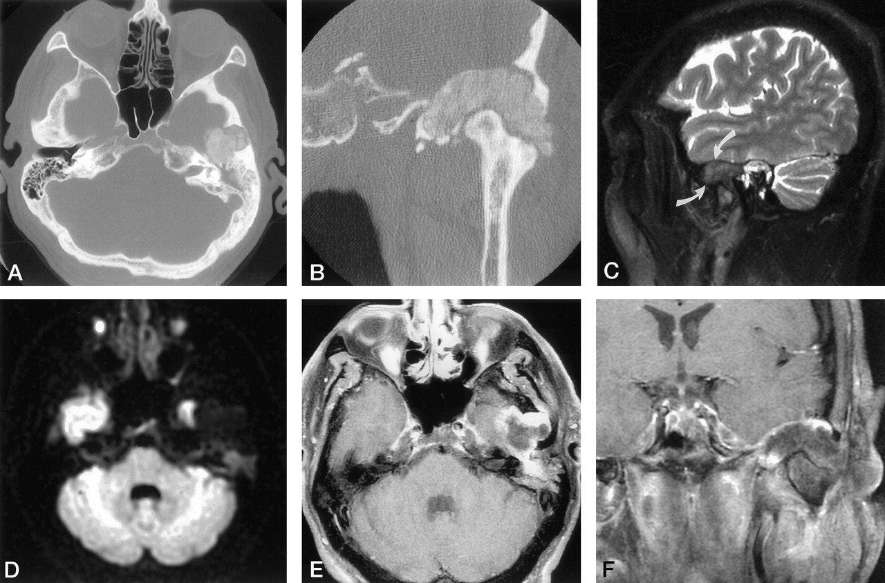
- Fig 4.
Axial (A)and coronal (B) CT images demonstrate a hyperattenuated mass eroding the roof of the glenoid fossa, abutting the Eustachian tube, and remodeling the mandibular condyle. Sagittal T2-weighted MR image (C) reveals the mass of low signal intensity (arrows) centered within the TMJ anterior to high signal intensity of fluid within the mastoid air cells (TR/TE/NEX = 3000/100/2). Diffusion-weighted image (D) demonstrates restricted diffusion. Axial (E) and coronal (F) contrast-enhanced T1-weighted images reveal a large lobulated mass eroding the squama, extending into the middle cranial fossa, with a superficial rim of enhancement (TR/TE/NEX = 600/20/2)


In this issue


Jump to section
Related Articles
Cited By...
- No citing articles found.