
Article Figures & Data
Figures
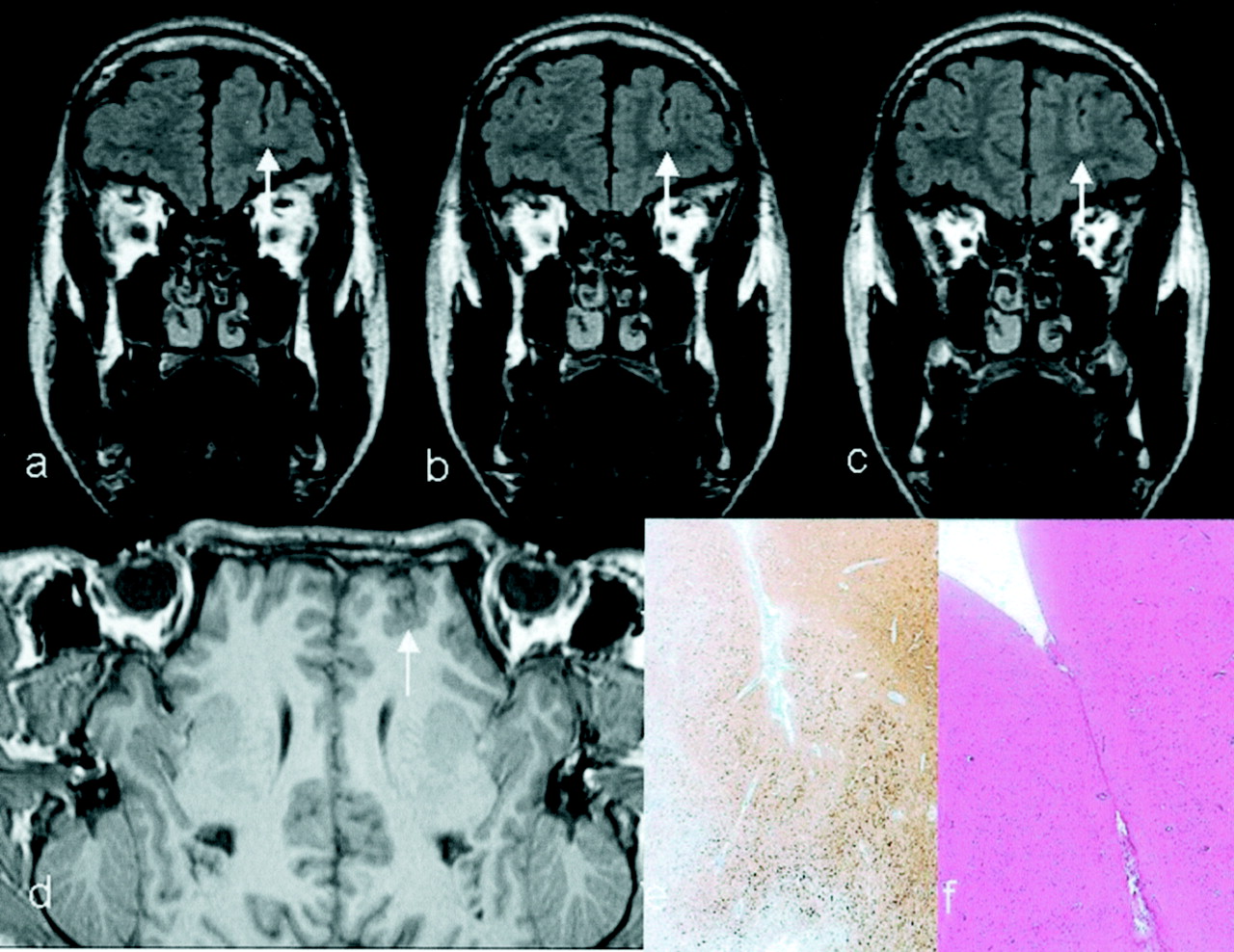
- Fig 1.
Scarring of the left superior frontal gyrus. Axial 5-mm-thick (a–c) and coronal 3-mm-thick (d) FLAIR fast spin-echo images show an atrophic and hyperintense lesion in the right superior frontal gyrus (solid-head arrows in a–d). Note also a tiny increase in signal intensity and the atrophy in the depth of the right frontal superior sulcus (open-head arrows in a, c, and d)
- Fig 2.
Schematic depiction of method used in MR imaging lesion detection.
- Fig 3.
Focal cortical dysplasia of the Taylor balloon cell type (FCDBC). Images in a and b show a small lesion in the depth of the left frontal superior sulcus (arrow). Image in c shows a huge lesion and a small lesion (arrow) in the right parietal and occipital lobe. Image in d shows a lesion under an atrophic left superior frontal gyrus. The MR imaging hallmark of FCDBC is a subcortical hyperintensity tapering toward the lateral ventricle. It is clearly visible (arrow), but because of atrophy of the superior frontal gyrus, the lesion was mistaken for gyral scarring.
- Fig 4.
Focal polymicrogyria in the left frontal lobe. Contiguous, 3-mm-thick, coronal FLAIR fast spin-echo sections (a–c) show a deep superior frontal sulcus with a slightly irregular contour and a normal signal intensity of the cortex (arrow). Planar surface-reconstructed view (d) generated from a 1.1-mm-thick, 3D, T1-weighted gradient-echo sequence confirms the distorted anatomy (arrow). Histopathologic sections show loss of neocortical architecture (MAP2 [e]) compared with that of adjacent six–layered cortex (nematoxylin–eosin straining [f]).
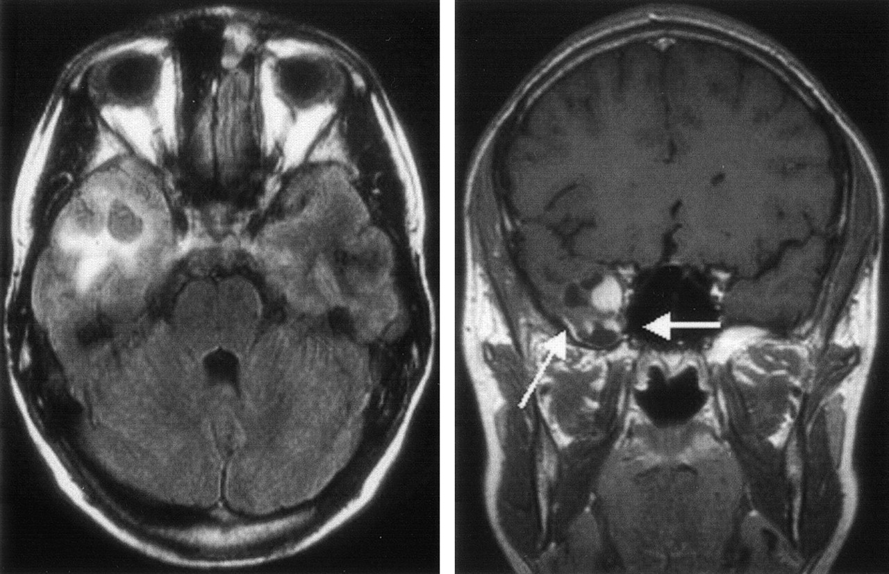
- Fig 5.
PXA of the right temporal lobe. The lesion shows typical meningeocerebral contrast enhancement (arrows), cystic parts, and surrounding edema.
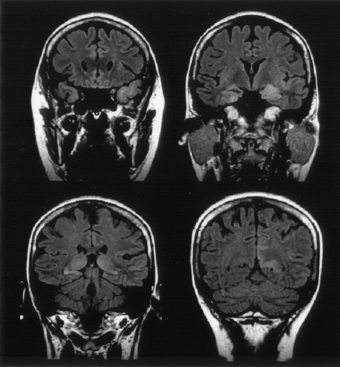
- Fig 6.
Anaplastic (limbic) astrocytoma of WHO grade III. Coronal 3-mm-thick FLAIR fast spin-echo sections show increased volume and signal intensity of both temporopolar cortices, the amygdala, hippocampi, thalami, and left parietal cortex. Because repeated CSF investigation showed a slightly increased protein content (54–95 mg/dL) and 6–8 cells per microliter, limbic encephalitis was suspected.
- Fig 7.
Schematic diagram depicting reasons not to operate on patients with MR imaging lesions.


Tables
Outcome/Engel IA Class MR with lesion Hippocampal sclerosis 104 (50%) 65/93 (70%) Ectopic white matter neurons 3 (1%) 2/3 (66%) FCD with balloon cells 17 (8%) 13/16 (81%) FCD without balloon cells 5 (2%) 2/4 (50%) Polymicrogyria 1 (0.5%) 0/1 (0%) Ganglioglioma 21 (10%) 15/21 (71%) DNT 10 (5%) 7/9 (78%) PXA 4 (2%) 3/3 (100%) Glioma II, III 8 (6%) 6/7 (86%) Cavernoma 15 (7%) 11/15 (73%) Hemimegalencephaly 2 (1%) 1/2 (50%) Meningeoangiomatosis 1 (0.5%) 1/1 (100%) Ulegyria, cortical, or glial scar 9 (4%) 3/9 (33%) Tuber cinereum hamartoma 2 (1%) 1/2 (50%) MR without lesion NAD 1 0/1 Reactive gliosis 6 3/6 Ectopic white matter neurons 2 2/2 Note.—NAD indicates nothing abnormal detected.
Histopathologic Findings MR Findings Hippocampal sclerosis 101 hippocampal sclerosis, 3 atrophy, no sclerosis Ectopic white matter neurons 2 poor gray-white matter demarcation, 1 FCD without BC, 2 NAD FCD with balloon cells (FCDBC) 14 FCDBC, 2 cortical/glial scar, 1 equivocal findings FCD without balloon cells 3 FCD without BC, 2 ganglioglioma Polymicrogyria 1 Polymicrogyria Ganglioglioma 16 ganglioglioma, 4 FCD without BC, 1 DNT DNT 8 DNT, 1 ganglioglioma, 1 FCD without BC PXA 1 PXA, 3 ganglioglioma Glioma II, III 5 glioma, 2 ganglioglioma, 1 limbic encephalitis Cavernoma 15 cavernoma Enzephalitis (Rasmussen, limbic) 5 encephalitis, 1 NAD Hemimegalencephaly 2 hemimegalencephaly Meningeoangiomatosis 1 ganglioglioma Ulegyria, cortical, or glial scar 7 cortical/glial scar, 1 ganglioglioma, 1 DNT Tuber cinereum hamartoma 2 tuber cinereum hamartoma Reactive gliosis 6 NAD Normal 1 NAD Note.—NAD indicates nothing abnormal detected.
In this issue


Jump to section
Related Articles
Cited By...
- MRI Interpretation Errors in Adult Patients with Medically Refractory Epilepsy
- Neural Fragility of Intracranial EEG Networks: Towards an EEG Fingerprint for the Seizure Onset Zone
- Using Network Analysis to Localize the Epileptogenic Zone from Invasive EEG Recordings in Intractable Focal Epilepsy
- MRI-identified pathology in adults with new-onset seizures
- Hippocampal resection length and memory outcome in selective epilepsy surgery