
Article Figures & Data
Figures
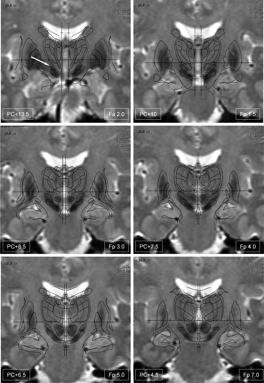
- Fig 1.
Fusion of the Schaltenbrand and Wahren atlas with the T2-weighted acquisition of a patient with Parkinson’s disease. The six anteroposterior levels of the atlas containing a section of the subthalamic nucleus (arrow in level Fa 2.0) are represented. Note that the subthalamic nucleus is hypointense at anterior levels (Fa 2.0−Fp 4.0) but only partly hypointense at level Fp 5.0 and not hypointense at the most posterior level (Fp 7.0). The contours of the Schaltenbrand and Wahren atlas are labeled in Figure 3.
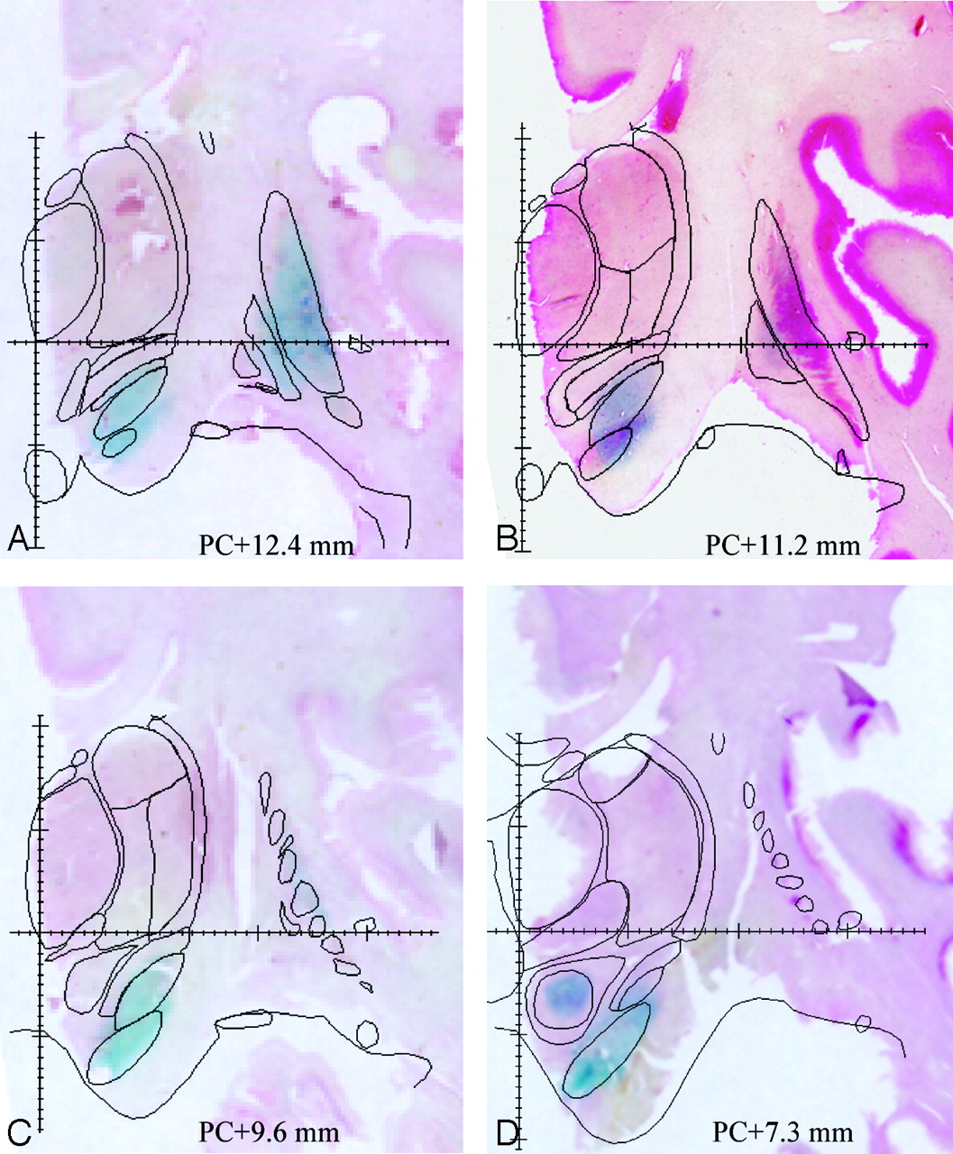
- Fig 2.
Four sections of an anatomicspecimen stained by the Perls method shows the blue stain revealing iron-rich regions. Sections have been counterstained by using neutral red. The contours of cerebral regions have been traced by using the counterstaining of the Perls stained section and Nissl staining of the adjacent sections. Note that the Perls reaction is positive in the anterior portion of the subthalamic nucleus (posterior commissure, +12.4 mm) but very weak in its posterior portion (posterior commissure, +7.3 mm).
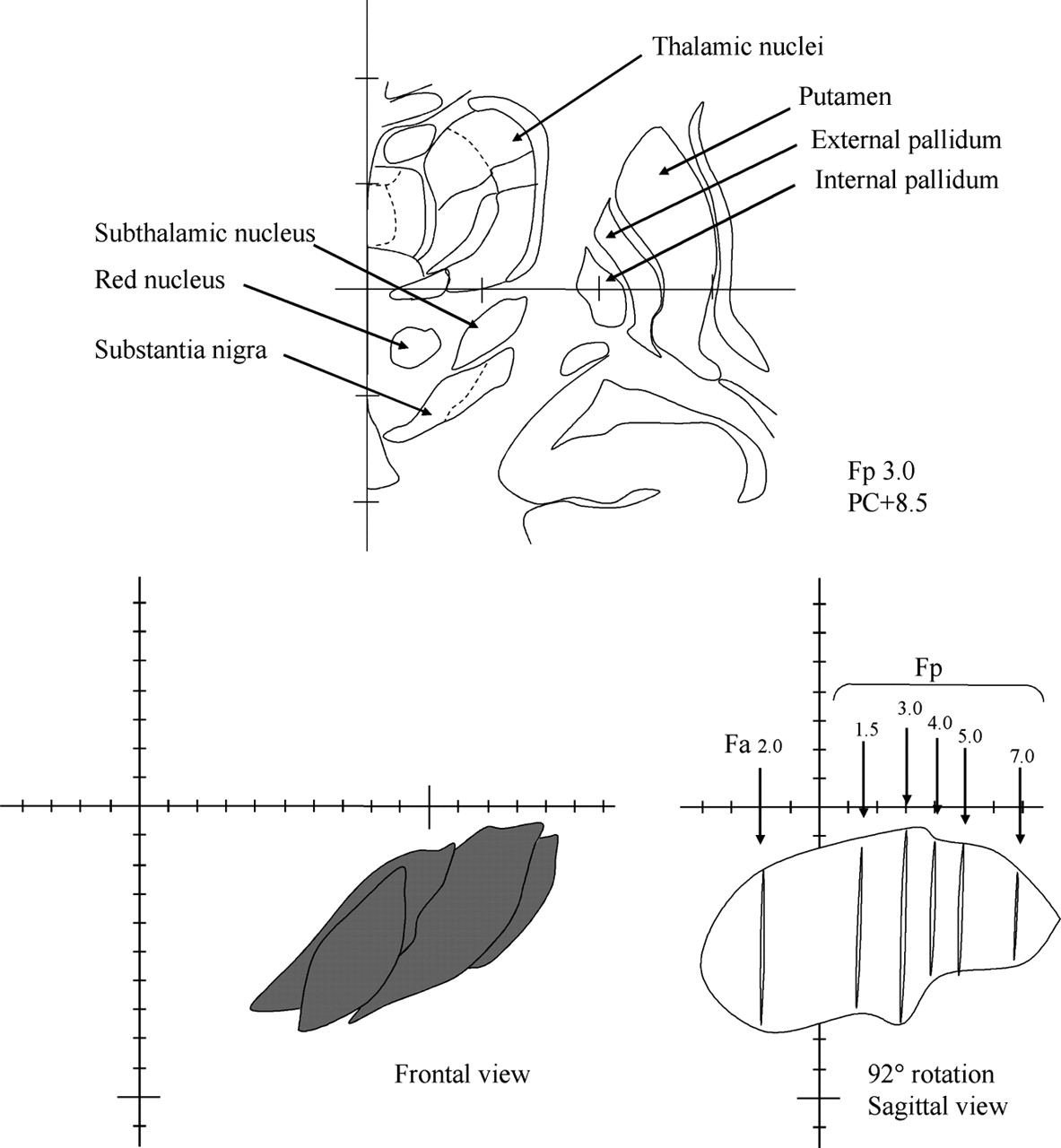
- Fig 3.
Position of the frontal sections on Schaltenbrand and Wahren atlas along the antero-posterior axis.
Top, Digitized frontal section of the Schaltenbrand and Wahren atlas (Fp 3.0). The main cerebral structures are indicated.
Bottom left, Frontal anterior view of the left subthalamic nucleus obtained by superimposing the six frontal sections of the Schaltenbrand and Wahren atlas containing the subthalamic nucleus.
Bottom right, Sagittal obtained by a 92-degree rotation of the same six frontal contours of the subthalamic nucleus. Note that frontal contours are irregularly spaced from the anterior level Fa 2.0 to the posterior level Fp 7.0, and that there are fewer sections located at the level of the anterior part of the nucleus than at the posterior part.


In this issue


Jump to section
- Article
- Abstract
- Methods
- Results
- Discussion
- MR Signal Intensity of the STN
- Atlas-MR Image Fusion Procedure
- Iron Distribution and MR Signal Intensity at the Level of the Basal Ganglia
- Anatomic Iron Distribution at the Levelof the STN
- Implications for the Stereotactic Treatment of Parkinson’s Disease
- Conclusion
- Footnotes
- References
- Figures & Data
- Info & Metrics
- Responses
- References
Related Articles
Cited By...
- Lateral Asymmetry and Spatial Difference of Iron Deposition in the Substantia Nigra of Patients with Parkinson Disease Measured with Quantitative Susceptibility Mapping
- Aging and Inhibitory Control of Action: Cortico-Subthalamic Connection Strength Predicts Stopping Performance
- MR Imaging of Ventral Thalamic Nuclei
- Anatomic Localization of Dyskinesia in Children with "Profound" Perinatal Hypoxic-Ischemic Injury
- Neuroimaging and Deep Brain Stimulation
- Localization of the Subthalamic Nucleus: Optimization with Susceptibility-Weighted Phase MR Imaging
- Pyramidal tract side effects induced by deep brain stimulation of the subthalamic nucleus
- Stimulation of subterritories of the subthalamic nucleus reveals its role in the integration of the emotional and motor aspects of behavior
- Triangulating a Cognitive Control Network Using Diffusion-Weighted Magnetic Resonance Imaging (MRI) and Functional MRI
- Cortical and Subcortical Contributions to Stop Signal Response Inhibition: Role of the Subthalamic Nucleus