Advertisement

Main menu

  • Home
  • Content
    • Current Issue
    • Accepted Manuscripts
    • Article Preview
    • Past Issue Archive
    • AJNR Case Collection
    • Case of the Week Archive
    • Classic Case Archive
    • Case of the Month Archive
  • Special Collections
    • Spinal CSF Leak Articles (Jan 2020-June 2024)
    • 2024 AJNR Journal Awards
    • Most Impactful AJNR Articles
  • Multimedia
    • AJNR Podcast
    • AJNR Scantastics
    • Video Articles
  • For Authors
    • Submit a Manuscript
    • Author Policies
    • Fast publishing of Accepted Manuscripts
    • Graphical Abstract Preparation
    • Manuscript Submission Guidelines
    • Imaging Protocol Submission
    • Submit a Case for the Case Collection
  • About Us
    • About AJNR
    • Editorial Board
  • More
    • Become a Reviewer/Academy of Reviewers
    • Subscribers
    • Permissions
    • Alerts
    • Feedback
    • Advertisers
    • ASNR Home
  • Other Publications
    • ajnr

User menu

  • Alerts
  • Log in

Search

  • Advanced search
American Journal of Neuroradiology

American Journal of Neuroradiology

ASHNR American Society of Functional Neuroradiology ASHNR American Society of Pediatric Neuroradiology ASSR
  • Alerts
  • Log in

Advanced Search

  • Home
  • Content
    • Current Issue
    • Accepted Manuscripts
    • Article Preview
    • Past Issue Archive
    • AJNR Case Collection
    • Case of the Week Archive
    • Classic Case Archive
    • Case of the Month Archive
  • Special Collections
    • Spinal CSF Leak Articles (Jan 2020-June 2024)
    • 2024 AJNR Journal Awards
    • Most Impactful AJNR Articles
  • Multimedia
    • AJNR Podcast
    • AJNR Scantastics
    • Video Articles
  • For Authors
    • Submit a Manuscript
    • Author Policies
    • Fast publishing of Accepted Manuscripts
    • Graphical Abstract Preparation
    • Manuscript Submission Guidelines
    • Imaging Protocol Submission
    • Submit a Case for the Case Collection
  • About Us
    • About AJNR
    • Editorial Board
  • More
    • Become a Reviewer/Academy of Reviewers
    • Subscribers
    • Permissions
    • Alerts
    • Feedback
    • Advertisers
    • ASNR Home
  • Follow AJNR on Twitter
  • Visit AJNR on Facebook
  • Follow AJNR on Instagram
  • Join AJNR on LinkedIn
  • RSS Feeds

Welcome to the new AJNR, Updated Hall of Fame, and more. Read the full announcements.


AJNR is seeking candidates for the position of Associate Section Editor, AJNR Case Collection. Read the full announcement.


OtherSPINE

A Technique to Circumvent Subcutaneous Cement Tracts during Percutaneous Vertebroplasty

Timothy J. Kaufmann, John T. Wald and David F. Kallmes
American Journal of Neuroradiology October 2004, 25 (9) 1595-1596;
Timothy J. Kaufmann
  • Find this author on Google Scholar
  • Find this author on PubMed
  • Search for this author on this site
John T. Wald
  • Find this author on Google Scholar
  • Find this author on PubMed
  • Search for this author on this site
David F. Kallmes
  • Find this author on Google Scholar
  • Find this author on PubMed
  • Search for this author on this site
  • Article
  • Figures & Data
  • Info & Metrics
  • Responses
  • References
  • PDF
Loading

Abstract

Summary: The deposition of a subcutaneous cement tract is a potential complication of percutaneous vertebroplasty. These tracts can be a source of pain and tenderness for the patient. We describe a case of symptomatic cement deposition within a needle tract in the subcutaneous tissues that required surgical removal, and we describe a technique to prevent this complication in a second patient, by using needle redirection to cut across the cement core.

Percutaneous vertebroplasty (PV) has become a widely employed therapy for painful vertebral compression fractures. Complications of the procedure are uncommon but are most frequently related to cement leakage (1). In this report, we describe a complication in which a tract of cement leading from the vertebral body to the subcutaneous tissue was created and required surgical removal. We also report a simple technique that can be used to avoid such a complication.

Case 1

A 57-year-old female patient who had been treated with corticosteroids for sarcoidosis developed osteoporosis and multiple thoracic and lumbar vertebral compression fractures. PV of the T9–T12 vertebrae was performed at the same sitting because of severe, persistent back pain and limitation in mobility that was refractory to conservative therapy.

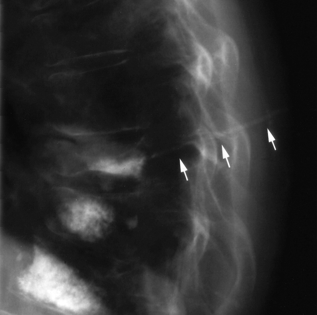
We typically employ a moderately viscous cement mixture of powdered Codman cranioplastic polymethylmethacrylate, gentamicin, barium (Parallax Medical, Scotts Valley, CA), and liquid Codman cranioplastic methylmethacrylate monomer in our PV cases (2). On withdrawal of the 13-gauge Osteo-Site needle (Cook Inc., Bloomington, IN) from the T9 vertebra, the cylindrical column of partially solidified cement that was yet in the needle remained in the needle tract from the site of instillation in the vertebral body, through the left pedicle, and into the subcutaneous tissues of the lower thorax, as depicted radiographically (Fig 1).

Fig 1.
  • Download figure
  • Open in new tab
  • Download powerpoint
Fig 1.

Cement is seen in the needle tract of the subcutaneous tissues of the lower thorax, extending dorsally from the T9 vertebral body cement deposition site (arrows).

The patient experienced persistent pain and tenderness at the site of this subcutaneous cement deposition, particularly when sitting on backed chairs. Two months after the procedure, the patient underwent surgical removal of 2.5 cm of the superficial portion of the subcutaneous cement. The surgery was effective in alleviating her pain.

Case 2

A 66-year-old female patient with multiple myeloma and osteoporosis developed multiple thoracic vertebral compression fractures. PV of the T8, T11, and T12 vertebrae was performed because of severe, persistent back pain and interference with activities of daily living.

We used 11-gauge Osteo-Site needles and the cement mixture described in case 1. Following cement instillation through the left T11 pedicle, we noticed that as the needle was being withdrawn with its tip still within the posterior aspect of the vertebral body, the core of partially solidified cement within the needle was being deposited in the needle tract (Fig 2A). Despite multiple attempts at breaking the cement core by rotating the needle and advancing and withdrawing the needle, the column of cement remained unbroken.

Fig 2.
  • Download figure
  • Open in new tab
  • Download powerpoint
Fig 2.

Deposition of a subncutaneous cement tract is averted.

A, Cement column, visualized in the posterior T11 vertebral body (arrow) as needle withdrawal is first begun.

B, The cement column inside the needle is broken at the tip of the needle (arrow) by pulling the needle hub inferiorly and advancing the tip of the needle toward the superior endplate.

C, The needle can then be withdrawn without further cement deposition in the needle tract (arrow).

To prevent the deposition of a subcutaneous cement tract, the needle tip was then redirected superiorly and advanced toward the superior endplate, which cut completely through the column of cement (Fig 2B). This maneuver disconnected the cement deposited in the vertebral body from that which remained in the needle and allowed the needle to be subsequently withdrawn completely without depositing cement within the needle tract (Fig 2C).

The patient tolerated the procedure well and is doing well in short-term follow-up.

Discussion

Occasionally during PV, the cement mixture partially solidifies before the introducing needle can be removed, particularly as the length of time between cement mixture and needle removal increases. This may be more common when multiple vertebral levels are treated in the same sitting by using one cement mixture or when complicating features occur which otherwise prolong the procedure. This cement solidification can result in symptomatic subcutaneous cement deposition in the needle tract, which requires surgical intervention, as presented in case 1. We have had one other similar case in our practice, which also required subcutaneous tract excision.

We describe a simple maneuver in case 2 that can avert this complication. This technique may be employed during needle removal as soon as it is recognized that cement deposition is occurring in the needle tract. As needle position within the vertebral body may differ in other cases, the needle may be advanced forward across the cement column to cut it in any direction that is safe, with care taken to remain within the vertebral body and to avoid transgression of a vertebral endplate. The need for this technique should not be frequent, but we have used it successfully in two patients since case 1.

There are potential limitations to this technique. It would be theoretically possible to fracture the pedicle if extreme repositioning of the needle trajectory were needed to cut through the cement tract. Risk of this complication might be mitigated in soft, osteoporotic bone. Further, redirection and advancement of the needle might injure adjacent tissue such as the vertebral endplate, which should be avoided.

References

  1. ↵
    Mathis JM. Percutaneous vertebroplasty: complication avoidance and technique optimization. AJNR Am J Neuroradiol 2003;24:1697–1706
    FREE Full Text
  2. ↵
    Jensen ME, Evans AJ, Mathis JM, et al. Percutaneous polymethylmethacrylate vertebroplasty in the treatment of osteoporotic vertebral body compression fractures: technical aspects. AJNR Am J Neuroradiol 1997;18:1897–1904
    Abstract
  • Received March 31, 2004.
  • Accepted after revision April 6, 2004.
  • Copyright © American Society of Neuroradiology
PreviousNext
Back to top

In this issue

American Journal of Neuroradiology: 25 (9)
American Journal of Neuroradiology
Vol. 25, Issue 9
1 Oct 2004
  • Table of Contents
  • Index by author
Advertisement
Print
Download PDF
Email Article

Thank you for your interest in spreading the word on American Journal of Neuroradiology.

NOTE: We only request your email address so that the person you are recommending the page to knows that you wanted them to see it, and that it is not junk mail. We do not capture any email address.

Enter multiple addresses on separate lines or separate them with commas.
A Technique to Circumvent Subcutaneous Cement Tracts during Percutaneous Vertebroplasty
(Your Name) has sent you a message from American Journal of Neuroradiology
(Your Name) thought you would like to see the American Journal of Neuroradiology web site.
CAPTCHA
This question is for testing whether or not you are a human visitor and to prevent automated spam submissions.
Cite this article
Timothy J. Kaufmann, John T. Wald, David F. Kallmes
A Technique to Circumvent Subcutaneous Cement Tracts during Percutaneous Vertebroplasty
American Journal of Neuroradiology Oct 2004, 25 (9) 1595-1596;

Citation Manager Formats

  • BibTeX
  • Bookends
  • EasyBib
  • EndNote (tagged)
  • EndNote 8 (xml)
  • Medlars
  • Mendeley
  • Papers
  • RefWorks Tagged
  • Ref Manager
  • RIS
  • Zotero
0 Responses
Respond to this article
Share
Bookmark this article
A Technique to Circumvent Subcutaneous Cement Tracts during Percutaneous Vertebroplasty
Timothy J. Kaufmann, John T. Wald, David F. Kallmes
American Journal of Neuroradiology Oct 2004, 25 (9) 1595-1596;
del.icio.us logo Twitter logo Facebook logo Mendeley logo
  • Tweet Widget
  • Facebook Like
  • Google Plus One

Jump to section

  • Article
    • Abstract
    • Case 1
    • Case 2
    • Discussion
    • References
  • Figures & Data
  • Info & Metrics
  • Responses
  • References
  • PDF

Related Articles

  • No related articles found.
  • PubMed
  • Google Scholar

Cited By...

  • Asymptomatic and Unrecognized Cement Pulmonary Embolism Commonly Occurs with Vertebroplasty
  • Efficacy of Percutaneous Vertebroplasty for Multiple Synchronous and Metachronous Vertebral Compression Fractures
  • Crossref
  • Google Scholar

This article has not yet been cited by articles in journals that are participating in Crossref Cited-by Linking.

More in this TOC Section

  • Bern Score Validity for SIH
  • MP2RAGE 7T in MS Lesions of the Cervical Spine
  • Deep Learning for STIR Spine MRI Quality
Show more Spine

Similar Articles

Advertisement

Indexed Content

  • Current Issue
  • Accepted Manuscripts
  • Article Preview
  • Past Issues
  • Editorials
  • Editors Choice
  • Fellow Journal Club
  • Letters to the Editor

Cases

  • Case Collection
  • Archive - Case of the Week
  • Archive - Case of the Month
  • Archive - Classic Case

Special Collections

  • Special Collections

Resources

  • News and Updates
  • Turn around Times
  • Submit a Manuscript
  • Author Policies
  • Manuscript Submission Guidelines
  • Evidence-Based Medicine Level Guide
  • Publishing Checklists
  • Graphical Abstract Preparation
  • Imaging Protocol Submission
  • Submit a Case
  • Become a Reviewer/Academy of Reviewers
  • Get Peer Review Credit from Publons

Multimedia

  • AJNR Podcast
  • AJNR SCANtastic
  • Video Articles

About Us

  • About AJNR
  • Editorial Board
  • Not an AJNR Subscriber? Join Now
  • Alerts
  • Feedback
  • Advertise with us
  • Librarian Resources
  • Permissions
  • Terms and Conditions

American Society of Neuroradiology

  • Not an ASNR Member? Join Now

© 2025 by the American Society of Neuroradiology All rights, including for text and data mining, AI training, and similar technologies, are reserved.
Print ISSN: 0195-6108 Online ISSN: 1936-959X

Powered by HighWire