Advertisement

Main menu

  • Home
  • Content
    • Current Issue
    • Accepted Manuscripts
    • Article Preview
    • Past Issue Archive
    • AJNR Case Collection
    • Case of the Week Archive
    • Classic Case Archive
    • Case of the Month Archive
  • Special Collections
    • Spinal CSF Leak Articles (Jan 2020-June 2024)
    • 2024 AJNR Journal Awards
    • Most Impactful AJNR Articles
  • Multimedia
    • AJNR Podcast
    • AJNR Scantastics
    • Video Articles
  • For Authors
    • Submit a Manuscript
    • Author Policies
    • Fast publishing of Accepted Manuscripts
    • Graphical Abstract Preparation
    • Manuscript Submission Guidelines
    • Imaging Protocol Submission
    • Submit a Case for the Case Collection
  • About Us
    • About AJNR
    • Editorial Board
  • More
    • Become a Reviewer/Academy of Reviewers
    • Subscribers
    • Permissions
    • Alerts
    • Feedback
    • Advertisers
    • ASNR Home
  • Other Publications
    • ajnr

User menu

  • Alerts
  • Log in

Search

  • Advanced search
American Journal of Neuroradiology

American Journal of Neuroradiology

ASHNR American Society of Functional Neuroradiology ASHNR American Society of Pediatric Neuroradiology ASSR
  • Alerts
  • Log in

Advanced Search

  • Home
  • Content
    • Current Issue
    • Accepted Manuscripts
    • Article Preview
    • Past Issue Archive
    • AJNR Case Collection
    • Case of the Week Archive
    • Classic Case Archive
    • Case of the Month Archive
  • Special Collections
    • Spinal CSF Leak Articles (Jan 2020-June 2024)
    • 2024 AJNR Journal Awards
    • Most Impactful AJNR Articles
  • Multimedia
    • AJNR Podcast
    • AJNR Scantastics
    • Video Articles
  • For Authors
    • Submit a Manuscript
    • Author Policies
    • Fast publishing of Accepted Manuscripts
    • Graphical Abstract Preparation
    • Manuscript Submission Guidelines
    • Imaging Protocol Submission
    • Submit a Case for the Case Collection
  • About Us
    • About AJNR
    • Editorial Board
  • More
    • Become a Reviewer/Academy of Reviewers
    • Subscribers
    • Permissions
    • Alerts
    • Feedback
    • Advertisers
    • ASNR Home
  • Follow AJNR on Twitter
  • Visit AJNR on Facebook
  • Follow AJNR on Instagram
  • Join AJNR on LinkedIn
  • RSS Feeds

Welcome to the new AJNR, Updated Hall of Fame, and more. Read the full announcements.


AJNR is seeking candidates for the position of Associate Section Editor, AJNR Case Collection. Read the full announcement.


Research ArticleBRAIN

Quantitative Proton MR Spectroscopic Findings of Cortical Reorganization in the Auditory Cortex of Musicians

Kubilay Aydin, Koray Ciftci, Ege Terzibasioglu, Mehmed Ozkan, Asli Demirtas, Serra Sencer and Ozenc Minareci
American Journal of Neuroradiology January 2005, 26 (1) 128-136;
Kubilay Aydin
  • Find this author on Google Scholar
  • Find this author on PubMed
  • Search for this author on this site
Koray Ciftci
  • Find this author on Google Scholar
  • Find this author on PubMed
  • Search for this author on this site
Ege Terzibasioglu
  • Find this author on Google Scholar
  • Find this author on PubMed
  • Search for this author on this site
Mehmed Ozkan
  • Find this author on Google Scholar
  • Find this author on PubMed
  • Search for this author on this site
Asli Demirtas
  • Find this author on Google Scholar
  • Find this author on PubMed
  • Search for this author on this site
Serra Sencer
  • Find this author on Google Scholar
  • Find this author on PubMed
  • Search for this author on this site
Ozenc Minareci
  • Find this author on Google Scholar
  • Find this author on PubMed
  • Search for this author on this site
  • Article
  • Figures & Data
  • Info & Metrics
  • Responses
  • References
  • PDF
Loading

Article Figures & Data

Figures

  • Tables
  • Fig 1.
    • Download figure
    • Open in new tab
    • Download powerpoint
    Fig 1.

    T1-weighted MR images (530/20/3) depict examples of voxel placement.

    A, Coronal MR image shows a voxel placed in the left planum temporale, which is located between the Heschl gyrus medially (arrow) and superior rim of the superior temporal gyrus.

    B, Sagittal T1-weighted image shows location of the voxel.

  • Fig 2.
    • Download figure
    • Open in new tab
    • Download powerpoint
    Fig 2.

    A, Image shows voxel placement for the phantom study.

    B, Spectrum obtained from the phantom shows the acetate peak and lactate doublet.

  • Fig 3.
    • Download figure
    • Open in new tab
    • Download powerpoint
    Fig 3.

    A–D, Proton spectra from the planum temporale in a musician, obtained with TE values of 30 ms (A), 48 ms (B), 135 ms (C), and 230 ms (D).

  • Fig 4.
    • Download figure
    • Open in new tab
    • Download powerpoint
    Fig 4.

    A–D, Proton spectra from the planum temporale in a non-musician control subject, obtained with TE values of 30 ms (A), 48 ms (B), 135 ms (C), and 230 ms (D).
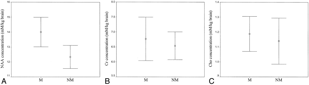
  • Fig 5.
    • Download figure
    • Open in new tab
    • Download powerpoint
    Fig 5.

    A–C, Graphs show metabolite concentrations in musicians (M) and the non-musician control group (NM) for NAA concentration (A), Cr concentration (B), and Cho concentration (C). Error bars show 95% confidence intervals for the means of the metabolite concentrations.

  • Fig 6.
    • Download figure
    • Open in new tab
    • Download powerpoint
    Fig 6.

    A, Graph demonstrates the relationship between NAA concentration in the musicians and total duration of musical training and activity. NAA concentration increases significantly as the duration of total musical training and activity increases (r = 0.733 [Pearson correlation coefficient], P = .016).

    B, NAA concentration aganist age of commencement (r = −0.463, P = .178).

    C, NAA concentration aganist time spent for musical activities per week (r = −0.327, P = .357).

Tables

  • Figures
    • View popup
    TABLE 1:

    Personal information and metabolite concentrations of the musicians

    Musician No./Age (y)/SexAge (y) at Commencement of Musical TrainingTotal Duration of Musical TrainingTime Spent on Musical Activity per week (h)Metabolite Concentration (mmol/kg brain)
    NAACrCho
    1/20/F6141814.526.921.27
    2/37/M7281615.587.391.42
    3/22/F7152212.787.671.14
    4/24/M5192413.345.321.14
    5/33/M7242015.866.330.97
    6/23/F5181415.225.911.04
    7/24/M6162813.828.351.24
    8/21/F8132411.267.460.94
    9/23/M6173613.725.341.34
    10/25/M7182013.916.961.32
    • View popup
    TABLE 2:

    Metabolite concentrations of study participants

    MetaboliteMetabolite Concentrations (mmol/kg brain)P Value
    MusiciansNonmusicians
    NAA14.00 ± 1.3812.33 ± 1.08.008
    Cr6.76 ± 1.016.54 ± 0.65.559
    Cho1.18 ± 0.161.14 ± 0.21.583
    • Note.—Data are mean ± standard deviation.

PreviousNext
Back to top

In this issue

American Journal of Neuroradiology: 26 (1)
American Journal of Neuroradiology
Vol. 26, Issue 1
1 Jan 2005
  • Table of Contents
  • Index by author
Advertisement
Print
Download PDF
Email Article

Thank you for your interest in spreading the word on American Journal of Neuroradiology.

NOTE: We only request your email address so that the person you are recommending the page to knows that you wanted them to see it, and that it is not junk mail. We do not capture any email address.

Enter multiple addresses on separate lines or separate them with commas.
Quantitative Proton MR Spectroscopic Findings of Cortical Reorganization in the Auditory Cortex of Musicians
(Your Name) has sent you a message from American Journal of Neuroradiology
(Your Name) thought you would like to see the American Journal of Neuroradiology web site.
CAPTCHA
This question is for testing whether or not you are a human visitor and to prevent automated spam submissions.
Cite this article
Kubilay Aydin, Koray Ciftci, Ege Terzibasioglu, Mehmed Ozkan, Asli Demirtas, Serra Sencer, Ozenc Minareci
Quantitative Proton MR Spectroscopic Findings of Cortical Reorganization in the Auditory Cortex of Musicians
American Journal of Neuroradiology Jan 2005, 26 (1) 128-136;

Citation Manager Formats

  • BibTeX
  • Bookends
  • EasyBib
  • EndNote (tagged)
  • EndNote 8 (xml)
  • Medlars
  • Mendeley
  • Papers
  • RefWorks Tagged
  • Ref Manager
  • RIS
  • Zotero
0 Responses
Respond to this article
Share
Bookmark this article
Quantitative Proton MR Spectroscopic Findings of Cortical Reorganization in the Auditory Cortex of Musicians
Kubilay Aydin, Koray Ciftci, Ege Terzibasioglu, Mehmed Ozkan, Asli Demirtas, Serra Sencer, Ozenc Minareci
American Journal of Neuroradiology Jan 2005, 26 (1) 128-136;
del.icio.us logo Twitter logo Facebook logo Mendeley logo
  • Tweet Widget
  • Facebook Like
  • Google Plus One

Jump to section

  • Article
    • Abstract
    • Methods
    • Results
    • Discussion
    • Conclusion
    • References
  • Figures & Data
  • Info & Metrics
  • Responses
  • References
  • PDF

Related Articles

  • No related articles found.
  • PubMed
  • Google Scholar

Cited By...

  • Mozart's movements and behaviour: a case of Tourette's syndrome?
  • Mozart's movements and behaviour: a case of Tourette's syndrome?
  • Crossref
  • Google Scholar

This article has not yet been cited by articles in journals that are participating in Crossref Cited-by Linking.

More in this TOC Section

  • Multimodal CT Provides Improved Performance for Lacunar Infarct Detection
  • Statin Therapy Does Not Affect the Radiographic and Clinical Profile of Patients with TIA and Minor Stroke
  • Optimal MRI Sequence for Identifying Occlusion Location in Acute Stroke: Which Value of Time-Resolved Contrast-Enhanced MRA?
Show more Brain

Similar Articles

Advertisement

Indexed Content

  • Current Issue
  • Accepted Manuscripts
  • Article Preview
  • Past Issues
  • Editorials
  • Editors Choice
  • Fellow Journal Club
  • Letters to the Editor

Cases

  • Case Collection
  • Archive - Case of the Week
  • Archive - Case of the Month
  • Archive - Classic Case

Special Collections

  • Special Collections

Resources

  • News and Updates
  • Turn around Times
  • Submit a Manuscript
  • Author Policies
  • Manuscript Submission Guidelines
  • Evidence-Based Medicine Level Guide
  • Publishing Checklists
  • Graphical Abstract Preparation
  • Imaging Protocol Submission
  • Submit a Case
  • Become a Reviewer/Academy of Reviewers
  • Get Peer Review Credit from Publons

Multimedia

  • AJNR Podcast
  • AJNR SCANtastic
  • Video Articles

About Us

  • About AJNR
  • Editorial Board
  • Not an AJNR Subscriber? Join Now
  • Alerts
  • Feedback
  • Advertise with us
  • Librarian Resources
  • Permissions
  • Terms and Conditions

American Society of Neuroradiology

  • Not an ASNR Member? Join Now

© 2025 by the American Society of Neuroradiology All rights, including for text and data mining, AI training, and similar technologies, are reserved.
Print ISSN: 0195-6108 Online ISSN: 1936-959X

Powered by HighWire