
Article Figures & Data
Figures
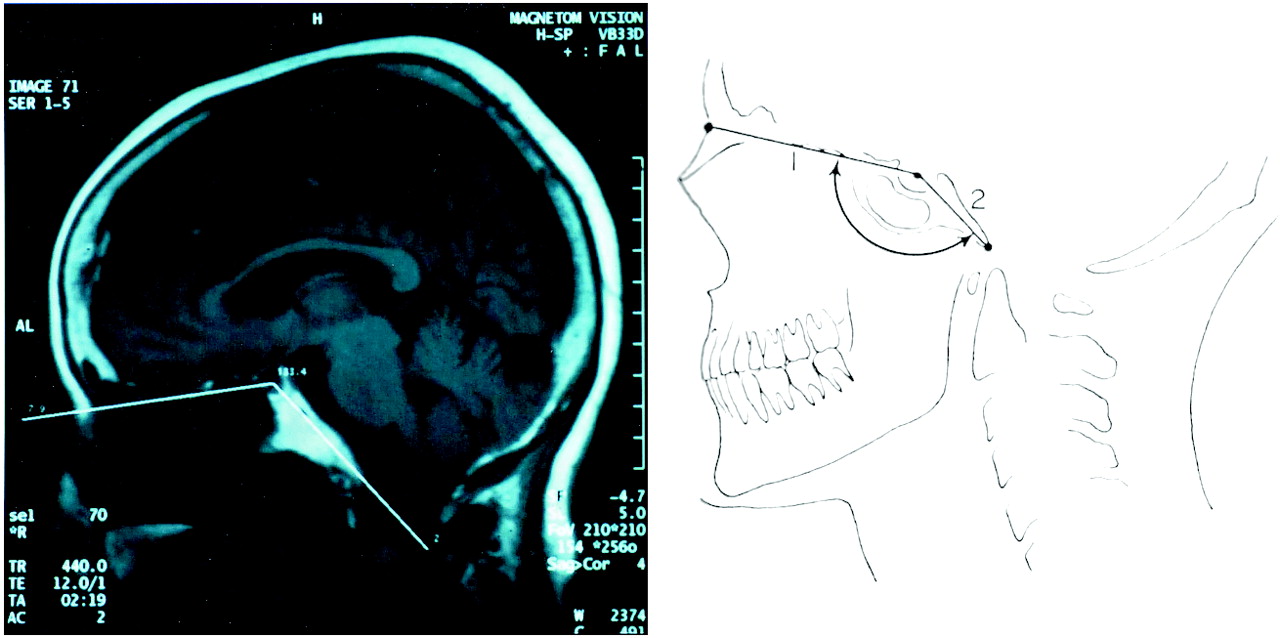
- Fig 1.
Standard MR imaging technique for measuring the basal angle. This technique involves measuring the angle formed by two lines—one joining the nasion with the center of the pituitary fossa and a second line joining the anterior border of the foramen magnum with the center of the pituitary fossa.
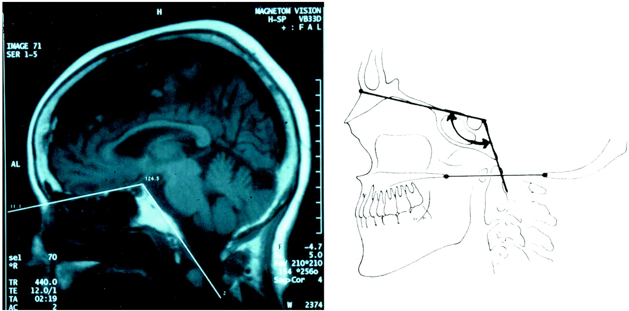
- Fig 2.
Modified MR imaging technique for measuring the basal angle. This method uses different landmarks—the angle formed by a line extending across the anterior cranial fossa to the tip to the dorsum sellae with a second, connecting line drawn along the posterior margin of the clivus.


Tables
- TABLE 1:
Comparison of basal angle measurements with standard radiographic and standard MR imaging techniques
Author Technique Mean Maximum Minimum 95% Confidence Limit* Brailsford7 Radiography 135° 149° 121° Not applicable McGregor2 (n = 203) Radiography 134° 148° 121° 141°, 127° Poppel et al3 (n = 102) Radiography 137° 152° 123° 147°, 127° Present series Adults MR imaging 129° ± 6° 143° 113° 130°, 128° Children MR imaging 127° ± 5° 136° 114° 128°, 126° Note.—Measurements are provided to nearest whole unit.
* Upper and lower 95% confidence limits for radiographic series are estimated on the basis of standard deviations and sample size.
Measurement Adults (n = 200) Children (n = 50) Mean 117 ± 6° 114° ± 5° Maximum 127° 125° Minimum 100° 103° 95% confidence limit 116°, 118° 113°, 115° Note.—Measurements are provided to nearest whole unit.


