Advertisement

Main menu

  • Home
  • Content
    • Current Issue
    • Accepted Manuscripts
    • Article Preview
    • Past Issue Archive
    • Video Articles
    • AJNR Case Collection
    • Case of the Week Archive
    • Case of the Month Archive
    • Classic Case Archive
  • Special Collections
    • AJNR Awards
    • Low-Field MRI
    • Alzheimer Disease
    • ASNR Foundation Special Collection
    • Photon-Counting CT
    • View All
  • Multimedia
    • AJNR Podcasts
    • AJNR SCANtastic
    • Trainee Corner
    • MRI Safety Corner
    • Imaging Protocols
  • For Authors
    • Submit a Manuscript
    • Submit a Video Article
    • Submit an eLetter to the Editor/Response
    • Manuscript Submission Guidelines
    • Statistical Tips
    • Fast Publishing of Accepted Manuscripts
    • Graphical Abstract Preparation
    • Imaging Protocol Submission
    • Author Policies
  • About Us
    • About AJNR
    • Editorial Board
    • Editorial Board Alumni
  • More
    • Become a Reviewer/Academy of Reviewers
    • Subscribers
    • Permissions
    • Alerts
    • Feedback
    • Advertisers
    • ASNR Home

User menu

  • Alerts
  • Log in

Search

  • Advanced search
American Journal of Neuroradiology

American Journal of Neuroradiology

ASHNR American Society of Functional Neuroradiology ASHNR American Society of Pediatric Neuroradiology ASSR
  • Alerts
  • Log in

Advanced Search

  • Home
  • Content
    • Current Issue
    • Accepted Manuscripts
    • Article Preview
    • Past Issue Archive
    • Video Articles
    • AJNR Case Collection
    • Case of the Week Archive
    • Case of the Month Archive
    • Classic Case Archive
  • Special Collections
    • AJNR Awards
    • Low-Field MRI
    • Alzheimer Disease
    • ASNR Foundation Special Collection
    • Photon-Counting CT
    • View All
  • Multimedia
    • AJNR Podcasts
    • AJNR SCANtastic
    • Trainee Corner
    • MRI Safety Corner
    • Imaging Protocols
  • For Authors
    • Submit a Manuscript
    • Submit a Video Article
    • Submit an eLetter to the Editor/Response
    • Manuscript Submission Guidelines
    • Statistical Tips
    • Fast Publishing of Accepted Manuscripts
    • Graphical Abstract Preparation
    • Imaging Protocol Submission
    • Author Policies
  • About Us
    • About AJNR
    • Editorial Board
    • Editorial Board Alumni
  • More
    • Become a Reviewer/Academy of Reviewers
    • Subscribers
    • Permissions
    • Alerts
    • Feedback
    • Advertisers
    • ASNR Home
  • Follow AJNR on Twitter
  • Visit AJNR on Facebook
  • Follow AJNR on Instagram
  • Join AJNR on LinkedIn
  • RSS Feeds

AJNR Awards, New Junior Editors, and more. Read the latest AJNR updates

Research ArticleBrain

Regional Brain Atrophy Evolves Differently in Patients with Multiple Sclerosis According to Clinical Phenotype

Elisabetta Pagani, Maria A. Rocca, Antonio Gallo, Marco Rovaris, Vittorio Martinelli, Giancarlo Comi and Massimo Filippi
American Journal of Neuroradiology February 2005, 26 (2) 341-346;
Elisabetta Pagani
  • Find this author on Google Scholar
  • Find this author on PubMed
  • Search for this author on this site
Maria A. Rocca
  • Find this author on Google Scholar
  • Find this author on PubMed
  • Search for this author on this site
Antonio Gallo
  • Find this author on Google Scholar
  • Find this author on PubMed
  • Search for this author on this site
Marco Rovaris
  • Find this author on Google Scholar
  • Find this author on PubMed
  • Search for this author on this site
Vittorio Martinelli
  • Find this author on Google Scholar
  • Find this author on PubMed
  • Search for this author on this site
Giancarlo Comi
  • Find this author on Google Scholar
  • Find this author on PubMed
  • Search for this author on this site
Massimo Filippi
  • Find this author on Google Scholar
  • Find this author on PubMed
  • Search for this author on this site
  • Article
  • Figures & Data
  • Info & Metrics
  • Responses
  • References
  • PDF
Loading

Article Figures & Data

Figures

  • Tables
  • Fig 1.
    • Download figure
    • Open in new tab
    • Download powerpoint
    Fig 1.

    Color-coded areas (SPMt) of brain atrophy development in RRMS overlaid on a template T1-weighted image. All sections show extensive involvement of the ventricular system. Images A and B show involvement of the pericerebellar spaces and cerebellar tentorium. Images A, C, and D show involvement of the putamen; corpus callosum; insula; cingulate sulcus; and frontal, parietal, temporal, and occipital cortex.

  • Fig 2.
    • Download figure
    • Open in new tab
    • Download powerpoint
    Fig 2.

    Color-coded areas (SPMt) of brain atrophy development in SPMS overlaid on a template T1-weighted image. Image A shows the involvement of the bilateral anterior orbital gyrus and left mammillary body. B shows the involvement of the caudate nuclei; left middle temporal gyrus; left thalamus; and frontal, parietal, temporal and occipital region. C shows the involvement of the cingulate sulcus and regions of frontal and parietal cortex.
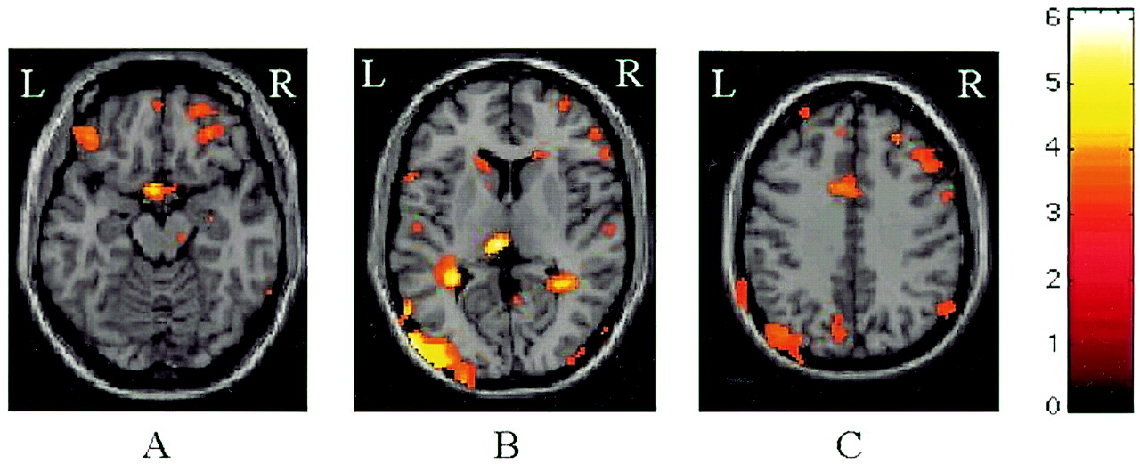
  • Fig 3.
    • Download figure
    • Open in new tab
    • Download powerpoint
    Fig 3.

    Color-coded areas (SPMt) of brain atrophy development in PPMS overlaid on a template T1-weighted image. A shows the involvement of the left middle occipital gyrus, bilateral parahippocampal gyri, and bilateral prepontine and quadrigeminal cisterns. B shows the involvement of the head of the left caudate nucleus, insula, and bilateral middle temporal gyrus. C shows the involvement of the bilateral central sulci (precentral and postcentral gyri) and regions of frontal and parietal cortex.

Tables

  • Figures
  • Demographic clinical characteristics and MR imaging findings at baseline and after 15 months

    Healthy SubjectsPatients
    RRMSSPMSPPMS
    M/F4/68/126/1312/19
    Mean age (years)42.6 (31–58)35.9 (25–43)49.3 (35–58)50.1 (25–68)
    Median EDSS scoreNA3.0 (0.0–5.0)6.0 (4.0–8.0)6.0 (2.5–7.0)
    Median disease duration (years)NA5 (0–20)16 (6–34)10 (2–26)
    Baseline lesion volume (mL)
        On T2-weighted imagesNA13.9 (10.6)21.7 (17.3)21.0 (16.9)
        On T1-weighted imagesNA3.1 (3.6)8.5 (7.3)8.2 (6.9)
    Lesion volume change (%)
        On T2-weighted imagesNA9.43.84.9
        On T1-weighted imagesNA3.411.04.3
    Baseline NBV (mL)1602 (80)1517 (70)1464 (90)1473 (70)
    • Note.—Data in parentheses are the range or standard deviation. NA indicates not applicable.

PreviousNext
Back to top

In this issue

American Journal of Neuroradiology: 26 (2)
American Journal of Neuroradiology
Vol. 26, Issue 2
1 Feb 2005
  • Table of Contents
  • Index by author
Advertisement
Print
Download PDF
Email Article

Thank you for your interest in spreading the word on American Journal of Neuroradiology.

NOTE: We only request your email address so that the person you are recommending the page to knows that you wanted them to see it, and that it is not junk mail. We do not capture any email address.

Enter multiple addresses on separate lines or separate them with commas.
Regional Brain Atrophy Evolves Differently in Patients with Multiple Sclerosis According to Clinical Phenotype
(Your Name) has sent you a message from American Journal of Neuroradiology
(Your Name) thought you would like to see the American Journal of Neuroradiology web site.
CAPTCHA
This question is for testing whether or not you are a human visitor and to prevent automated spam submissions.
Cite this article
Elisabetta Pagani, Maria A. Rocca, Antonio Gallo, Marco Rovaris, Vittorio Martinelli, Giancarlo Comi, Massimo Filippi
Regional Brain Atrophy Evolves Differently in Patients with Multiple Sclerosis According to Clinical Phenotype
American Journal of Neuroradiology Feb 2005, 26 (2) 341-346;

Citation Manager Formats

  • BibTeX
  • Bookends
  • EasyBib
  • EndNote (tagged)
  • EndNote 8 (xml)
  • Medlars
  • Mendeley
  • Papers
  • RefWorks Tagged
  • Ref Manager
  • RIS
  • Zotero
0 Responses
Respond to this article
Share
Bookmark this article
Regional Brain Atrophy Evolves Differently in Patients with Multiple Sclerosis According to Clinical Phenotype
Elisabetta Pagani, Maria A. Rocca, Antonio Gallo, Marco Rovaris, Vittorio Martinelli, Giancarlo Comi, Massimo Filippi
American Journal of Neuroradiology Feb 2005, 26 (2) 341-346;
del.icio.us logo Twitter logo Facebook logo Mendeley logo
  • Tweet Widget
  • Facebook Like
  • Google Plus One

Jump to section

  • Article
    • Abstract
    • Methods
    • Results
    • Discussion
    • Conclusion
    • References
  • Figures & Data
  • Info & Metrics
  • Responses
  • References
  • PDF

Related Articles

  • No related articles found.
  • PubMed
  • Google Scholar

Cited By...

  • Five dominant dimensions of brain aging are identified via deep learning: associations with clinical, lifestyle, and genetic measures
  • Similar neural pathways link psychological stress and brain health in health and multiple sclerosis
  • Accumulation of meningeal lymphocytes, but not myeloid cells, correlates with subpial cortical demyelination and white matter lesion activity in progressive MS patients
  • Predicting disability progression and cognitive worsening in multiple sclerosis using patterns of grey matter volumes
  • Predicting disability progression and cognitive worsening in multiple sclerosis using grey matter network measures
  • Reduced Global Efficiency and Random Network Features in Patients with Relapsing-Remitting Multiple Sclerosis with Cognitive Impairment
  • Early central atrophy rate predicts 5 year clinical outcome in multiple sclerosis
  • Assessing brain atrophy rates in a large population of untreated multiple sclerosis subtypes
  • Cortical lesions in primary progressive multiple sclerosis: A 2-year longitudinal MR study
  • Thalamic atrophy and cognition in multiple sclerosis
  • Progression of non-age-related callosal brain atrophy in multiple sclerosis: a 9-year longitudinal MRI study representing four decades of disease development
  • Ventricular enlargement in MS: One-year change at various stages of disease
  • Crossref
  • Google Scholar

This article has not yet been cited by articles in journals that are participating in Crossref Cited-by Linking.

More in this TOC Section

  • Usefulness of Quantitative Susceptibility Mapping for the Diagnosis of Parkinson Disease
  • White Matter Alterations in the Brains of Patients with Active, Remitted, and Cured Cushing Syndrome: A DTI Study
  • Qualitative and Quantitative Analysis of MR Imaging Findings in Patients with Middle Cerebral Artery Stroke Implanted with Mesenchymal Stem Cells
Show more Brain

Similar Articles

Advertisement

Indexed Content

  • Current Issue
  • Accepted Manuscripts
  • Article Preview
  • Past Issues
  • Editorials
  • Editor's Choice
  • Fellows' Journal Club
  • Letters to the Editor
  • Video Articles

Cases

  • Case Collection
  • Archive - Case of the Week
  • Archive - Case of the Month
  • Archive - Classic Case

More from AJNR

  • Trainee Corner
  • Imaging Protocols
  • MRI Safety Corner
  • Book Reviews

Multimedia

  • AJNR Podcasts
  • AJNR Scantastics

Resources

  • Turnaround Time
  • Submit a Manuscript
  • Submit a Video Article
  • Submit an eLetter to the Editor/Response
  • Manuscript Submission Guidelines
  • Statistical Tips
  • Fast Publishing of Accepted Manuscripts
  • Graphical Abstract Preparation
  • Imaging Protocol Submission
  • Evidence-Based Medicine Level Guide
  • Publishing Checklists
  • Author Policies
  • Become a Reviewer/Academy of Reviewers
  • News and Updates

About Us

  • About AJNR
  • Editorial Board
  • Editorial Board Alumni
  • Alerts
  • Permissions
  • Not an AJNR Subscriber? Join Now
  • Advertise with Us
  • Librarian Resources
  • Feedback
  • Terms and Conditions
  • AJNR Editorial Board Alumni

American Society of Neuroradiology

  • Not an ASNR Member? Join Now

© 2025 by the American Society of Neuroradiology All rights, including for text and data mining, AI training, and similar technologies, are reserved.
Print ISSN: 0195-6108 Online ISSN: 1936-959X

Powered by HighWire