Advertisement

Main menu

  • Home
  • Content
    • Current Issue
    • Accepted Manuscripts
    • Article Preview
    • Past Issue Archive
    • AJNR Case Collection
    • Case of the Week Archive
    • Classic Case Archive
    • Case of the Month Archive
  • Special Collections
    • Spinal CSF Leak Articles (Jan 2020-June 2024)
    • 2024 AJNR Journal Awards
    • Most Impactful AJNR Articles
  • Multimedia
    • AJNR Podcast
    • AJNR Scantastics
    • Video Articles
  • For Authors
    • Submit a Manuscript
    • Author Policies
    • Fast publishing of Accepted Manuscripts
    • Graphical Abstract Preparation
    • Manuscript Submission Guidelines
    • Imaging Protocol Submission
    • Submit a Case for the Case Collection
  • About Us
    • About AJNR
    • Editorial Board
  • More
    • Become a Reviewer/Academy of Reviewers
    • Subscribers
    • Permissions
    • Alerts
    • Feedback
    • Advertisers
    • ASNR Home
  • Other Publications
    • ajnr

User menu

  • Alerts
  • Log in

Search

  • Advanced search
American Journal of Neuroradiology

American Journal of Neuroradiology

ASHNR American Society of Functional Neuroradiology ASHNR American Society of Pediatric Neuroradiology ASSR
  • Alerts
  • Log in

Advanced Search

  • Home
  • Content
    • Current Issue
    • Accepted Manuscripts
    • Article Preview
    • Past Issue Archive
    • AJNR Case Collection
    • Case of the Week Archive
    • Classic Case Archive
    • Case of the Month Archive
  • Special Collections
    • Spinal CSF Leak Articles (Jan 2020-June 2024)
    • 2024 AJNR Journal Awards
    • Most Impactful AJNR Articles
  • Multimedia
    • AJNR Podcast
    • AJNR Scantastics
    • Video Articles
  • For Authors
    • Submit a Manuscript
    • Author Policies
    • Fast publishing of Accepted Manuscripts
    • Graphical Abstract Preparation
    • Manuscript Submission Guidelines
    • Imaging Protocol Submission
    • Submit a Case for the Case Collection
  • About Us
    • About AJNR
    • Editorial Board
  • More
    • Become a Reviewer/Academy of Reviewers
    • Subscribers
    • Permissions
    • Alerts
    • Feedback
    • Advertisers
    • ASNR Home
  • Follow AJNR on Twitter
  • Visit AJNR on Facebook
  • Follow AJNR on Instagram
  • Join AJNR on LinkedIn
  • RSS Feeds

Welcome to the new AJNR, Updated Hall of Fame, and more. Read the full announcements.


AJNR is seeking candidates for the position of Associate Section Editor, AJNR Case Collection. Read the full announcement.


OtherBRAIN

Pallidoreticular Damage in Acute Carbon Monoxide Poisoning: Diffusion-Weighted MR Imaging Findings

Toshibumi Kinoshita, Shuji Sugihara, Eiji Matsusue, Shinya Fujii, Motoki Ametani and Toshihide Ogawa
American Journal of Neuroradiology August 2005, 26 (7) 1845-1848;
Toshibumi Kinoshita
  • Find this author on Google Scholar
  • Find this author on PubMed
  • Search for this author on this site
Shuji Sugihara
  • Find this author on Google Scholar
  • Find this author on PubMed
  • Search for this author on this site
Eiji Matsusue
  • Find this author on Google Scholar
  • Find this author on PubMed
  • Search for this author on this site
Shinya Fujii
  • Find this author on Google Scholar
  • Find this author on PubMed
  • Search for this author on this site
Motoki Ametani
  • Find this author on Google Scholar
  • Find this author on PubMed
  • Search for this author on this site
Toshihide Ogawa
  • Find this author on Google Scholar
  • Find this author on PubMed
  • Search for this author on this site
  • Article
  • Figures & Data
  • Info & Metrics
  • Responses
  • References
  • PDF
Loading

Article Figures & Data

Figures

  • Fig 1.
    • Download figure
    • Open in new tab
    • Download powerpoint
    Fig 1.

    T2-weighted image obtained 5 days after exposure to carbon monoxide (A) shows symmetrical high-signal-intensity lesions in the bilateral globi pallidi. Diffusion-weighted image (B) reveals symmetrical high-signal-intensity lesions in the globi pallidi. Corresponding ADC map (C) demonstrates low-signal-intensity lesions with high-signal-intensity rims in the bilateral globi pallidi.
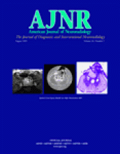
  • Fig 2.
    • Download figure
    • Open in new tab
    • Download powerpoint
    Fig 2.

    T1-weighted image obtained 12 days after exposure to carbon monoxide (A) shows slightly high-signal-intensity lesions with relatively low-intensity rim in the bilateral globi pallidi. T2-weighted image (B) shows slightly low-signal-intensity lesions with high-signal-intensity rim in the bilateral globi pallidi. Coronal T2-weighted image (C) reveals high-signal-intensity lesions in the bilateral substantia nigra, in addition to high-signal-intensity pallidal lesions. Diffusion-weighted image (D) reveals high-signal-intensity lesions in the pars reticulata of the bilateral substantia nigra. Corresponding ADC map (E) demonstrates low signal intensity in the bilateral substantia nigra.
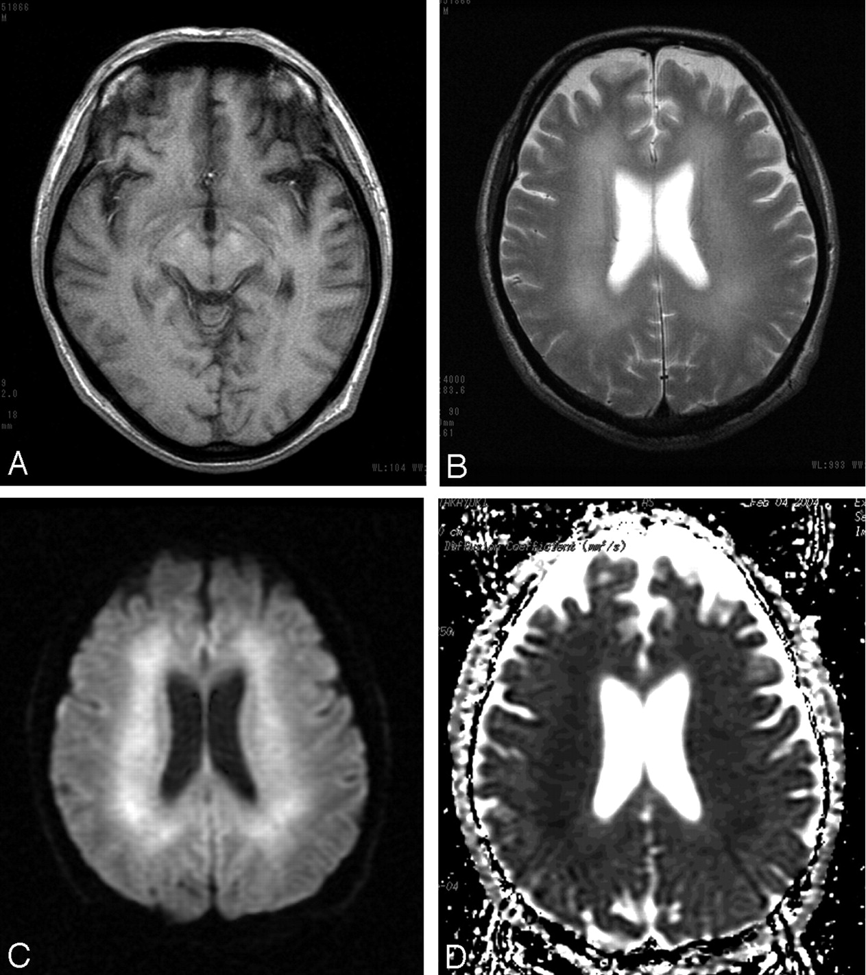
  • Fig 3.
    • Download figure
    • Open in new tab
    • Download powerpoint
    Fig 3.

    T1-weighted image obtained 2 months after exposure to carbon monoxide (A) shows slightly high-signal-intensity lesions in the bilateral substantia nigra. T2-weighted image (B) shows confluent high-signal-intensity lesions in the bilateral periventricular deep white matter. Diffusion-weighted image (C) reveals diffuse high-signal-intensity lesions in the bilateral periventricular deep white matter. Corresponding ADC map (D) demonstrates low-signal-intensity in the bilateral periventricular deep white matter.

PreviousNext
Back to top

In this issue

American Journal of Neuroradiology: 26 (7)
American Journal of Neuroradiology
Vol. 26, Issue 7
1 Aug 2005
  • Table of Contents
  • Index by author
Advertisement
Print
Download PDF
Email Article

Thank you for your interest in spreading the word on American Journal of Neuroradiology.

NOTE: We only request your email address so that the person you are recommending the page to knows that you wanted them to see it, and that it is not junk mail. We do not capture any email address.

Enter multiple addresses on separate lines or separate them with commas.
Pallidoreticular Damage in Acute Carbon Monoxide Poisoning: Diffusion-Weighted MR Imaging Findings
(Your Name) has sent you a message from American Journal of Neuroradiology
(Your Name) thought you would like to see the American Journal of Neuroradiology web site.
CAPTCHA
This question is for testing whether or not you are a human visitor and to prevent automated spam submissions.
Cite this article
Toshibumi Kinoshita, Shuji Sugihara, Eiji Matsusue, Shinya Fujii, Motoki Ametani, Toshihide Ogawa
Pallidoreticular Damage in Acute Carbon Monoxide Poisoning: Diffusion-Weighted MR Imaging Findings
American Journal of Neuroradiology Aug 2005, 26 (7) 1845-1848;

Citation Manager Formats

  • BibTeX
  • Bookends
  • EasyBib
  • EndNote (tagged)
  • EndNote 8 (xml)
  • Medlars
  • Mendeley
  • Papers
  • RefWorks Tagged
  • Ref Manager
  • RIS
  • Zotero
0 Responses
Respond to this article
Share
Bookmark this article
Pallidoreticular Damage in Acute Carbon Monoxide Poisoning: Diffusion-Weighted MR Imaging Findings
Toshibumi Kinoshita, Shuji Sugihara, Eiji Matsusue, Shinya Fujii, Motoki Ametani, Toshihide Ogawa
American Journal of Neuroradiology Aug 2005, 26 (7) 1845-1848;
del.icio.us logo Twitter logo Facebook logo Mendeley logo
  • Tweet Widget
  • Facebook Like
  • Google Plus One

Jump to section

  • Article
    • Abstract
    • Case Report
    • Discussion
    • References
  • Figures & Data
  • Info & Metrics
  • Responses
  • References
  • PDF

Related Articles

  • No related articles found.
  • PubMed
  • Google Scholar

Cited By...

  • Cortical abnormalities on MRI: what a neurologist should know
  • The Role of MR Imaging in Assessment of Brain Damage from Carbon Monoxide Poisoning: A Review of the Literature
  • Chronic carbon monoxide poisoning resulting in bilateral cataracts and a cystic globus pallidus lesion
  • White Matter Damage in Carbon Monoxide Intoxication Assessed in Vivo Using Diffusion Tensor MR Imaging
  • Crossref
  • Google Scholar

This article has not yet been cited by articles in journals that are participating in Crossref Cited-by Linking.

More in this TOC Section

  • Evaluating the Effects of White Matter Multiple Sclerosis Lesions on the Volume Estimation of 6 Brain Tissue Segmentation Methods
  • White Matter Alterations in the Brains of Patients with Active, Remitted, and Cured Cushing Syndrome: A DTI Study
  • Enhanced Axonal Metabolism during Early Natalizumab Treatment in Relapsing-Remitting Multiple Sclerosis
Show more Brain

Similar Articles

Advertisement

Indexed Content

  • Current Issue
  • Accepted Manuscripts
  • Article Preview
  • Past Issues
  • Editorials
  • Editors Choice
  • Fellow Journal Club
  • Letters to the Editor

Cases

  • Case Collection
  • Archive - Case of the Week
  • Archive - Case of the Month
  • Archive - Classic Case

Special Collections

  • Special Collections

Resources

  • News and Updates
  • Turn around Times
  • Submit a Manuscript
  • Author Policies
  • Manuscript Submission Guidelines
  • Evidence-Based Medicine Level Guide
  • Publishing Checklists
  • Graphical Abstract Preparation
  • Imaging Protocol Submission
  • Submit a Case
  • Become a Reviewer/Academy of Reviewers
  • Get Peer Review Credit from Publons

Multimedia

  • AJNR Podcast
  • AJNR SCANtastic
  • Video Articles

About Us

  • About AJNR
  • Editorial Board
  • Not an AJNR Subscriber? Join Now
  • Alerts
  • Feedback
  • Advertise with us
  • Librarian Resources
  • Permissions
  • Terms and Conditions

American Society of Neuroradiology

  • Not an ASNR Member? Join Now

© 2025 by the American Society of Neuroradiology All rights, including for text and data mining, AI training, and similar technologies, are reserved.
Print ISSN: 0195-6108 Online ISSN: 1936-959X

Powered by HighWire