Advertisement

Main menu

  • Home
  • Content
    • Current Issue
    • Accepted Manuscripts
    • Article Preview
    • Past Issue Archive
    • AJNR Case Collection
    • Case of the Week Archive
    • Classic Case Archive
    • Case of the Month Archive
  • Special Collections
    • Spinal CSF Leak Articles (Jan 2020-June 2024)
    • 2024 AJNR Journal Awards
    • Most Impactful AJNR Articles
  • Multimedia
    • AJNR Podcast
    • AJNR Scantastics
    • Video Articles
  • For Authors
    • Submit a Manuscript
    • Author Policies
    • Fast publishing of Accepted Manuscripts
    • Graphical Abstract Preparation
    • Manuscript Submission Guidelines
    • Imaging Protocol Submission
    • Submit a Case for the Case Collection
  • About Us
    • About AJNR
    • Editorial Board
  • More
    • Become a Reviewer/Academy of Reviewers
    • Subscribers
    • Permissions
    • Alerts
    • Feedback
    • Advertisers
    • ASNR Home
  • Other Publications
    • ajnr

User menu

  • Alerts
  • Log in

Search

  • Advanced search
American Journal of Neuroradiology

American Journal of Neuroradiology

ASHNR American Society of Functional Neuroradiology ASHNR American Society of Pediatric Neuroradiology ASSR
  • Alerts
  • Log in

Advanced Search

  • Home
  • Content
    • Current Issue
    • Accepted Manuscripts
    • Article Preview
    • Past Issue Archive
    • AJNR Case Collection
    • Case of the Week Archive
    • Classic Case Archive
    • Case of the Month Archive
  • Special Collections
    • Spinal CSF Leak Articles (Jan 2020-June 2024)
    • 2024 AJNR Journal Awards
    • Most Impactful AJNR Articles
  • Multimedia
    • AJNR Podcast
    • AJNR Scantastics
    • Video Articles
  • For Authors
    • Submit a Manuscript
    • Author Policies
    • Fast publishing of Accepted Manuscripts
    • Graphical Abstract Preparation
    • Manuscript Submission Guidelines
    • Imaging Protocol Submission
    • Submit a Case for the Case Collection
  • About Us
    • About AJNR
    • Editorial Board
  • More
    • Become a Reviewer/Academy of Reviewers
    • Subscribers
    • Permissions
    • Alerts
    • Feedback
    • Advertisers
    • ASNR Home
  • Follow AJNR on Twitter
  • Visit AJNR on Facebook
  • Follow AJNR on Instagram
  • Join AJNR on LinkedIn
  • RSS Feeds

Welcome to the new AJNR, Updated Hall of Fame, and more. Read the full announcements.


AJNR is seeking candidates for the position of Associate Section Editor, AJNR Case Collection. Read the full announcement.


OtherHEAD AND NECK

Chondrosarcoma of the Hyoid Bone: Imaging, Surgical, and Histopathologic Correlation

S. Hirunpat, K. Prueganusak, Y. Sinruk and W. Tanomkiat
American Journal of Neuroradiology January 2006, 27 (1) 123-125;
S. Hirunpat
  • Find this author on Google Scholar
  • Find this author on PubMed
  • Search for this author on this site
K. Prueganusak
  • Find this author on Google Scholar
  • Find this author on PubMed
  • Search for this author on this site
Y. Sinruk
  • Find this author on Google Scholar
  • Find this author on PubMed
  • Search for this author on this site
W. Tanomkiat
  • Find this author on Google Scholar
  • Find this author on PubMed
  • Search for this author on this site
  • Article
  • Figures & Data
  • Info & Metrics
  • Responses
  • References
  • PDF
Loading

Abstract

SUMMARY: Chondrosarcoma of the hyoid bone is rare. Only 14 cases have been reported in the literature, to date. We present a case of chondrosarcoma of the hyoid bone with imaging, surgical, and pathologic correlation, and a review of the literature.

Chondrosarcomas constitute approximately 11% of all primary bone tumors.1 Although it is the third-most-common primary malignant bone tumor (following osteosarcoma and multiple myeloma)2 and the second-most-common sarcoma arising in bone,1 most cases occur in the pelvic bones, proximal femur, proximal humerus, distal femur, and the ribs. Less frequently (range, 1%–12%)1 chondrosarcoma originates in the head and neck region, with the skull base, maxilla, and larynx more commonly involved.

Case Report

A 30-year-old woman presented with a painless, nontender, hard, fixed 7-cm submental mass that had increased in size over a 7-month period and had no signs of inflammation.

Ultrasonography demonstrated a well-circumscribed solid submental mass with calcifications (Fig 1).

Fig 1.
  • Download figure
  • Open in new tab
  • Download powerpoint
Fig 1.

An inhomogeneous solid echoic mass within the submental area, slightly more prominent on the left side.

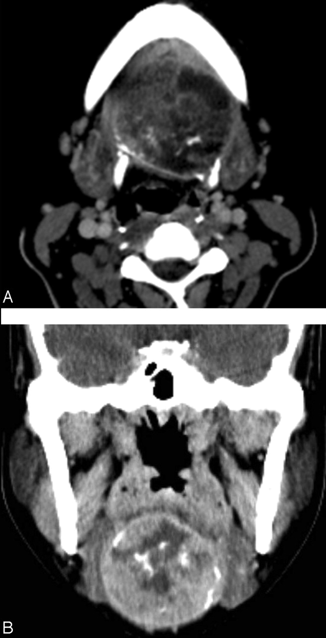
The CT scan revealed a heterogeneous mass at the level of, and replacing, the body of the hyoid bone, which was expanded to the left lesser cornu (hyoid bone consists of 5 segments: a body, 2 greater cornua and 2 lesser cornua). The location of the mass just inferior to the floor of the mouth, with peripheral rim calcification and internal chondroid calcification, suggested a cartilaginous tumor of the hyoid (Fig 2).

Fig 2.
  • Download figure
  • Open in new tab
  • Download powerpoint
Fig 2.

A, Contrast-enhanced axial CT scan at the level of hyoid bone revealed a well- defined mixed attenuation mass that has expanded to the left lesser cornu and has replaced the normal body of the hyoid bone with peripheral rim calcification and internal chondroid calcification. The right lesser cornu cannot be demonstrated.

B, Contrast-enhanced coronal CT scan revealed a round well-defined inhomogeneous attenuation mass with peripheral rim calcification and internal chondroid calcification below the floor of the mouth.

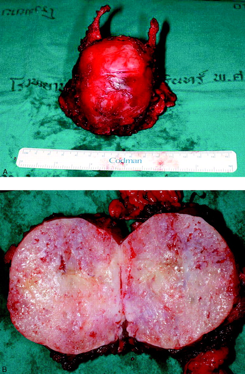
The operative finding revealed a well-capsulated mass originating form the hyoid bone (Fig 3). The mass adhered to the submandibular glands, strap, and mylohyoid muscles, but without definite invasion.

Fig 3.
  • Download figure
  • Open in new tab
  • Download powerpoint
Fig 3.

A, The surgical specimen revealed a well-defined, almost-round mass originated from the body, both lesser cornua of the hyoid bone, more prominent on the left side The appearance was concordant with the CT finding.

B, The cut surface of the resected specimen revealed an encapsulated firm, gray-white tissue tumor measuring 7 cm in diameter.

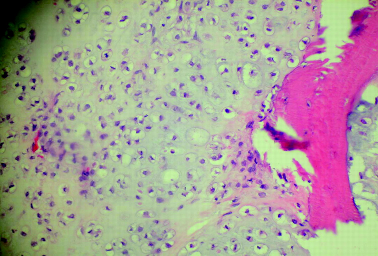
Histopathologically, the neoplasm was composed of mature chondrocytes with minimal nuclear and cytoplasmic atypia compatible with grade I chondrosarcoma (Fig 4, Table).3

Fig 4.
  • Download figure
  • Open in new tab
  • Download powerpoint
Fig 4.

Histopathology revealed grade 1 chondrosarcoma seen as mature chondrocytes with minimal nuclear and cytoplasmic atypia.

View this table:
  • View inline
  • View popup

Histological grades of chondrosarcoma28 in correlation with 5-year survival rate29

Discussion

The diagnosis of a chondrosarcoma of the hyoid bone may be missed because of its infrequent occurrence. Solid primary lesions of the hyoid bone are exceedingly rare and the reported cases have included plasmacytoma, osteosarcoma, giant cell tumor, aneurysmal bone cysts, osteoma, chondroma, and chondrosarcoma.3–11 Only 14 cases of chondrosarcoma of the hyoid bone have been reported in the literature.3,12–21

Chondrosarcoma may involve any part of the hyoid bone. Involvement of the body, greater or lesser cornus have already been reported in the literature.3,12–21 The size of the lesions varied from 0.5 cm to 7 cm. The age of patients also varied between 30 and 82 years (average age, 51.69 years).

Ultrasonography can help determine the texture of the mass, but often not the site of the origin. The common associated calcifications may be detected by ultrasonography, but not as well as with a CT scan. It was difficult and may be impossible to suggest the correct diagnosis by sonography.

A CT scan is the imaging technique of choice because it can better clarify a tumor’s extension and origin. The location beneath the mylohyoid muscles and the expansion of the hyoid bone with chondroid calcification should suggest the correct diagnosis. Approximately 75% of chondrosarcomas will demonstrate intrinsic calcification.3,17 The multiplanar capabilities and better tissue contrast of MR imaging can more precisely define the extension of the tumor and its relationship to surrounding structures and aid in planning the surgical approach.17 Chondrosarcoma appears as an inhomogeneous area of low signal intensity on a T1-weighted image and a more inhomogenous mixed high–low signal intensity on T2-weighted image. The calcifications revealed as areas of signal intensity void.3,12 A strongly enhancing mosaic appearance within the high-signal-intensity area on T2-weighted image may be an important diagnostic clue for chondrosarcoma of the hyoid bone; however, osteogenic tumor and aneurysmal bone cysts may have a similar MR imaging appearance.12

It is very difficult and may be impossible to distinguish chondroma from low-grade chondrosarcoma radiographically.2,3,22 Rare cases of chondroma have been reported.23–26 Geirnaerdt et al27 reported that fast contrast-enhanced MR imaging may assist in differentiation between benign and malignant cartilaginous tumors. Early enhancement was seen in chondrosarcoma, not seen in chondroma, and seen in osteochondroma only when growth plates were unfused. Pathologic differentiation between low-grade chondrosarcoma and chondroma can be also very difficult. Higher-grade chondrosarcomas have an increased cellularity, more nuclear pleomorphism, and identifiable mitotic figures.

Chrondrosarcoma can be histologically graded from 1 to 3 on the basis of the mitotic rate, cellularity, and nuclear size (Table).28

Surgical excision is the treatment of choice for chondrosarcoma. Neither radiation therapy nor chemotherapy plays a significant role in either primary or adjuvant treatment. Chondrosarcoma is radioresistant, though a trial of radiation therapy may be considered for patients who decline or are not surgical candidates.12,17

The hyoid bone is the insertion of the musculature of the tongue and also is attached to the thyroid cartilage via the thyrohyoid membrane and the extrinsic muscles of the larynx. Total hyoidectomy to completely excise the tumor may cause abnormal phonation and difficulties in swallowing and respiration. Our patient, however, did not have any abnormal symptoms and was able to return to her normal life and work.

References

  1. ↵
    Koch BB, Karnell LH, Hoffman HT, et al. National cancer database report on chondrosarcoma of the head and neck. Head Neck 2000;22:408–25
    CrossRefPubMed
  2. ↵
    Manaster BJ. Skeletal radiology: handbooks in radiology. Chicago: Year Book Medical Publishers;1989
  3. ↵
    Demeyere A, De Somer F, Perdieus D, et al. Chondrosarcoma of the hyoid bone. Eur Radiol 2000;10:308–309
    CrossRefPubMed
  4. Anderson TD, Kearney JJ. Osteosarcoma of the hyoid bone. Otolaryngol Head Neck Surg 2002;126:81–82
    CrossRefPubMed
  5. Commins DJ, O’Malley S, Athanasou NA, et al. Giant cell tumour of the hyoid: first reported case. J Laryngol Otol 1999;113:566–68
    PubMed
  6. Danaci M, Belet U, Akan H, et al. Solitary plasmacytoma of the hyoid bone. Otolaryngol Head Neck Surg 1999;121:834–35
    Abstract/FREE Full Text
  7. Goel S, Moorjani V, Kulkarni P, et al. Plasmacytoma of the hyoid. J Laryngol Otol 1994;108:604–606
    PubMed
  8. Iype EM, Abraham EK, Kumar K, et al. Giant cell tumour of hyoid bone: case report. Br J Oral Maxillofac Surg 2000;38:610–11
    CrossRefPubMed
  9. Khademi B, Gandomi B. Giant cell reparative granuloma of the hyoid bone. Otolaryngol Head Neck Surg 2001;124:117–18
    FREE Full Text
  10. Shadaba A, Zaidi S. Aneurysmal bone cyst of the hyoid. J Laryngol Otol 1992;106:71–72
    PubMed
  11. ↵
    Weber AL, Loewenheim HM. Osteoma arising from the stylohyoid chain and manifesting as a neck and oropharyngeal mass. Ann Otol Rhinol Laryngol 1993;102:399–401
    FREE Full Text
  12. ↵
    Itoh K, Nobori T, Fukuda K, et al. Chondrosarcoma of the hyoid bone. J Laryngol Otol 1993;107:642–46
    PubMed
  13. Blandamura S, Rampazzo F, Altavilla G, et al. [Chondrosarcoma of the hyoid bone: follow-up and cytological study of a case]. Pathologica 1992;84:235–41
    PubMed
  14. Dalla Palma P, Piazza M. Peripheral chondrosarcoma of the hyoid bone: report of a case. Appl Pathol 1983;1:333–38
    PubMed
  15. Greer JA Jr, Devine KD, Dahlin DC. Gardner’s syndrome and chondrosarcoma of the hyoid bone. Arch Otolaryngol 1977;103:425–27
    CrossRefPubMed
  16. Hasan S, Kannan V, Shenoy AM, et al. Chondrosarcoma of the hyoid. J Laryngol Otol 1992;106:273–76
    PubMed
  17. ↵
    Hediger R, McEniff N, Karmody C, et al. Recurrent chondrosarcoma of the hyoid bone. Clin Imaging 1997;21:69–72
    CrossRefPubMed
  18. Joo M, Kang YK, Kim HS, et al. Mesenchymal chondrosarcoma of the hyoid bone: a case report. J Korean Med Sci 1998;13:696–700
    PubMed
  19. Ozer E, Kanlikama M, Bayazit YA, et al. Chondrosarcoma of the hyoid bone. Otolaryngol Head Neck Surg 2004;131:787–88
    FREE Full Text
  20. Umekita Y, Yoshida A, Yoshida H. Parosteal chondrosarcoma attached to the hyoid bone: a case report. Int J Oral Maxillofac Surg 1998;27:215–16
    CrossRefPubMed
  21. ↵
    Hoffer O, Baccioni R. [Invasion of the tongue by a rare malignancy (chondrosarcoma of the hyoid bone, Ewing’s tumor of the cranial base), with reference to Garcin’s syndrome]. Arch De Vecchi Anat Patol 1959;29:393–408
    PubMed
  22. ↵
    Baatenburg de Jong RJ, van Lent S, Hogendoorn PC. Chondroma and chondrosarcoma of the larynx. Curr Opin Otolaryngol Head Neck Surg 2004;12:98–105
    CrossRefPubMed
  23. ↵
    Fedorova MA. [Chondroma of the hyoid bone.]. Vestn Otorinolaringol 1953;15:90–91
    PubMed
  24. Grayson A, Bain M. Juxtacortical chondroma of the hyoid bone: report of a case. Arch Otolaryngol 1967;86:679–80
    CrossRefPubMed
  25. Nakagawa Y, Ishibashi K, Asada K, et al. Chondroma of the hyoid bone: a case report. Oral Surg Oral Med Oral Pathol Oral Radiol Endod 1999;88:333–36
    CrossRefPubMed
  26. ↵
    Tamura H, Sato T. [A case of cystic enchondroma of the hyoid bone.]. Hno 1961;9:275–77
    PubMed
  27. ↵
    Geirnaerdt MJ, Hogendoorn PC, Bloem JL, et al. Cartilaginous tumors: fast contrast-enhanced MR imaging. Radiology 2000;214:539–46
    PubMed
  28. ↵
    Evans HL, Ayala AG, Romsdahl MM. Prognostic factors in chondrosarcoma of bone. Cancer 1977;40:818–31
    CrossRefPubMed
  29. ↵
    Finn DG, Goepfert H, Batsakis JG. Chondrosarcoma of the head and neck. Laryngoscope 1984;94:1539–44
    PubMed
  • Received November 16, 2004.
  • Accepted after revision February 24, 2005.
  • Copyright © American Society of Neuroradiology
View Abstract
PreviousNext
Back to top

In this issue

American Journal of Neuroradiology: 27 (1)
American Journal of Neuroradiology
Vol. 27, Issue 1
January, 2006
  • Table of Contents
  • Index by author
Advertisement
Print
Download PDF
Email Article

Thank you for your interest in spreading the word on American Journal of Neuroradiology.

NOTE: We only request your email address so that the person you are recommending the page to knows that you wanted them to see it, and that it is not junk mail. We do not capture any email address.

Enter multiple addresses on separate lines or separate them with commas.
Chondrosarcoma of the Hyoid Bone: Imaging, Surgical, and Histopathologic Correlation
(Your Name) has sent you a message from American Journal of Neuroradiology
(Your Name) thought you would like to see the American Journal of Neuroradiology web site.
CAPTCHA
This question is for testing whether or not you are a human visitor and to prevent automated spam submissions.
Cite this article
S. Hirunpat, K. Prueganusak, Y. Sinruk, W. Tanomkiat
Chondrosarcoma of the Hyoid Bone: Imaging, Surgical, and Histopathologic Correlation
American Journal of Neuroradiology Jan 2006, 27 (1) 123-125;

Citation Manager Formats

  • BibTeX
  • Bookends
  • EasyBib
  • EndNote (tagged)
  • EndNote 8 (xml)
  • Medlars
  • Mendeley
  • Papers
  • RefWorks Tagged
  • Ref Manager
  • RIS
  • Zotero
0 Responses
Respond to this article
Share
Bookmark this article
Chondrosarcoma of the Hyoid Bone: Imaging, Surgical, and Histopathologic Correlation
S. Hirunpat, K. Prueganusak, Y. Sinruk, W. Tanomkiat
American Journal of Neuroradiology Jan 2006, 27 (1) 123-125;
del.icio.us logo Twitter logo Facebook logo Mendeley logo
  • Tweet Widget
  • Facebook Like
  • Google Plus One

Jump to section

  • Article
    • Abstract
    • Case Report
    • Discussion
    • References
  • Figures & Data
  • Info & Metrics
  • Responses
  • References
  • PDF

Related Articles

  • No related articles found.
  • PubMed
  • Google Scholar

Cited By...

  • Chondrosarcoma of the hyoid bone: a case of mistaken identity in the submandibular region
  • Chondrosarcoma of the hyoid bone: an atypical site of a sarcoma of the head and the neck
  • Osteoblastoma of the Hyoid Bone
  • Crossref
  • Google Scholar

This article has not yet been cited by articles in journals that are participating in Crossref Cited-by Linking.

More in this TOC Section

  • Correlation of Apparent Diffusion Coefficient at 3T with Prognostic Parameters of Retinoblastoma
  • Parathyroid Lesions: Characterization with Dual-Phase Arterial and Venous Enhanced CT of the Neck
  • Efficacy of Diffusion-Weighted Imaging for the Differentiation between Lymphomas and Carcinomas of the Nasopharynx and Oropharynx: Correlations of Apparent Diffusion Coefficients and Histologic Features
Show more Head and Neck

Similar Articles

Advertisement

Indexed Content

  • Current Issue
  • Accepted Manuscripts
  • Article Preview
  • Past Issues
  • Editorials
  • Editors Choice
  • Fellow Journal Club
  • Letters to the Editor

Cases

  • Case Collection
  • Archive - Case of the Week
  • Archive - Case of the Month
  • Archive - Classic Case

Special Collections

  • Special Collections

Resources

  • News and Updates
  • Turn around Times
  • Submit a Manuscript
  • Author Policies
  • Manuscript Submission Guidelines
  • Evidence-Based Medicine Level Guide
  • Publishing Checklists
  • Graphical Abstract Preparation
  • Imaging Protocol Submission
  • Submit a Case
  • Become a Reviewer/Academy of Reviewers
  • Get Peer Review Credit from Publons

Multimedia

  • AJNR Podcast
  • AJNR SCANtastic
  • Video Articles

About Us

  • About AJNR
  • Editorial Board
  • Not an AJNR Subscriber? Join Now
  • Alerts
  • Feedback
  • Advertise with us
  • Librarian Resources
  • Permissions
  • Terms and Conditions

American Society of Neuroradiology

  • Not an ASNR Member? Join Now

© 2025 by the American Society of Neuroradiology All rights, including for text and data mining, AI training, and similar technologies, are reserved.
Print ISSN: 0195-6108 Online ISSN: 1936-959X

Powered by HighWire