Advertisement

Main menu

  • Home
  • Content
    • Current Issue
    • Accepted Manuscripts
    • Article Preview
    • Past Issue Archive
    • AJNR Case Collection
    • Case of the Week Archive
    • Classic Case Archive
    • Case of the Month Archive
  • Special Collections
    • Spinal CSF Leak Articles (Jan 2020-June 2024)
    • 2024 AJNR Journal Awards
    • Most Impactful AJNR Articles
  • Multimedia
    • AJNR Podcast
    • AJNR Scantastics
    • Video Articles
  • For Authors
    • Submit a Manuscript
    • Author Policies
    • Fast publishing of Accepted Manuscripts
    • Graphical Abstract Preparation
    • Manuscript Submission Guidelines
    • Imaging Protocol Submission
    • Submit a Case for the Case Collection
  • About Us
    • About AJNR
    • Editorial Board
  • More
    • Become a Reviewer/Academy of Reviewers
    • Subscribers
    • Permissions
    • Alerts
    • Feedback
    • Advertisers
    • ASNR Home
  • Other Publications
    • ajnr

User menu

  • Alerts
  • Log in

Search

  • Advanced search
American Journal of Neuroradiology

American Journal of Neuroradiology

ASHNR American Society of Functional Neuroradiology ASHNR American Society of Pediatric Neuroradiology ASSR
  • Alerts
  • Log in

Advanced Search

  • Home
  • Content
    • Current Issue
    • Accepted Manuscripts
    • Article Preview
    • Past Issue Archive
    • AJNR Case Collection
    • Case of the Week Archive
    • Classic Case Archive
    • Case of the Month Archive
  • Special Collections
    • Spinal CSF Leak Articles (Jan 2020-June 2024)
    • 2024 AJNR Journal Awards
    • Most Impactful AJNR Articles
  • Multimedia
    • AJNR Podcast
    • AJNR Scantastics
    • Video Articles
  • For Authors
    • Submit a Manuscript
    • Author Policies
    • Fast publishing of Accepted Manuscripts
    • Graphical Abstract Preparation
    • Manuscript Submission Guidelines
    • Imaging Protocol Submission
    • Submit a Case for the Case Collection
  • About Us
    • About AJNR
    • Editorial Board
  • More
    • Become a Reviewer/Academy of Reviewers
    • Subscribers
    • Permissions
    • Alerts
    • Feedback
    • Advertisers
    • ASNR Home
  • Follow AJNR on Twitter
  • Visit AJNR on Facebook
  • Follow AJNR on Instagram
  • Join AJNR on LinkedIn
  • RSS Feeds

Welcome to the new AJNR, Updated Hall of Fame, and more. Read the full announcements.


AJNR is seeking candidates for the position of Associate Section Editor, AJNR Case Collection. Read the full announcement.


OtherHEAD AND NECK

Chondrosarcoma of the Hyoid Bone: Imaging, Surgical, and Histopathologic Correlation

S. Hirunpat, K. Prueganusak, Y. Sinruk and W. Tanomkiat
American Journal of Neuroradiology January 2006, 27 (1) 123-125;
S. Hirunpat
  • Find this author on Google Scholar
  • Find this author on PubMed
  • Search for this author on this site
K. Prueganusak
  • Find this author on Google Scholar
  • Find this author on PubMed
  • Search for this author on this site
Y. Sinruk
  • Find this author on Google Scholar
  • Find this author on PubMed
  • Search for this author on this site
W. Tanomkiat
  • Find this author on Google Scholar
  • Find this author on PubMed
  • Search for this author on this site
  • Article
  • Figures & Data
  • Info & Metrics
  • Responses
  • References
  • PDF
Loading

Article Figures & Data

Figures

  • Tables
  • Fig 1.
    • Download figure
    • Open in new tab
    • Download powerpoint
    Fig 1.

    An inhomogeneous solid echoic mass within the submental area, slightly more prominent on the left side.
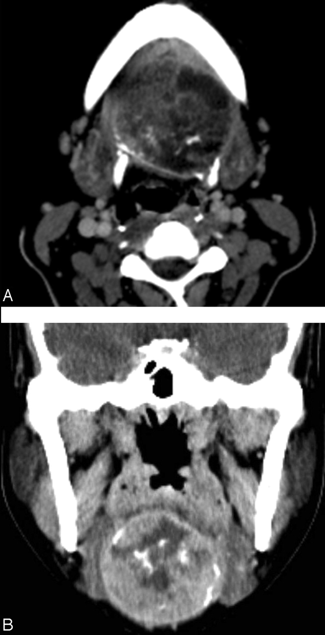
  • Fig 2.
    • Download figure
    • Open in new tab
    • Download powerpoint
    Fig 2.

    A, Contrast-enhanced axial CT scan at the level of hyoid bone revealed a well- defined mixed attenuation mass that has expanded to the left lesser cornu and has replaced the normal body of the hyoid bone with peripheral rim calcification and internal chondroid calcification. The right lesser cornu cannot be demonstrated.

    B, Contrast-enhanced coronal CT scan revealed a round well-defined inhomogeneous attenuation mass with peripheral rim calcification and internal chondroid calcification below the floor of the mouth.
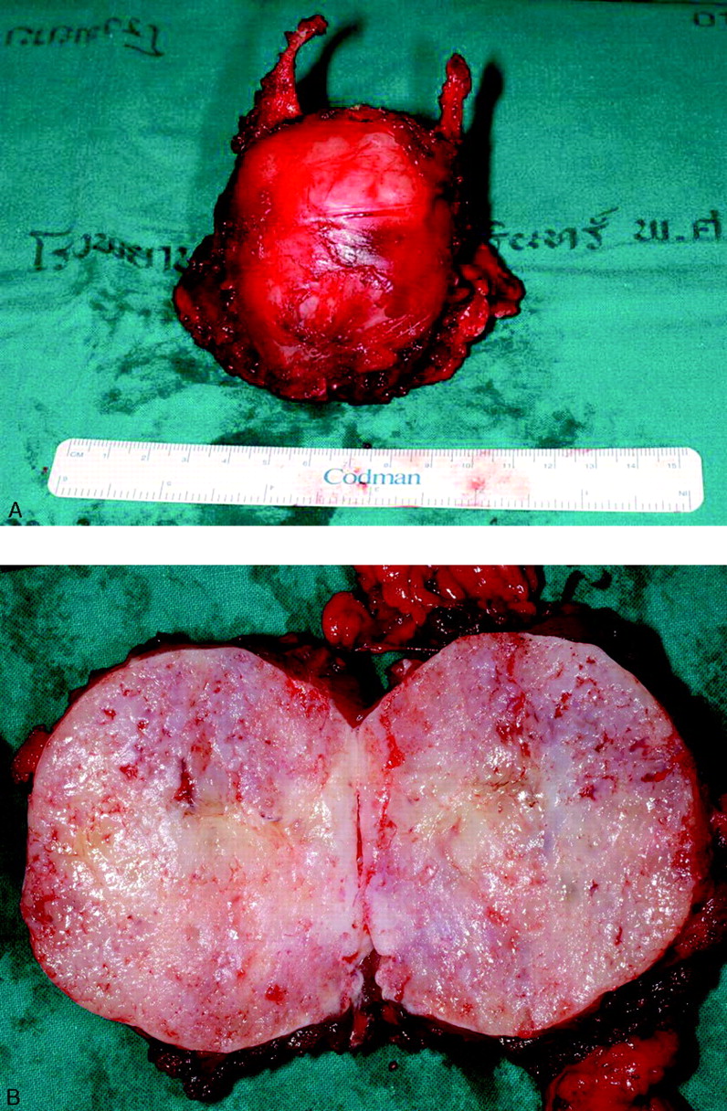
  • Fig 3.
    • Download figure
    • Open in new tab
    • Download powerpoint
    Fig 3.

    A, The surgical specimen revealed a well-defined, almost-round mass originated from the body, both lesser cornua of the hyoid bone, more prominent on the left side The appearance was concordant with the CT finding.

    B, The cut surface of the resected specimen revealed an encapsulated firm, gray-white tissue tumor measuring 7 cm in diameter.
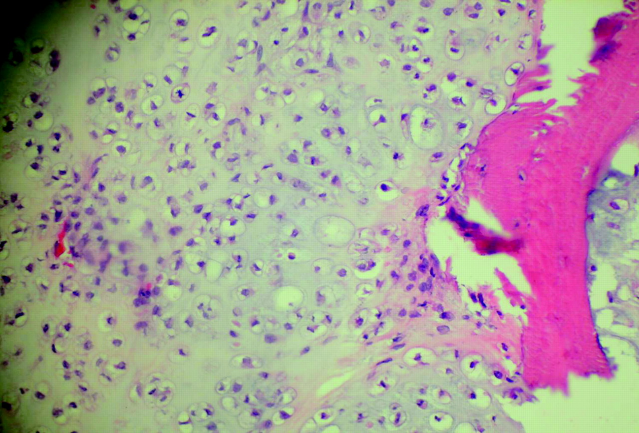
  • Fig 4.
    • Download figure
    • Open in new tab
    • Download powerpoint
    Fig 4.

    Histopathology revealed grade 1 chondrosarcoma seen as mature chondrocytes with minimal nuclear and cytoplasmic atypia.

Tables

  • Figures
  • Histological grades of chondrosarcoma28 in correlation with 5-year survival rate29

    GradeHistology5-Year Survival Rate (%)
    1Lesion exhibits a preponderance of small, densely stained nuclei90
    2Lesion contains areas with moderate-sized nuclei but with a low mitotic rate81
    3Lesion has large nuclei, with foci of dense cellularity and a high mitotic rate43
PreviousNext
Back to top

In this issue

American Journal of Neuroradiology: 27 (1)
American Journal of Neuroradiology
Vol. 27, Issue 1
January, 2006
  • Table of Contents
  • Index by author
Advertisement
Print
Download PDF
Email Article

Thank you for your interest in spreading the word on American Journal of Neuroradiology.

NOTE: We only request your email address so that the person you are recommending the page to knows that you wanted them to see it, and that it is not junk mail. We do not capture any email address.

Enter multiple addresses on separate lines or separate them with commas.
Chondrosarcoma of the Hyoid Bone: Imaging, Surgical, and Histopathologic Correlation
(Your Name) has sent you a message from American Journal of Neuroradiology
(Your Name) thought you would like to see the American Journal of Neuroradiology web site.
CAPTCHA
This question is for testing whether or not you are a human visitor and to prevent automated spam submissions.
Cite this article
S. Hirunpat, K. Prueganusak, Y. Sinruk, W. Tanomkiat
Chondrosarcoma of the Hyoid Bone: Imaging, Surgical, and Histopathologic Correlation
American Journal of Neuroradiology Jan 2006, 27 (1) 123-125;

Citation Manager Formats

  • BibTeX
  • Bookends
  • EasyBib
  • EndNote (tagged)
  • EndNote 8 (xml)
  • Medlars
  • Mendeley
  • Papers
  • RefWorks Tagged
  • Ref Manager
  • RIS
  • Zotero
0 Responses
Respond to this article
Share
Bookmark this article
Chondrosarcoma of the Hyoid Bone: Imaging, Surgical, and Histopathologic Correlation
S. Hirunpat, K. Prueganusak, Y. Sinruk, W. Tanomkiat
American Journal of Neuroradiology Jan 2006, 27 (1) 123-125;
del.icio.us logo Twitter logo Facebook logo Mendeley logo
  • Tweet Widget
  • Facebook Like
  • Google Plus One

Jump to section

  • Article
    • Abstract
    • Case Report
    • Discussion
    • References
  • Figures & Data
  • Info & Metrics
  • Responses
  • References
  • PDF

Related Articles

  • No related articles found.
  • PubMed
  • Google Scholar

Cited By...

  • Chondrosarcoma of the hyoid bone: a case of mistaken identity in the submandibular region
  • Chondrosarcoma of the hyoid bone: an atypical site of a sarcoma of the head and the neck
  • Osteoblastoma of the Hyoid Bone
  • Crossref
  • Google Scholar

This article has not yet been cited by articles in journals that are participating in Crossref Cited-by Linking.

More in this TOC Section

  • Correlation of Apparent Diffusion Coefficient at 3T with Prognostic Parameters of Retinoblastoma
  • Parathyroid Lesions: Characterization with Dual-Phase Arterial and Venous Enhanced CT of the Neck
  • Efficacy of Diffusion-Weighted Imaging for the Differentiation between Lymphomas and Carcinomas of the Nasopharynx and Oropharynx: Correlations of Apparent Diffusion Coefficients and Histologic Features
Show more Head and Neck

Similar Articles

Advertisement

Indexed Content

  • Current Issue
  • Accepted Manuscripts
  • Article Preview
  • Past Issues
  • Editorials
  • Editors Choice
  • Fellow Journal Club
  • Letters to the Editor

Cases

  • Case Collection
  • Archive - Case of the Week
  • Archive - Case of the Month
  • Archive - Classic Case

Special Collections

  • Special Collections

Resources

  • News and Updates
  • Turn around Times
  • Submit a Manuscript
  • Author Policies
  • Manuscript Submission Guidelines
  • Evidence-Based Medicine Level Guide
  • Publishing Checklists
  • Graphical Abstract Preparation
  • Imaging Protocol Submission
  • Submit a Case
  • Become a Reviewer/Academy of Reviewers
  • Get Peer Review Credit from Publons

Multimedia

  • AJNR Podcast
  • AJNR SCANtastic
  • Video Articles

About Us

  • About AJNR
  • Editorial Board
  • Not an AJNR Subscriber? Join Now
  • Alerts
  • Feedback
  • Advertise with us
  • Librarian Resources
  • Permissions
  • Terms and Conditions

American Society of Neuroradiology

  • Not an ASNR Member? Join Now

© 2025 by the American Society of Neuroradiology All rights, including for text and data mining, AI training, and similar technologies, are reserved.
Print ISSN: 0195-6108 Online ISSN: 1936-959X

Powered by HighWire