
Abstract
SUMMARY: The existence of the vein of the foramen caecum (VFC) in humans is still controversial. We present 2 patients with intracranial drainage of the nasal mucosa by a frontal cortical vein into a superior sagittal sinus, demonstrated by digital subtraction angiography. In both, the position of the intracranial passage was found to be slightly paramedian. An analogy to the VFC is made.
The vein of the foramen caecum (VFC), is described as a vein connecting the nasal and paranasal mucosa to the superior sagittal sinus, through the foramen caecum.1 Although this vein may be found in lower mammals such as the mole,2 its existence in humans is questioned by various authors.2,3 We present 2 cases of intracranial drainage of the nasal mucosa by a frontal cortical vein into a superior sagittal sinus (SSS), demonstrated by digital subtraction angiography (DSA).
Case Descriptions
Case 1
A 31-year-old woman with adult polycystic kidney disease underwent routine screening for intracranial aneurysm disease by MR imaging. The discovery of a small aneurysm at the right middle cerebral artery (MCA) bifurcation by MR angiography prompted further investigation by DSA. Four-vessel cerebral DSA confirmed the existence of a single 4-mm saccular aneurysm at the MCA bifurcation.
An unusual intracranial drainage pathway for the septal nasal mucosa was incidentally documented angiographically. The septal nasal mucosa was drained by a thin ascending nasal draining vein, which passed through the floor of the anterior cranial fossa and continued its course intracranially as a left frontal cortical vein. This left frontal vein received several pial tributaries from the superior frontal gyrus, before ending its course into the SSS (Figs 1–3). The ascending nasal vein was located in the anterior-most aspect of the floor of the anterior cranial fossa and occupied a slightly paramedian position. This disposition was only found on the left side. The anterior portion of the SSS was hypoplastic, being replaced by bilateral longitudinal frontal veins.

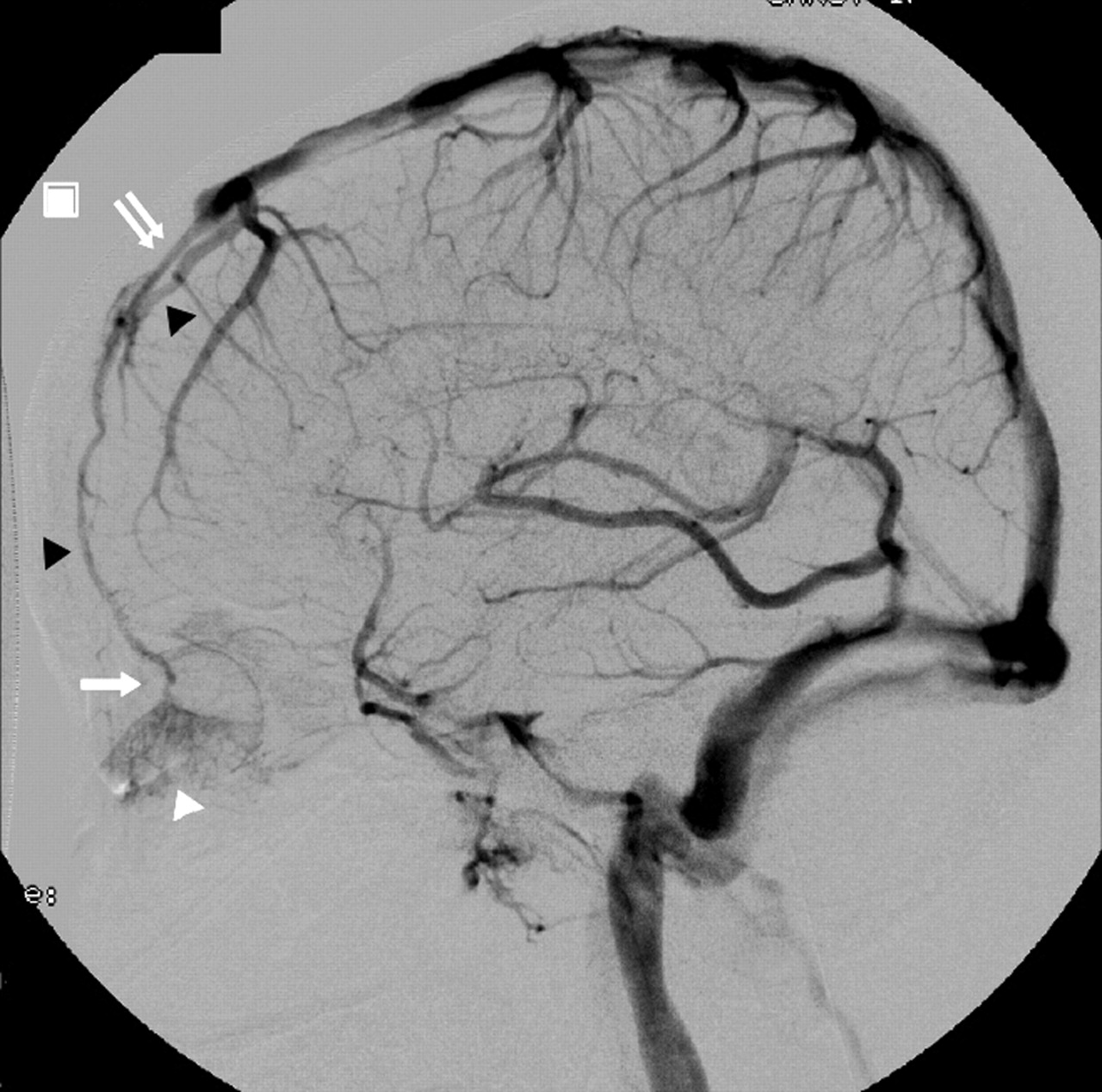
Case 1. DSA, left ICA injection, venous phase varying from early to late venous filling (Figs. 1–3). Lateral view. The left septal nasal mucosa (white arrowhead) is drained by a thin ascending nasal vein (white arrow) at the anterior-most aspect of the anterior cranial fossa. Intracranially, the thin vein continues its course as a frontal cortical vein (black arrowheads) and drains ultimately into the SSS. Note how the anterior third of the SSS is hypoplastic (double white arrow).

Case 1. DSA anteroposterior projection, intermediate venous phase of left ICA injection, with (A) and without subtraction (B), as in Fig 1. The medial left septal nasal mucosa appears as a slitlike opacity. The thin ascending nasal vein (black arrow) occupies a slightly paramedian position, which is better appreciated relative to bony landmarks in the nonsubtracted images (B). In conjunction with the lateral views, the position of the ascending nasal vein is compatible with the position of a foramen of the cribriform plate. The frontal cortical vein ascends toward the SSS.

Case 2
A 39-year-old woman underwent follow-up cerebral DSA after surgical clipping of an anterior communicating artery aneurysm.
DSA documented a small patch of nasal mucosa draining in the same fashion as in case 1 (Fig 4). This drainage variant was not found in the right side. The anterior third of the SSS was also hypoplastic, being replaced by bilateral longitudinal frontal veins.

Case 2. DSA, left ICA injection, early and intermediary venous phase. Anteroposterior view (as Figs 1–3). There is gradual filling of the left septal nasal mucosa (white arrowhead), which drains in the same fashion as case 1. Note the presence of a hypoplastic SSS, as in case 1. Black arrowheads indicate frontal cortical vein.
Discussion
The existence in humans of a VFC, that is, a vein connecting the nasal and paranasal mucosa to the SSS, has often been mentioned in classic anatomy texts.1,4,5 Zuckerkandl,6 however, could not demonstrate, by using injection and corrosion techniques, any direct venous communication between the nasal mucosa and the SSS through the foramen caecum. He did, by contrast, observe venous drainage pathways passing through the cribriform plate before entering the SSS, the venous plexus surrounding the olfactory tract, or the superficial cerebral veins of the frontal lobe.
The existence of the VFC has also been refuted by Kaplan et al,7 who found no venous structure passing through the foramen caecum in a series of 201 autopsy cases that included subjects of all age categories. Although the foramen caecum was patent in all of the cases, it was filled with a fibrous material. Boyd,3 studying the inside of 212 dry skulls, found only 3 cases in which the foramen caecum was permeable, and he described them as being so narrow that they could only give way to a fine hair. In 2 dissected specimens, Boyd found that the foramen caecum formed a pit filled with a rootlike extension of dura mater and was unable to find a connection with the lumen of the SSS. The same was observed by Zuckerkandl.6 Although these reports are contradictory regarding the patency of the foramen caecum, they are consistent with the concept that, even when the foramen caecum is permeable, its small size or its fibrous content do not allow the passage of a macroscopically visible vein.
The situation may be different in neonates and during intrauterine life. Theile8 believed that the VFC was present in neonates. Zuckerkandl6 described a case in which blood vessels in the foramen caecum connected the SSS with veins of the soft tissues of the face, including the nose. Luschka9 also reported the case of a neonate baby, in whom a large vein could be followed through the foramen caecum as far as to the veins of the face. Finally, in his treatise on cerebral veins, Hédon reported the observation of VFC in infants, while admitting that it was rarely found in adult specimens.5
We present the angiographic observation of 2 patients in whom a portion of the nasal mucosa drained intracranially via a thin ascending nasal vein that crossed the floor of the anterior cranial fossa to continue as a left cortical frontal vein. In both cases, a hypoplastic anterior portion of the SSS was replaced by bilateral longitudinal frontal cortical veins, which on the left side, drained the nasal septal mucosa. Hypoplasia or absence of the anterior segment of the SSS and its replacement by longitudinal cortical veins is a relatively common anatomic variation. On the other hand, ascending intracranial drainage of the nasal mucosa is exceptional, and has, to the best of our knowledge, not been previously reported from an angiographic standpoint. In both cases the ascending nasal vein was found to be in a slightly paramedian position on the anteroposterior views, and occupied the anterior-most aspect of the floor of the anterior cranial fossa on the lateral views. The exact site of passage of the ascending nasal vein through the anterior cranial fossa is difficult to ascertain on the basis of the available angiographic data, but the paramedian location suggests passage through a foramen of the cribriform plate, rather than through a foramen caecum. Thus, the intracranial drainage of the nasal mucosa observed here differed from the classic description of a VFC in that it joined a frontal cortical vein before connecting to the SSS, and intracranial passage most likely occurred through a foramen of the cribriform plate. Functionally, however, it is analogous to a VFC.
As mentioned above, the available literature on the foramen caecum and VFC remains controversial but seems to offer at least one consistent line of thought: the VFC, if it exists, is more likely to be found in fetuses or neonate babies than later in life. This suggests a regressive process, not unlike the disappearance of other embryonic venous structures during the late fetal stages or early postnatal period. Again, such a hypothesis appears consistent with the paucity of reported observations of VFC in adults and, a fortiori, the absence of angiographic demonstration of the variant so far. In our 2 patients, intracranial drainage of the nasal mucosa most likely occurred through a foramen of the cribriform plate. Foramina of the cribriform remain patent with age, thus, allowing for this connection to persist in adult life.
The anomalous venous drainage through the cribriform plate presented here has both intra- and extracranial components but should not be considered a cerebral emissary vein, because as it does not offer an extracranial drainage route for encephalic blood, but rather an intracranial drainage route for the nasal mucosa. Finally, from a clinical perspective, the VFC and its cribriform plate equivalent represent a potential pathway for intracranial spread of infectious or tumoral processes originating from the nasal cavity. Furthermore, an anatomic disposition such as the one presented here could also provide the anatomic substratum for the development of dural arteriovenous fistulas of the cribriform plate region with frontal cortical drainage.
- Received December 10, 2004.
- Accepted after revision February 24, 2005.
- Copyright © American Society of Neuroradiology
In this issue


Jump to section
Related Articles
Cited By...
- No citing articles found.