
Article Figures & Data
Figures
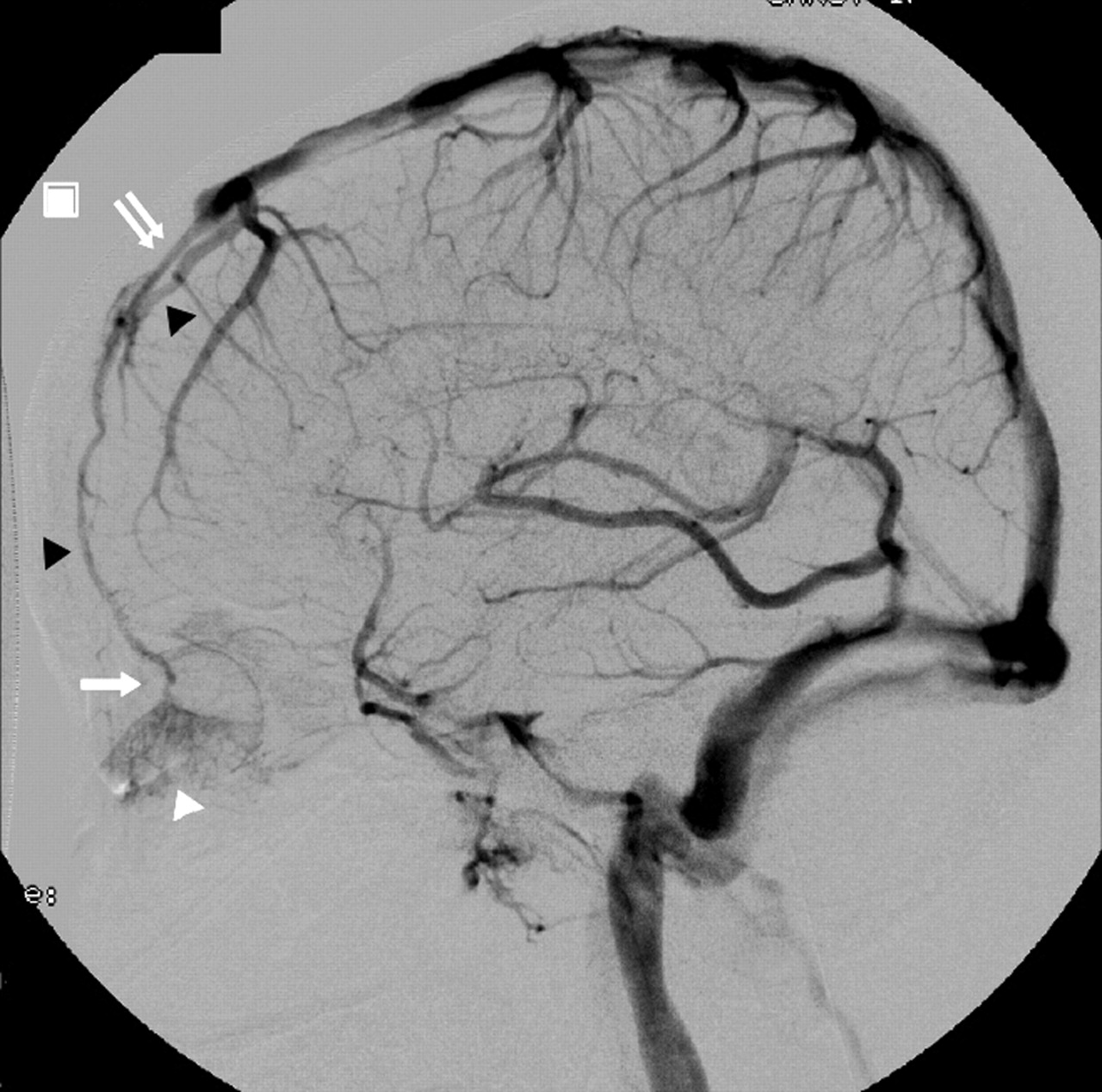
- Fig 1.
Case 1. DSA, left ICA injection, venous phase varying from early to late venous filling (Figs. 1–3). Lateral view. The left septal nasal mucosa (white arrowhead) is drained by a thin ascending nasal vein (white arrow) at the anterior-most aspect of the anterior cranial fossa. Intracranially, the thin vein continues its course as a frontal cortical vein (black arrowheads) and drains ultimately into the SSS. Note how the anterior third of the SSS is hypoplastic (double white arrow).
- Fig 2.
Case 1. DSA anteroposterior projection, intermediate venous phase of left ICA injection, with (A) and without subtraction (B), as in Fig 1. The medial left septal nasal mucosa appears as a slitlike opacity. The thin ascending nasal vein (black arrow) occupies a slightly paramedian position, which is better appreciated relative to bony landmarks in the nonsubtracted images (B). In conjunction with the lateral views, the position of the ascending nasal vein is compatible with the position of a foramen of the cribriform plate. The frontal cortical vein ascends toward the SSS.
- Fig 4.
Case 2. DSA, left ICA injection, early and intermediary venous phase. Anteroposterior view (as Figs 1–3). There is gradual filling of the left septal nasal mucosa (white arrowhead), which drains in the same fashion as case 1. Note the presence of a hypoplastic SSS, as in case 1. Black arrowheads indicate frontal cortical vein.


In this issue


Jump to section
Related Articles
Cited By...
- No citing articles found.