
Article Figures & Data
Figures
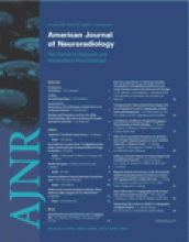
- Fig 1.
Photomicrographs illustrating neck healing. All sections were stained with H&E.
A, Representative section (magnification ×25) with the neck healing score = 0, showing unorganized laminated thrombus along the entire aneurysm neck in this section.
B, Representative section (magnification ×40) with the neck healing score = 1, illustrating unorganized fibrin across the aneurysm neck.
C, Representative section with the neck healing score = 2, showing both organized tissue and unorganized fibrin at the aneurysm neck (magnification ×50).
D, Representative section with the neck healing score = 3, showing a thin layer of completely organized tissue across the entire aneurysm neck (magnification ×100).
E, Representative section with the neck healing score = 4, showing a thick layer of completely organized tissue traversing the entire aneurysm neck (magnification ×60).
- Fig 2.
Photomicrographs illustrating dome healing. All sections were stained with H&E.
A, Representative section (magnification ×20) of a dome score = 0, showing that the aneurysm cavity is completely filled with unorganized thrombus.
B, Representative section (magnification ×15) when the dome score = 1, where less than one third of aneurysm cavity is filled with organized tissue.
C, Representative section (magnification ×20) with dome score = 2, in that approximately one half of the aneurysm cavity is filled with organized tissue.
D, Representative section (magnification ×15) with a dome score = 3, most the aneurysm cavity filled with organized tissue; a residual small area of unorganized thrombus is also evident.
E, Representative section (magnification ×15) with the dome score = 4, showing the aneurysm cavity to be entirely filled with loose organized tissue.
F, Representative section (magnification ×40) with the dome score = 5. Staining in this section reveals the aneurysm cavity is completely filled with attenuated cellular tissue.
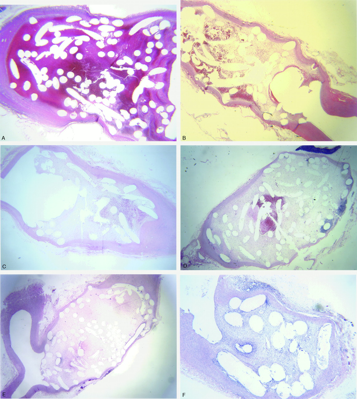
- Fig 3.
Photomicrographs illustrating histologic coil compaction scoring. All sections were stained with H&E.
A, Representative section for neck compaction score = 3, showing the tissue across the aneurysm neck is convex to the aneurysm cavity (magnification ×20).
B, Representative section for a neck compaction score = 2, showing the tissue at the neck is flat (magnification ×60).
C, Representative section for a neck compaction score = 1, showing the tissue at the neck slightly concave to the aneurysm cavity (magnification ×15).
D, Representative section for a neck compaction score = 0, showing the tissue at the interface between the aneurysm cavity and parent artery is markedly concave to the aneurysm cavity (magnification ×15).
- Fig 4.
Mean and SD for scoring categories, in addition to Total Score, over survival time in weeks. Different colored bars represent the mean ± SD for each variable in the ordinal scale at 2 weeks (n = 5), 4 weeks (n = 8), 10 weeks (n = 5), 16 weeks (n = 6), and 24 weeks (n = 6). Values for 2 weeks are significantly less (P < .05) than for 4 weeks, 16 weeks, and 24 weeks. Ten-week values are significantly less (P < .05) than 24-week values.
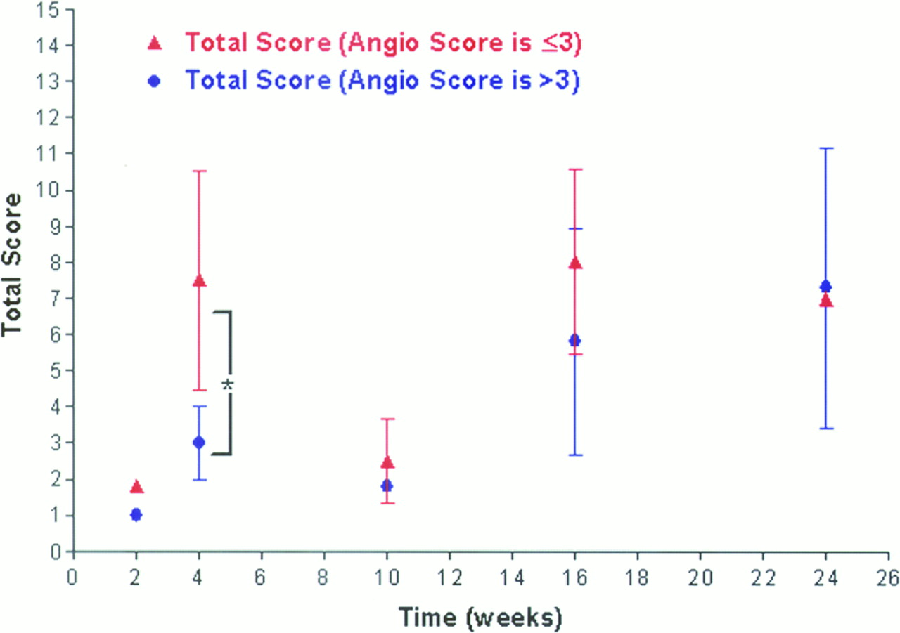
- Fig 5.
A comparison of healing (total score) over time between broad-necked (closed blue circle; >3) and small-necked (closed red triangle; ≤3) aneurysms. Data are represented by mean ± SD. There was a significant difference between the 2 groups (indicated by an asterisk) at the 4-week time period.


Tables
Scale Number Grading Components Gross Neck Micro Neck Neck Compaction Dome 0 No coverage Unorganized clot Macro-concave Blood clot (all) 1 <50% coverage Fibrin Micro-concave Organized tissue in <1/3 of dome area 2 50–75% coverage Fibrin with organized tissue Flat Organized tissue in 1/3–2/3 dome area 3 >75% coverage Completely organized tissue, thickness ≤1/3 coil diameter Convex Organized tissue in >2/3 of dome area 4 N/A Completely organized tissue, thickness >1/3 of coil diameter N/A Completely organized loose tissue 5 N/A N/A N/A Completely organized dense fibrous or cellular tissue Duration (weeks) Neck (mm) Width (mm) Height (mm) 2 3.1 ± 1.2 4.2 ± 0.8 9.8 ± 1.3 4 3.4 ± 1.0 4.4 ± 0.7 9.2 ± 1.8 10 4.0 ± 1.3 4.9 ± 1.1 9.1 ± 1.0 16 3.2 ± 0.9 4.0 ± 1.3 8.2 ± 1.9 24 3.2 ± 0.7 3.8 ± 0.8 8.4 ± 2.0 Note:—Data are represented as the mean ± SD. There were no significant differences between time points.
Gross Neck Micro Neck Compaction Dome Total Score 2 wk (n = 5) 0.0 ± 0.0 0.2 ± 0.3 0.8 ± 0.8 0.5 ± 0.4 1.4 ± 0.8 4 wk (n = 7) 1.3 ± 1.3 1.7 ± 1.0 0.9 ± 0.7 2.4 ± 1.0* 4.7 ± 1.8* 10 wk (n = 5) 0.8 ± 1.3 0.9 ± 1.2 0.4 ± 0.5 2.8 ± 1.4* 4.1 ± 2.5 16 wk (n = 4) 1.5 ± 0.6 2.1 ± 0.9* 1.1 ± 0.7 3.8 ± 0.4*† 6.5 ± 1.0* 24 wk (n = 6) 1.7 ± 1.2 2.4 ± 1.0* 1.2 ± 0.8 4.0 ± 0.0*† 7.2 ± 1.6*£ Note:—Data are represented as the mean ± SD.
* Variable is significantly different from the 2-week mean.
† Variable is significantly different from the 4-week mean.
£ Variable is significantly different from the 10-week mean.
In this issue


Jump to section
Related Articles
Cited By...
- WEB shape modifications: angiography-histopathology correlations in rabbits
- WEB shape modifications: angiography-histopathology correlations in rabbits
- Rabbit Elastase Aneurysm Model Mimics the Recurrence Rate of Human Intracranial Aneurysms following Platinum Coil Embolization
- WEB Device Shape Changes in Elastase-Induced Aneurysms in Rabbits
- Histologic and Biomolecular Similarities in Healing between Aneurysms and Cutaneous Skin Wounds
- Autologous adipose-derived mesenchymal stem cells improve healing of coiled experimental saccular aneurysms: an angiographic and histopathological study
- Combined endovascular coiling and intra-aneurysmal allogeneic mesenchymal stromal cell therapy for intracranial aneurysms in a rabbit model: a proof-of-concept study
- Statins are not associated with short-term improved aneurysm healing in a rabbit model of unruptured aneurysms
- From bench to bedside: utility of the rabbit elastase aneurysm model in preclinical studies of intracranial aneurysm treatment
- Healing of saccular aneurysms following platinum coil embolization: lack of improved efficacy with vitamin C supplementation
- 1-Hexyl n-cyanoacrylate compound (Neucrylate™ AN), a new berry aneurysm treatment. II. Rabbit implant studies: technique and histology
- Evaluation of a Second-Generation Self-Expanding Variable-Porosity Flow Diverter in a Rabbit Elastase Aneurysm Model
- Preliminary Results of the Luna Aneurysm Embolization System in a Rabbit Model: A New Intrasaccular Aneurysm Occlusion Device
- In Vivo Experimental Intracranial Aneurysm Models: A Systematic Review
- Control of Aneurysm Volume by Adjusting the Position of Ligation During Creation of Elastase-Induced Aneurysms: A Prospective Study
- Endovascular Treatment of Experimental Aneurysms by Use of Fibroblast-Coated Platinum Coils: An Angiographic and Histopathologic Study