Advertisement

Main menu

  • Home
  • Content
    • Current Issue
    • Accepted Manuscripts
    • Article Preview
    • Past Issue Archive
    • AJNR Case Collection
    • Case of the Week Archive
    • Classic Case Archive
    • Case of the Month Archive
  • Special Collections
    • Spinal CSF Leak Articles (Jan 2020-June 2024)
    • 2024 AJNR Journal Awards
    • Most Impactful AJNR Articles
  • Multimedia
    • AJNR Podcast
    • AJNR Scantastics
    • Video Articles
  • For Authors
    • Submit a Manuscript
    • Author Policies
    • Fast publishing of Accepted Manuscripts
    • Graphical Abstract Preparation
    • Manuscript Submission Guidelines
    • Imaging Protocol Submission
    • Submit a Case for the Case Collection
  • About Us
    • About AJNR
    • Editorial Board
  • More
    • Become a Reviewer/Academy of Reviewers
    • Subscribers
    • Permissions
    • Alerts
    • Feedback
    • Advertisers
    • ASNR Home
  • Other Publications
    • ajnr

User menu

  • Alerts
  • Log in

Search

  • Advanced search
American Journal of Neuroradiology

American Journal of Neuroradiology

ASHNR American Society of Functional Neuroradiology ASHNR American Society of Pediatric Neuroradiology ASSR
  • Alerts
  • Log in

Advanced Search

  • Home
  • Content
    • Current Issue
    • Accepted Manuscripts
    • Article Preview
    • Past Issue Archive
    • AJNR Case Collection
    • Case of the Week Archive
    • Classic Case Archive
    • Case of the Month Archive
  • Special Collections
    • Spinal CSF Leak Articles (Jan 2020-June 2024)
    • 2024 AJNR Journal Awards
    • Most Impactful AJNR Articles
  • Multimedia
    • AJNR Podcast
    • AJNR Scantastics
    • Video Articles
  • For Authors
    • Submit a Manuscript
    • Author Policies
    • Fast publishing of Accepted Manuscripts
    • Graphical Abstract Preparation
    • Manuscript Submission Guidelines
    • Imaging Protocol Submission
    • Submit a Case for the Case Collection
  • About Us
    • About AJNR
    • Editorial Board
  • More
    • Become a Reviewer/Academy of Reviewers
    • Subscribers
    • Permissions
    • Alerts
    • Feedback
    • Advertisers
    • ASNR Home
  • Follow AJNR on Twitter
  • Visit AJNR on Facebook
  • Follow AJNR on Instagram
  • Join AJNR on LinkedIn
  • RSS Feeds

Welcome to the new AJNR, Updated Hall of Fame, and more. Read the full announcements.


AJNR is seeking candidates for the position of Associate Section Editor, AJNR Case Collection. Read the full announcement.


Research ArticleINTERVENTIONAL

In Vitro Evaluation of 2D-Digital Subtraction Angiography versus 3D-Time-of-Flight in Assessment of Intracranial Cerebral Aneurysm Filling after Endovascular Therapy

V. Costalat, E. Lebars, L. Sarry, A. Defasque, E. Barbotte, H. Brunel, G. Bourbotte and A. Bonafé
American Journal of Neuroradiology January 2006, 27 (1) 177-184;
V. Costalat
  • Find this author on Google Scholar
  • Find this author on PubMed
  • Search for this author on this site
E. Lebars
  • Find this author on Google Scholar
  • Find this author on PubMed
  • Search for this author on this site
L. Sarry
  • Find this author on Google Scholar
  • Find this author on PubMed
  • Search for this author on this site
A. Defasque
  • Find this author on Google Scholar
  • Find this author on PubMed
  • Search for this author on this site
E. Barbotte
  • Find this author on Google Scholar
  • Find this author on PubMed
  • Search for this author on this site
H. Brunel
  • Find this author on Google Scholar
  • Find this author on PubMed
  • Search for this author on this site
G. Bourbotte
  • Find this author on Google Scholar
  • Find this author on PubMed
  • Search for this author on this site
A. Bonafé
  • Find this author on Google Scholar
  • Find this author on PubMed
  • Search for this author on this site
  • Article
  • Figures & Data
  • Info & Metrics
  • Responses
  • References
  • PDF
Loading

Article Figures & Data

Figures

  • Tables
  • Fig 1.
    • Download figure
    • Open in new tab
    • Download powerpoint
    Fig 1.

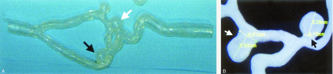
    A, silicon model of a left internal carotid artery with a total of 3 aneurysms including a wide-necked aneurysm (white arrow) and a narrow-necked (black arrow) aneurysm. B, 3D-DSA imaging of the model showing neck size measurements.

  • Fig 2.
    • Download figure
    • Open in new tab
    • Download powerpoint
    Fig 2.

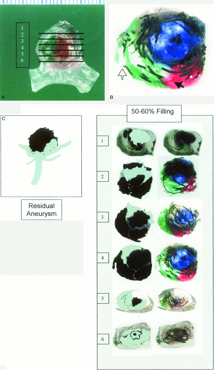
    A, Wide-necked aneurysm filled with colored silicon and coil. Two-micron-thick section preparations (numbered 1–6) were studied via transillumination. B, Enlargement of section 4 (coils, white arrow; colored silicon, black arrow). C, For example, at the level of filling 50%–60%, blue, green, and black silicon were injected (compare Table 2). We then darkened those colors and denoted the open space with gray. At this filling level, the aneurysmal residue on the reference was rated “residual aneurysm” according to the Jean Raymond grading scale.

  • Fig 3.
    • Download figure
    • Open in new tab
    • Download powerpoint
    Fig 3.

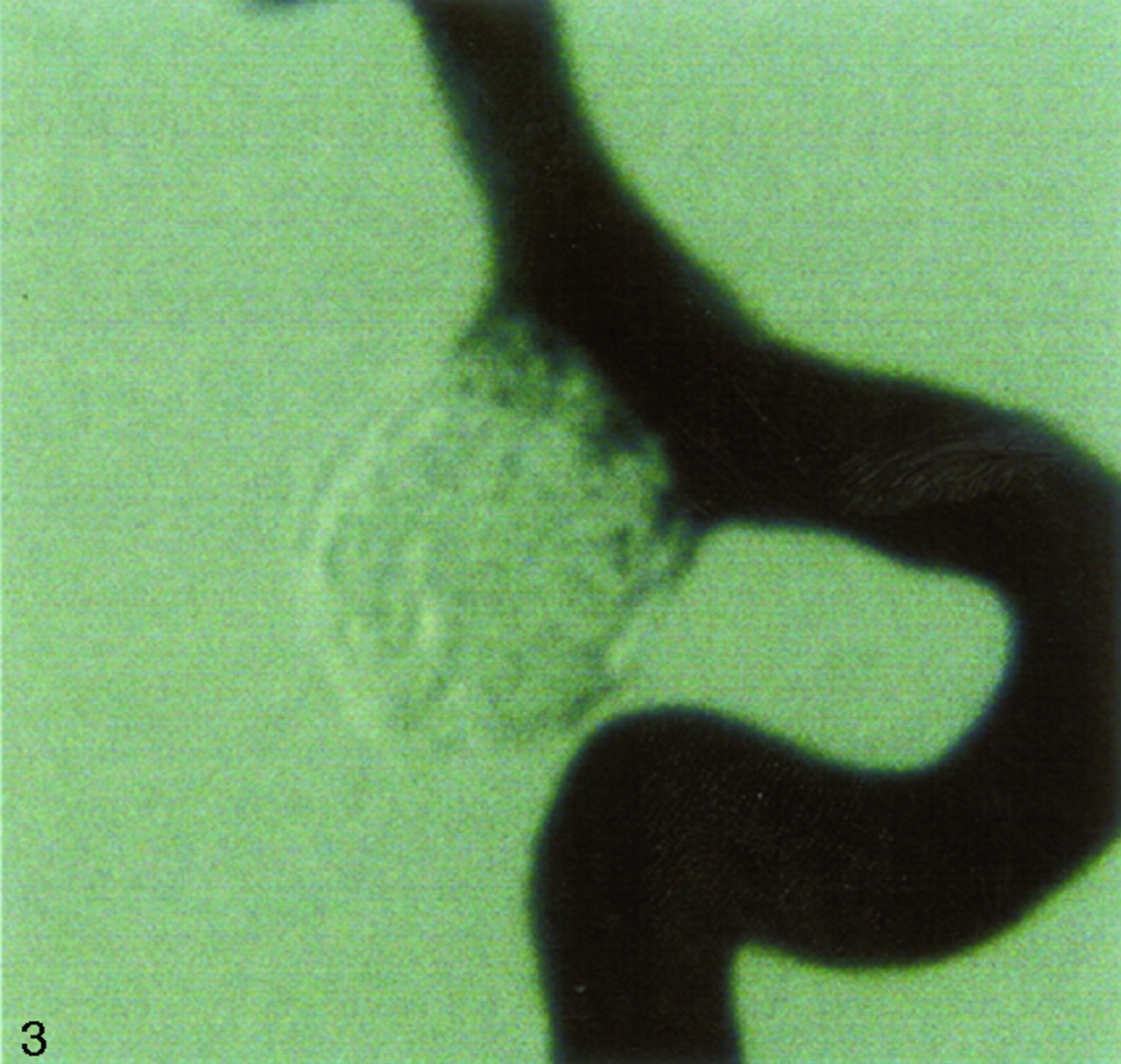
    2D-DSA, wide-neck aneurysm with a partial treatment (residual aneurysm, 50%–60% filling, on the reference) classified as a “residual neck” or “dog ear” by the 2 experts.
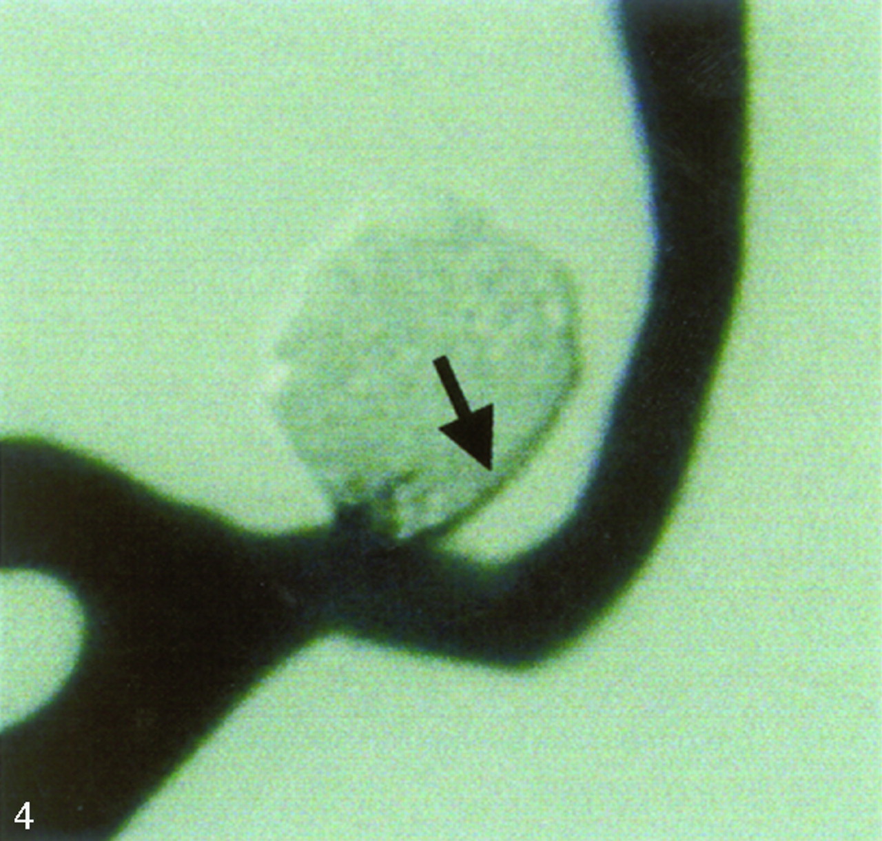
  • Fig 4.
    • Download figure
    • Open in new tab
    • Download powerpoint
    Fig 4.

    2D-DSA imaging of the narrow-necked aneurysm with 90%–100% filling (residual neck on the reference; compare Table 1). A black border is observed around the aneurysm (black arrow). Arterial pulsation may be responsible for misregistration of the mask and the injected series and can mimic a residual neck.
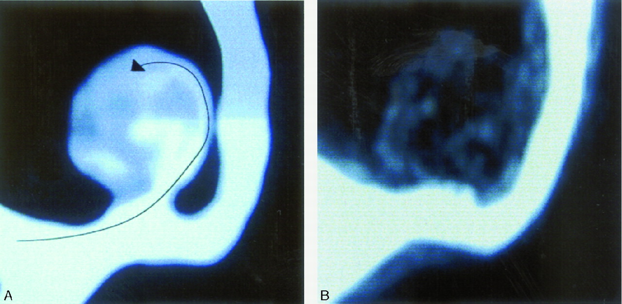
  • Fig 5.
    • Download figure
    • Open in new tab
    • Download powerpoint
    Fig 5.
    • Download figure
    • Open in new tab
    • Download powerpoint
    Fig 5.

    MR 3D-TOF, wide-necked aneurysm with a 30% partial filling (residual aneurysm on the reference; compare Table 2) showing residual flow (arrow) on MIP view (A) and axial reconstructed view (B). C, Residual flow in gray on the reference (arrow) is not observed on 2D-DSA imaging (D).
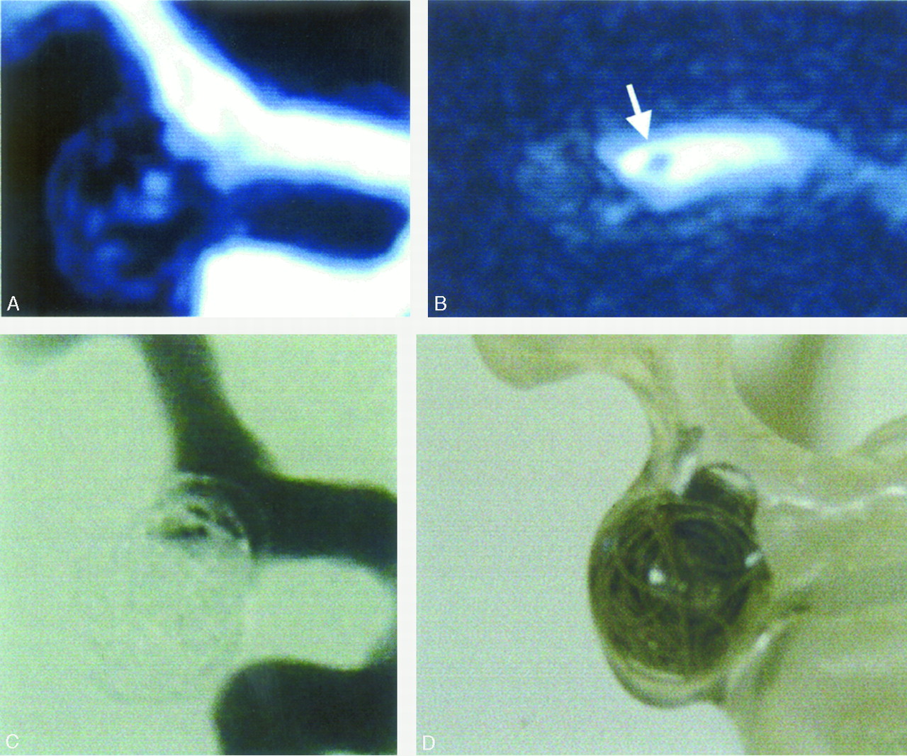
  • Fig 6.
    • Download figure
    • Open in new tab
    • Download powerpoint
    Fig 6.

    MR imaging of the wide-necked aneurysm with a loop of coil into the parent artery. The loop is difficult to recognize on MIP view (A) and on source images (B; arrow). 2D-DSA imaging (C) is closely concordant with the reference (D).

  • Fig 7.
    • Download figure
    • Open in new tab
    • Download powerpoint
    Fig 7.

    Narrow-necked aneurysm (filling 70%–80%, “residual neck,” no silicon protrusion on the reference). A, MR imaging showing the residual neck (white arrow) and the loss of signal intensity around the packing (black arrows). B, 2D-DSA imaging of the same aneurysm.
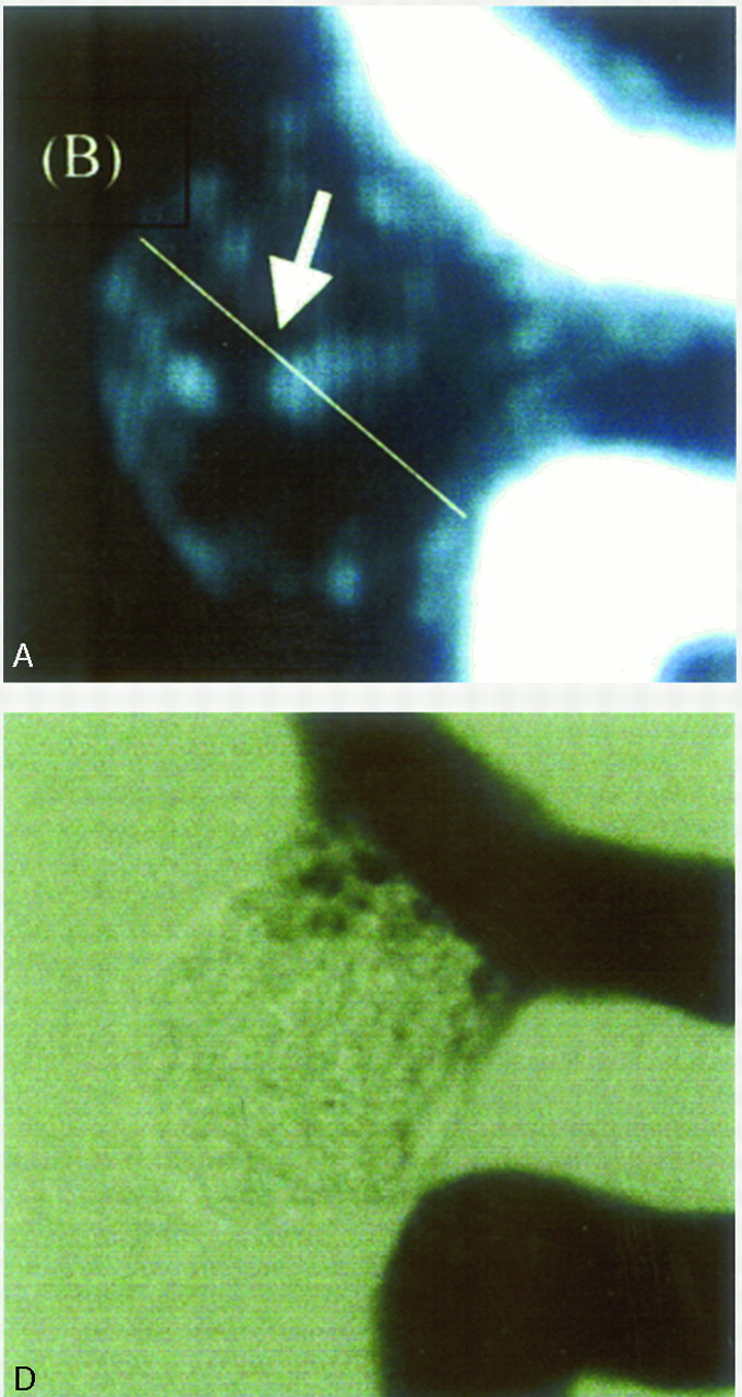
  • Fig 8.
    • Download figure
    • Open in new tab
    • Download powerpoint
    Fig 8.

    A, Narrow-necked aneurysm in 3D-TOF. Loss of signal intensity is observed in the aneurysm sac because of disturbed flow predominant in the center and proximal border of the sac and neck. B, The same aneurysm coiled with a 20% VER without silicon. Coils cause a major disturbance in the flow and a major loss of signal intensity explaining the overestimation of filling with 3D-TOF evaluations.

Tables

  • Figures
    • View popup
    Table 1:

    Filling of the narrow-necked aneurysm

    Aneurysm Filling (%)Aneurysmal Residue* (Reference)Silicon Protrusion
    20 Coils onlyResidual aneurysmNo
    40–50Residual aneurysmNo
    50–60Residual aneurysmNo
    60–70Residual aneurysmNo
    70–80Residual neckNo
    80–90Residual neckNo
    90–100Residual neckNo
    >100CompleteYes
    • * According to Jean Raymond grading scale.

    • View popup
    Table 2:

    Filling of the wide-necked aneurysm

    Aneurysm Filling (%)Color of Silicone InjectionAneurysmal Residue* ReferenceLoop in the Parent Artery
    BlueGreenRedGrayBlackPinkViolet
    Coils only———————Residual aneurysmYes
    30Residual aneurysmNo
    40–50Residual aneurysmYes
    50–60Residual aneurysmNo
    60–70Residual aneurysmNo
    70–80Residual neckNo
    80–90Residual neckNo
    100CompleteNo
    • * According to modified Jean Raymond grading scale.

    • View popup
    Table 3:

    κ value of agreement between the reference and the evaluation by percentage

    ClassκSEProb > Zb_infb_sup
    2D-DSA<70−0.0300.121−0.2490.598−0.267
    70%–90%−0.0570.121−0.4760.683−0.295
    90%–100%0.0360.1210.3040.380−0.200
    3D-TOF<70%−0.2720.125−2.180.985−0.517
    70%–90%−0.3610.125−2.890.998−0.606
    90%–100%−0.3610.125−2.890.998−0.606
    • View popup
    Table 4:

    κ value of agreement between 2D-DSA evaluation (using Jean Raymond grading scale) and the reference

    2D-DSAκSEZProb > ZB_infB_sup
    Residual neck or dog ear0.1140.1210.9450.172−0.1220.352
    Partial (residual aneurysm)0.1450.1211.1980.115−0.0920.383
    Complete0.7160.1215.909<.00010.478.954
    Global agreement0.2320.0942.4500.00710.0460.418
    • View popup
    Table 5:

    κ value of agreement between 3D-TOF evaluation (using Jean Raymond grading scale) and the reference

    3D-TOFκSEZProb > Zb_infb-sup
    Residual neck or dog ear0.3530.1252.8290.0020.1080.598
    Partial (residual aneurysm)0.5*0.1254<.00010.250.745
    Complete0.4830.1253.869<.00010.2380.728
    Global agreement0.4470.0904.928<.00010.2690.625
    • View popup
    Table 6:

    Agreement between 3D-TOF and 2D-DSA evaluation (using Jean Raymond grading scale) and the reference on the wide-necked aneurysm

    Wide Neck κ and SE (*)Residual Neck or Dog EarPartial (Residual Aneurysm)CompleteGlobal Agreement
    2D-DSA0.1 (0.166)0.041 (0.166)0.306 (0.166)0.138 (0.119)
    3D-TOF0.39 (0.176)0.679 (0.176)0.589 (0.176)0.565 (0.129)
    P value.282.018*.294.028
    • View popup
    Table 7:

    Agreement between 3D-TOF and 2D-DSA evaluation (using Jean Raymond grading scale) and the reference on the narrow-necked aneurysm

    Narrow Neck κ and SE (*)Residual Neck or Dog EarPartial (Residual Aneurysm)CompleteGlobal Agreement
    2D-DSA0.066 (0.176)0.186 (0.176)0.612 (0.176)0.225 (0.134)
    3D-TOF0.307 (0.176)0.295 (0.176)0.390 (0.176)0.325 (0.129)
    P value.385.69.40.628
    • View popup
    Table 8:

    Difference of therapeutic decision depending on the acquisition mode

    IRM Additional Treatment Decision2D-DSATotal
    Frequency PercentageYesNo
    Yes314
    4.691.566.25
    No65460
    9.3884.3893.7
    Total95564
    14.0685.9100
    Frequency Missing = 4
PreviousNext
Back to top

In this issue

American Journal of Neuroradiology: 27 (1)
American Journal of Neuroradiology
Vol. 27, Issue 1
January, 2006
  • Table of Contents
  • Index by author
Advertisement
Print
Download PDF
Email Article

Thank you for your interest in spreading the word on American Journal of Neuroradiology.

NOTE: We only request your email address so that the person you are recommending the page to knows that you wanted them to see it, and that it is not junk mail. We do not capture any email address.

Enter multiple addresses on separate lines or separate them with commas.
In Vitro Evaluation of 2D-Digital Subtraction Angiography versus 3D-Time-of-Flight in Assessment of Intracranial Cerebral Aneurysm Filling after Endovascular Therapy
(Your Name) has sent you a message from American Journal of Neuroradiology
(Your Name) thought you would like to see the American Journal of Neuroradiology web site.
CAPTCHA
This question is for testing whether or not you are a human visitor and to prevent automated spam submissions.
Cite this article
V. Costalat, E. Lebars, L. Sarry, A. Defasque, E. Barbotte, H. Brunel, G. Bourbotte, A. Bonafé
In Vitro Evaluation of 2D-Digital Subtraction Angiography versus 3D-Time-of-Flight in Assessment of Intracranial Cerebral Aneurysm Filling after Endovascular Therapy
American Journal of Neuroradiology Jan 2006, 27 (1) 177-184;

Citation Manager Formats

  • BibTeX
  • Bookends
  • EasyBib
  • EndNote (tagged)
  • EndNote 8 (xml)
  • Medlars
  • Mendeley
  • Papers
  • RefWorks Tagged
  • Ref Manager
  • RIS
  • Zotero
0 Responses
Respond to this article
Share
Bookmark this article
In Vitro Evaluation of 2D-Digital Subtraction Angiography versus 3D-Time-of-Flight in Assessment of Intracranial Cerebral Aneurysm Filling after Endovascular Therapy
V. Costalat, E. Lebars, L. Sarry, A. Defasque, E. Barbotte, H. Brunel, G. Bourbotte, A. Bonafé
American Journal of Neuroradiology Jan 2006, 27 (1) 177-184;
del.icio.us logo Twitter logo Facebook logo Mendeley logo
  • Tweet Widget
  • Facebook Like
  • Google Plus One

Jump to section

  • Article
    • Abstract
    • Materials and Methods
    • Results
    • Discussion
    • Acknowledgments
    • References
  • Figures & Data
  • Info & Metrics
  • Responses
  • References
  • PDF

Related Articles

  • No related articles found.
  • PubMed
  • Google Scholar

Cited By...

  • Three-dimensional printing of anatomically accurate, patient specific intracranial aneurysm models
  • Outcomes of Endovascular Treatments of Aneurysms: Observer Variability and Implications for Interpreting Case Series and Planning Randomized Trials
  • Stent-Assisted Coiling of Complex Middle Cerebral Artery Aneurysms: Initial and Midterm Results
  • Neuroform Stent-Assisted Coiling of Unruptured Intracranial Aneurysms: Short- and Midterm Results from a Single-Center Experience with 68 Patients
  • Evaluation of the Occlusion Status of Coiled Intracranial Aneurysms with MR Angiography at 3T: Is Contrast Enhancement Necessary?
  • Crossref
  • Google Scholar

This article has not yet been cited by articles in journals that are participating in Crossref Cited-by Linking.

More in this TOC Section

  • SAVE vs. Solumbra Techniques for Thrombectomy
  • CT Perfusion&Reperfusion in Acute Ischemic Stroke
  • Delayed Reperfusion Post-Thrombectomy&Thrombolysis
Show more Interventional

Similar Articles

Advertisement

Indexed Content

  • Current Issue
  • Accepted Manuscripts
  • Article Preview
  • Past Issues
  • Editorials
  • Editors Choice
  • Fellow Journal Club
  • Letters to the Editor

Cases

  • Case Collection
  • Archive - Case of the Week
  • Archive - Case of the Month
  • Archive - Classic Case

Special Collections

  • Special Collections

Resources

  • News and Updates
  • Turn around Times
  • Submit a Manuscript
  • Author Policies
  • Manuscript Submission Guidelines
  • Evidence-Based Medicine Level Guide
  • Publishing Checklists
  • Graphical Abstract Preparation
  • Imaging Protocol Submission
  • Submit a Case
  • Become a Reviewer/Academy of Reviewers
  • Get Peer Review Credit from Publons

Multimedia

  • AJNR Podcast
  • AJNR SCANtastic
  • Video Articles

About Us

  • About AJNR
  • Editorial Board
  • Not an AJNR Subscriber? Join Now
  • Alerts
  • Feedback
  • Advertise with us
  • Librarian Resources
  • Permissions
  • Terms and Conditions

American Society of Neuroradiology

  • Not an ASNR Member? Join Now

© 2025 by the American Society of Neuroradiology All rights, including for text and data mining, AI training, and similar technologies, are reserved.
Print ISSN: 0195-6108 Online ISSN: 1936-959X

Powered by HighWire