Advertisement

Main menu

  • Home
  • Content
    • Current Issue
    • Accepted Manuscripts
    • Article Preview
    • Past Issue Archive
    • AJNR Case Collection
    • Case of the Week Archive
    • Classic Case Archive
    • Case of the Month Archive
  • Special Collections
    • Spinal CSF Leak Articles (Jan 2020-June 2024)
    • 2024 AJNR Journal Awards
    • Most Impactful AJNR Articles
  • Multimedia
    • AJNR Podcast
    • AJNR Scantastics
    • Video Articles
  • For Authors
    • Submit a Manuscript
    • Author Policies
    • Fast publishing of Accepted Manuscripts
    • Graphical Abstract Preparation
    • Manuscript Submission Guidelines
    • Imaging Protocol Submission
    • Submit a Case for the Case Collection
  • About Us
    • About AJNR
    • Editorial Board
  • More
    • Become a Reviewer/Academy of Reviewers
    • Subscribers
    • Permissions
    • Alerts
    • Feedback
    • Advertisers
    • ASNR Home
  • Other Publications
    • ajnr

User menu

  • Alerts
  • Log in

Search

  • Advanced search
American Journal of Neuroradiology

American Journal of Neuroradiology

ASHNR American Society of Functional Neuroradiology ASHNR American Society of Pediatric Neuroradiology ASSR
  • Alerts
  • Log in

Advanced Search

  • Home
  • Content
    • Current Issue
    • Accepted Manuscripts
    • Article Preview
    • Past Issue Archive
    • AJNR Case Collection
    • Case of the Week Archive
    • Classic Case Archive
    • Case of the Month Archive
  • Special Collections
    • Spinal CSF Leak Articles (Jan 2020-June 2024)
    • 2024 AJNR Journal Awards
    • Most Impactful AJNR Articles
  • Multimedia
    • AJNR Podcast
    • AJNR Scantastics
    • Video Articles
  • For Authors
    • Submit a Manuscript
    • Author Policies
    • Fast publishing of Accepted Manuscripts
    • Graphical Abstract Preparation
    • Manuscript Submission Guidelines
    • Imaging Protocol Submission
    • Submit a Case for the Case Collection
  • About Us
    • About AJNR
    • Editorial Board
  • More
    • Become a Reviewer/Academy of Reviewers
    • Subscribers
    • Permissions
    • Alerts
    • Feedback
    • Advertisers
    • ASNR Home
  • Follow AJNR on Twitter
  • Visit AJNR on Facebook
  • Follow AJNR on Instagram
  • Join AJNR on LinkedIn
  • RSS Feeds

Welcome to the new AJNR, Updated Hall of Fame, and more. Read the full announcements.


AJNR is seeking candidates for the position of Associate Section Editor, AJNR Case Collection. Read the full announcement.


Research ArticleBRAIN

First-Pass Quantitative CT Perfusion Identifies Thresholds for Salvageable Penumbra in Acute Stroke Patients Treated with Intra-arterial Therapy

P.W. Schaefer, L. Roccatagliata, C. Ledezma, B. Hoh, L.H. Schwamm, W. Koroshetz, R.G. Gonzalez and M.H. Lev
American Journal of Neuroradiology January 2006, 27 (1) 20-25;
P.W. Schaefer
  • Find this author on Google Scholar
  • Find this author on PubMed
  • Search for this author on this site
L. Roccatagliata
  • Find this author on Google Scholar
  • Find this author on PubMed
  • Search for this author on this site
C. Ledezma
  • Find this author on Google Scholar
  • Find this author on PubMed
  • Search for this author on this site
B. Hoh
  • Find this author on Google Scholar
  • Find this author on PubMed
  • Search for this author on this site
L.H. Schwamm
  • Find this author on Google Scholar
  • Find this author on PubMed
  • Search for this author on this site
W. Koroshetz
  • Find this author on Google Scholar
  • Find this author on PubMed
  • Search for this author on this site
R.G. Gonzalez
  • Find this author on Google Scholar
  • Find this author on PubMed
  • Search for this author on this site
M.H. Lev
  • Find this author on Google Scholar
  • Find this author on PubMed
  • Search for this author on this site
  • Article
  • Figures & Data
  • Info & Metrics
  • Responses
  • References
  • PDF
Loading

Article Figures & Data

Figures

  • Tables
  • Fig 1.
    • Download figure
    • Open in new tab
    • Download powerpoint
    Fig 1.

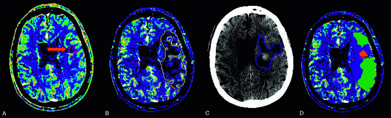
    A 61-year-old male patient with right hemiparesis imaged at 2.3 hours and 3 days. Abnormal areas are outlined on CBV (A), CBF (B), and follow-up CT (C) images. After coregistration, we defined 3 regions on CBF maps (D): region 1 (red), “infarct core”—abnormal on CBV, CBF, and follow-up CT images; region 2 (blue), “penumbra that infarcts”—normal on CBV but abnormal on CBF and follow-up CT images; and region 3 (green) “penumbra that recovers”—abnormal on CBF but normal on CBV and follow-up CT images.

  • Fig 2.
    • Download figure
    • Open in new tab
    • Download powerpoint
    Fig 2.

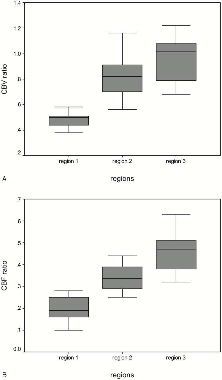
    Box and Whisker graphs of normalized CBV (A) and CBF ratios (B) in regions 1–3.

  • Fig 3.
    • Download figure
    • Open in new tab
    • Download powerpoint
    Fig 3.

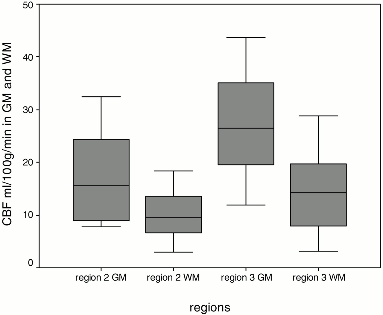
    Box and Whisker graphs of mean GM and WM quantitative CBF values for each patient in regions 2 and 3.

Tables

  • Figures
    • View popup
    Table 1:

    Demographic data and treatment characteristics

    Patient No./Sex/Age (y)Admission NIHSSTime to CTP (h)Occ SiteTime to trtmnt (h)TherapyTime to F/U (d)Mori ScalemRS at DischargeCBF ratio Reg 1CBF ratio Reg 2CBF ratio Reg 3CBV ratio Reg 1CBV ratio Reg 2CBV ratio Reg 3
    1/F/80271.7M13.5IA rtPA12240.100.290.440.380.560.69
    2/F/81153.7M14Mechanical1420.160.250.400.510.881.04
    3/F/74102.1M13.3IV + IA rtPA5320.120.300.340.310.680.88
    4/F/74181.2ICA3.5IA rtPA7230.280.330.490.520.700.70
    5/F/81250.9ICA2IA rtPA4140.270.390.630.500.831.18
    6/F/69224.8M26IV rtPA + Mechanical3020.190.250.320.581.161.09
    7/M/7790.9M13.3IV + IA rtPA5310.250.390.460.500.700.68
    8/M/61112.3ICA, MI4.4IA rtPA3410.200.320.360.651.081.05
    9/M/46203.3M13.2IV rtPA + IA rtPA3160.260.400.550.480.911.22
    10/F/44131.7M13IV rtPA + IA rtPA5400.170.250.520.440.810.91
    11/F/82162.4M14.3Mechanical326NP0.40NPNP0.68NP
    12/F/71195.4ICA7Mechanical5140.200.340.500.511.031.07
    13/M/7563.8M25.5Mechanical3020.180.39NP0.450.81NP
    14/F/71192.4ICA3.6IA rtPA1260.130.440.480.390.910.99
    • Note:— CTP indicates computed tomography perfusion; F/U, follow-up; mechanical, clot disruption/retrieval; NP, region not present; mRS, modified Rankin Score; trtmnt, treatment; reg, region; pt, patient; occ, occlusion.

    • View popup
    Table 2:

    CTP ratios and absolute values for regions 1–3

    ParameterRegion 1 Mean Ratio ± SDRegion 2 Mean Ratio ± SDRegion 3 Mean Ratio ± SDP Value (Region 1 vs 2)P Value (Region 2 vs 3)
    CBV ratio0.48 ± 0.090.84 ± 0.170.96 ± 0.19<0.00010.03
    CBF ratio0.19 ± 0.060.34 ± 0.060.46 ± 0.09<0.0001<0.0001
    CBV value (ml/100 g)1.47 ± 0.42.83 ± 0.672.94 ± 0.45<0.00010.05
    CBF value (ml/100 g/min)8.88 ± 2.316.08 ± 5.7117.92 ± 4.0<0.00010.1
    • Note:— CBF indicates cerebral blood flow; CBV, cerebral blood volume.

    • View popup
    Table 3:

    CTP absolute values for penumbra that infarcts (region 2) and penumbra that recovers (region 3) in gray and white matter

    Region 2 Mean ratio ± SDRegion 3 Mean ratio ± SDP Value
    GM CBV (ml/100 g)3.54 ± 1.333.87 ± 0.610.34
    WM CBV (ml/100 g)2.64 ± 1.382.27 ± 0.930.46
    GM CBF (ml/100 g/min)17.17 ± 8.6727.22 ± 9.740.01
    WM CBF (ml/100 g/min)10.12 ± 4.7714.01 ± 7.610.06
    • Note:— CBF indicates cerebral blood flow; CBV, cerebral blood volume; GM, gray matter; WM, white matter.

PreviousNext
Back to top

In this issue

American Journal of Neuroradiology: 27 (1)
American Journal of Neuroradiology
Vol. 27, Issue 1
January, 2006
  • Table of Contents
  • Index by author
Advertisement
Print
Download PDF
Email Article

Thank you for your interest in spreading the word on American Journal of Neuroradiology.

NOTE: We only request your email address so that the person you are recommending the page to knows that you wanted them to see it, and that it is not junk mail. We do not capture any email address.

Enter multiple addresses on separate lines or separate them with commas.
First-Pass Quantitative CT Perfusion Identifies Thresholds for Salvageable Penumbra in Acute Stroke Patients Treated with Intra-arterial Therapy
(Your Name) has sent you a message from American Journal of Neuroradiology
(Your Name) thought you would like to see the American Journal of Neuroradiology web site.
CAPTCHA
This question is for testing whether or not you are a human visitor and to prevent automated spam submissions.
Cite this article
P.W. Schaefer, L. Roccatagliata, C. Ledezma, B. Hoh, L.H. Schwamm, W. Koroshetz, R.G. Gonzalez, M.H. Lev
First-Pass Quantitative CT Perfusion Identifies Thresholds for Salvageable Penumbra in Acute Stroke Patients Treated with Intra-arterial Therapy
American Journal of Neuroradiology Jan 2006, 27 (1) 20-25;

Citation Manager Formats

  • BibTeX
  • Bookends
  • EasyBib
  • EndNote (tagged)
  • EndNote 8 (xml)
  • Medlars
  • Mendeley
  • Papers
  • RefWorks Tagged
  • Ref Manager
  • RIS
  • Zotero
0 Responses
Respond to this article
Share
Bookmark this article
First-Pass Quantitative CT Perfusion Identifies Thresholds for Salvageable Penumbra in Acute Stroke Patients Treated with Intra-arterial Therapy
P.W. Schaefer, L. Roccatagliata, C. Ledezma, B. Hoh, L.H. Schwamm, W. Koroshetz, R.G. Gonzalez, M.H. Lev
American Journal of Neuroradiology Jan 2006, 27 (1) 20-25;
del.icio.us logo Twitter logo Facebook logo Mendeley logo
  • Tweet Widget
  • Facebook Like
  • Google Plus One

Jump to section

  • Article
    • Abstract
    • Materials and Methods
    • Results
    • Discussion
    • Conclusion
    • References
  • Figures & Data
  • Info & Metrics
  • Responses
  • References
  • PDF

Related Articles

  • No related articles found.
  • PubMed
  • Google Scholar

Cited By...

  • Reducing False-Positives in CT Perfusion Infarct Core Segmentation Using Contralateral Local Normalization
  • Mechanical thrombectomy with the Trevo ProVue device in ischemic stroke patients: does improved visibility translate into a clinical benefit?
  • Clinical Significance of Fluid-Attenuated Inversion Recovery Vascular Hyperintensities in Borderzone Infarcts
  • Limited Reliability of Computed Tomographic Perfusion Acute Infarct Volume Measurements Compared With Diffusion-Weighted Imaging in Anterior Circulation Stroke
  • Six-Minute Magnetic Resonance Imaging Protocol for Evaluation of Acute Ischemic Stroke: Pushing the Boundaries
  • Role of EPI-FLAIR in Patients with Acute Stroke: A Comparative Analysis with FLAIR
  • Guidelines for the Early Management of Patients With Acute Ischemic Stroke: A Guideline for Healthcare Professionals From the American Heart Association/American Stroke Association
  • C-Arm CT Measurement of Cerebral Blood Volume Using Intra-Arterial Injection of Contrast Medium: An Experimental Study in Canines
  • Contrast Delay on Perfusion CT as a Predictor of New, Incident Infarct: A Retrospective Cohort Study
  • Acute Stroke Imaging: CT with CT Angiography and CT Perfusion before Management Decisions
  • Reperfusion by Combined Thrombolysis and Mechanical Thrombectomy in Acute Stroke: Effect of Collateralization, Mismatch, and Time to and Grade of Recanalization on Clinical and Tissue Outcome
  • Cerebral Blood Flow Is the Optimal CT Perfusion Parameter for Assessing Infarct Core
  • CT Cerebral Blood Flow Maps Optimally Correlate With Admission Diffusion-Weighted Imaging in Acute Stroke but Thresholds Vary by Postprocessing Platform
  • Multimodal Imaging Does Not Delay Intravenous Thrombolytic Therapy in Acute Stroke
  • Cerebral Blood Flow Thresholds for Tissue Infarction in Patients with Acute Ischemic Stroke Treated with Intra-Arterial Revascularization Therapy Depend on Timing of Reperfusion
  • Regional Ischemic Vulnerability of the Brain to Hypoperfusion: The Need for Location Specific Computed Tomography Perfusion Thresholds in Acute Stroke Patients
  • Predicting Language Improvement in Acute Stroke Patients Presenting with Aphasia: A Multivariate Logistic Model Using Location-Weighted Atlas-Based Analysis of Admission CT Perfusion Scans
  • Diagnostic Threshold Values of Cerebral Perfusion Measured With Computed Tomography for Delayed Cerebral Ischemia After Aneurysmal Subarachnoid Hemorrhage
  • C-Arm CT Measurement of Cerebral Blood Volume in Ischemic Stroke: An Experimental Study in Canines
  • FDA Investigates the Safety of Brain Perfusion CT
  • Recommendations for Imaging of Acute Ischemic Stroke: A Scientific Statement From the American Heart Association
  • Hemodynamic Factors and Perfusion Abnormalities in Early Neurological Deterioration
  • Theoretic Basis and Technical Implementations of CT Perfusion in Acute Ischemic Stroke, Part 2: Technical Implementations
  • Perfusion CT in Patients with Acute Ischemic Stroke Treated with Intra-Arterial Thrombolysis: Predictive Value of Infarct Core Size on Clinical Outcome
  • Cortical Regional Hyperperfusion in Nonconvulsive Status Epilepticus Measured by Dynamic Brain Perfusion CT
  • CT Angiography Clot Burden Score and Collateral Score: Correlation with Clinical and Radiologic Outcomes in Acute Middle Cerebral Artery Infarct
  • Quantitative Assessment of Core/Penumbra Mismatch in Acute Stroke: CT and MR Perfusion Imaging Are Strongly Correlated When Sufficient Brain Volume Is Imaged
  • The MRA-DWI Mismatch Identifies Patients With Stroke Who Are Likely to Benefit From Reperfusion
  • Global Hemispheric CT Hypoperfusion May Differentiate Headache With Associated Neurological Deficits and Lymphocytosis From Acute Stroke
  • CT/NIHSS Mismatch for Detection of Salvageable Brain in Acute Stroke Triage Beyond the 3-Hour Time Window: Overrated or Undervalued?
  • Critical Care and Emergency Medicine
  • Cerebral Blood Flow Thresholds in Acute Stroke Triage
  • Response to Letters by Lee et al and Lev et al
  • Crossref
  • Google Scholar

This article has not yet been cited by articles in journals that are participating in Crossref Cited-by Linking.

More in this TOC Section

  • Fast Contrast-Enhanced 4D MRA and 4D Flow MRI Using Constrained Reconstruction (HYPRFlow): Potential Applications for Brain Arteriovenous Malformations
  • Quiet PROPELLER MRI Techniques Match the Quality of Conventional PROPELLER Brain Imaging Techniques
  • Predictors of Reperfusion in Patients with Acute Ischemic Stroke
Show more Brain

Similar Articles

Advertisement

Indexed Content

  • Current Issue
  • Accepted Manuscripts
  • Article Preview
  • Past Issues
  • Editorials
  • Editors Choice
  • Fellow Journal Club
  • Letters to the Editor

Cases

  • Case Collection
  • Archive - Case of the Week
  • Archive - Case of the Month
  • Archive - Classic Case

Special Collections

  • Special Collections

Resources

  • News and Updates
  • Turn around Times
  • Submit a Manuscript
  • Author Policies
  • Manuscript Submission Guidelines
  • Evidence-Based Medicine Level Guide
  • Publishing Checklists
  • Graphical Abstract Preparation
  • Imaging Protocol Submission
  • Submit a Case
  • Become a Reviewer/Academy of Reviewers
  • Get Peer Review Credit from Publons

Multimedia

  • AJNR Podcast
  • AJNR SCANtastic
  • Video Articles

About Us

  • About AJNR
  • Editorial Board
  • Not an AJNR Subscriber? Join Now
  • Alerts
  • Feedback
  • Advertise with us
  • Librarian Resources
  • Permissions
  • Terms and Conditions

American Society of Neuroradiology

  • Not an ASNR Member? Join Now

© 2025 by the American Society of Neuroradiology All rights, including for text and data mining, AI training, and similar technologies, are reserved.
Print ISSN: 0195-6108 Online ISSN: 1936-959X

Powered by HighWire