Advertisement

Main menu

  • Home
  • Content
    • Current Issue
    • Accepted Manuscripts
    • Article Preview
    • Past Issue Archive
    • AJNR Case Collection
    • Case of the Week Archive
    • Classic Case Archive
    • Case of the Month Archive
  • Special Collections
    • Spinal CSF Leak Articles (Jan 2020-June 2024)
    • 2024 AJNR Journal Awards
    • Most Impactful AJNR Articles
  • Multimedia
    • AJNR Podcast
    • AJNR Scantastics
    • Video Articles
  • For Authors
    • Submit a Manuscript
    • Author Policies
    • Fast publishing of Accepted Manuscripts
    • Graphical Abstract Preparation
    • Manuscript Submission Guidelines
    • Imaging Protocol Submission
    • Submit a Case for the Case Collection
  • About Us
    • About AJNR
    • Editorial Board
  • More
    • Become a Reviewer/Academy of Reviewers
    • Subscribers
    • Permissions
    • Alerts
    • Feedback
    • Advertisers
    • ASNR Home
  • Other Publications
    • ajnr

User menu

  • Alerts
  • Log in

Search

  • Advanced search
American Journal of Neuroradiology

American Journal of Neuroradiology

ASHNR American Society of Functional Neuroradiology ASHNR American Society of Pediatric Neuroradiology ASSR
  • Alerts
  • Log in

Advanced Search

  • Home
  • Content
    • Current Issue
    • Accepted Manuscripts
    • Article Preview
    • Past Issue Archive
    • AJNR Case Collection
    • Case of the Week Archive
    • Classic Case Archive
    • Case of the Month Archive
  • Special Collections
    • Spinal CSF Leak Articles (Jan 2020-June 2024)
    • 2024 AJNR Journal Awards
    • Most Impactful AJNR Articles
  • Multimedia
    • AJNR Podcast
    • AJNR Scantastics
    • Video Articles
  • For Authors
    • Submit a Manuscript
    • Author Policies
    • Fast publishing of Accepted Manuscripts
    • Graphical Abstract Preparation
    • Manuscript Submission Guidelines
    • Imaging Protocol Submission
    • Submit a Case for the Case Collection
  • About Us
    • About AJNR
    • Editorial Board
  • More
    • Become a Reviewer/Academy of Reviewers
    • Subscribers
    • Permissions
    • Alerts
    • Feedback
    • Advertisers
    • ASNR Home
  • Follow AJNR on Twitter
  • Visit AJNR on Facebook
  • Follow AJNR on Instagram
  • Join AJNR on LinkedIn
  • RSS Feeds

Welcome to the new AJNR, Updated Hall of Fame, and more. Read the full announcements.


AJNR is seeking candidates for the position of Associate Section Editor, AJNR Case Collection. Read the full announcement.


Research ArticleBRAIN

Minimizing Clip Artifacts in Multi CT Angiography of Clipped Patients

I. van der Schaaf, M. van Leeuwen, A. Vlassenbroek and B. Velthuis
American Journal of Neuroradiology January 2006, 27 (1) 60-66;
I. van der Schaaf
  • Find this author on Google Scholar
  • Find this author on PubMed
  • Search for this author on this site
M. van Leeuwen
  • Find this author on Google Scholar
  • Find this author on PubMed
  • Search for this author on this site
A. Vlassenbroek
  • Find this author on Google Scholar
  • Find this author on PubMed
  • Search for this author on this site
B. Velthuis
  • Find this author on Google Scholar
  • Find this author on PubMed
  • Search for this author on this site
  • Article
  • Figures & Data
  • Info & Metrics
  • Responses
  • References
  • PDF
Loading

Article Figures & Data

Figures

  • Tables
  • Fig 1.
    • Download figure
    • Open in new tab
    • Download powerpoint
    Fig 1.

    Phantom. A cylindrical PMMA phantom (A) with a cylinder containing a clip (B).
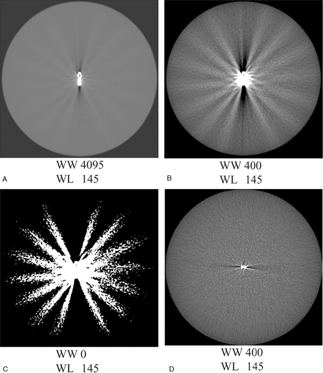
  • Fig 2.
    • Download figure
    • Open in new tab
    • Download powerpoint
    Fig 2.

    Metal artifact of Cobalt clips scanned at pitch 1 positioned in the scanplance (A–C) and perpendicular to the scan plane (D). The metal artifact consists of a black-and-white component. The aspect of the artifact changes with decreasing window width: widest width (A); window width as used in clinical practice (B); and binary picture (C). The artifact volume largely depends on the position of the clip to the scan plane, with much less artifact for a clip perpendicular to the scan plane (D) compared to a clip in the scan plane (B).

  • Fig 3.
    • Download figure
    • Open in new tab
    • Download powerpoint
    Fig 3.

    Artifact quantification in patients: quantification of the black-and-white component of the artifact in patients in CTA source images.

    A, Clip artifact in clinical window setting, with background of 32 HU and an SD of 21.

    B, Area of the black component of the artifact enclosing all pixels with HU < 31 (±3 SD).

    C, Area of the white component of the artifact enclosing all pixels with HU > 95 (±3 SD).
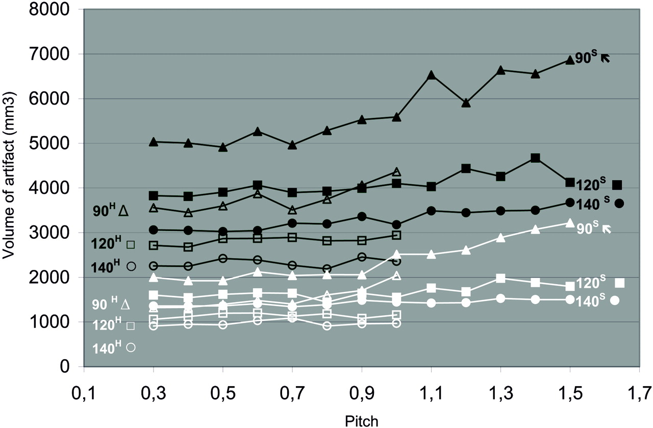
  • Fig 4.
    • Download figure
    • Open in new tab
    • Download powerpoint
    Fig 4.

    Artifact volume of the Cobalt containing alloly and titanium clip at increasing pitch. Volume of clip artifact at increasing pitch for cobalt-containing alloy (upper, black lines) and titanium (lower, white lines) clip. Both clips were scanned in the high mode (superscript H, open symbols) up to pitch 1, and standard mode (superscript S, closed symbols) up to pitch 1.5 at 90 (triangles), 120 (squares), and 140 (circles) kVp.

  • Fig 5.
    • Download figure
    • Open in new tab
    • Download powerpoint
    Fig 5.

    Number of white streaks for the cobalt-containing alloy (upper, black lines) and titanium clips (lower, white lines) scanned in high mode (H) (open symbols, up to P 1) and standard mode (S) (closed symbols, up to P 1.5).

  • Fig 6.
    • Download figure
    • Open in new tab
    • Download powerpoint
    Fig 6.

    Aspect of a cobalt clip artifact, positioned in the scanplane, at increasing pitch. The numbers of white streaks increase with increasing pitch.

Tables

  • Figures
    • View popup
    Table 1:

    Influence of kVp, CBR, slip composition, and orientation on artifact volume

    Ratio
    KVp*
        90 vs 120, High mode1.3
        120 vs 140, High mode1.2
        90 vs 120, Standard mode1.4
        120 vs 140, Standard mode1.2
    Conebeam reconstruction vs linear interpolation*1.1
    Cobalt-containing alloy vs Titanium clip#
        High mode1.3
        Standard mode1.5
    In plane vs perpendicular‡1.4
    • Note:—CBR indicates conebeam reconstruction.

    • * for titanium- and cobalt-containing alloy clip.

    • # for all kVp/mAs combinations.

    • ‡ for both clips in high and standard mode.

    • View popup
    Table 2:

    Clip orientation and diagnostic quality at the clip site

    Position of Clip to Scan PlaneCompletely DiagnosticClinically UsefulNot Diagnostic
    In plane3 (6.3%)25 (52%)20 (42%)
        (24 clips, 48 evaluations*)
    Diagonal6 (15%)25 (63%)9 (23%)
        (20 clips, 40 evaluations)
    Perpendicular3 (25%)8 (67%)1 (8.3%)
        (6 clips, 12 evaluations)
    Total (all positions)12 (12%)58 (58%)30 (30%)
        (50 clips, 100 evaluations)
    • * A double reading per clip site results in twice as many evaluations as clip sites.

PreviousNext
Back to top

In this issue

American Journal of Neuroradiology: 27 (1)
American Journal of Neuroradiology
Vol. 27, Issue 1
January, 2006
  • Table of Contents
  • Index by author
Advertisement
Print
Download PDF
Email Article

Thank you for your interest in spreading the word on American Journal of Neuroradiology.

NOTE: We only request your email address so that the person you are recommending the page to knows that you wanted them to see it, and that it is not junk mail. We do not capture any email address.

Enter multiple addresses on separate lines or separate them with commas.
Minimizing Clip Artifacts in Multi CT Angiography of Clipped Patients
(Your Name) has sent you a message from American Journal of Neuroradiology
(Your Name) thought you would like to see the American Journal of Neuroradiology web site.
CAPTCHA
This question is for testing whether or not you are a human visitor and to prevent automated spam submissions.
Cite this article
I. van der Schaaf, M. van Leeuwen, A. Vlassenbroek, B. Velthuis
Minimizing Clip Artifacts in Multi CT Angiography of Clipped Patients
American Journal of Neuroradiology Jan 2006, 27 (1) 60-66;

Citation Manager Formats

  • BibTeX
  • Bookends
  • EasyBib
  • EndNote (tagged)
  • EndNote 8 (xml)
  • Medlars
  • Mendeley
  • Papers
  • RefWorks Tagged
  • Ref Manager
  • RIS
  • Zotero
0 Responses
Respond to this article
Share
Bookmark this article
Minimizing Clip Artifacts in Multi CT Angiography of Clipped Patients
I. van der Schaaf, M. van Leeuwen, A. Vlassenbroek, B. Velthuis
American Journal of Neuroradiology Jan 2006, 27 (1) 60-66;
del.icio.us logo Twitter logo Facebook logo Mendeley logo
  • Tweet Widget
  • Facebook Like
  • Google Plus One

Jump to section

  • Article
    • Abstract
    • Materials and Methods
    • Results
    • Discussion
    • Conclusion
    • References
  • Figures & Data
  • Info & Metrics
  • Responses
  • References
  • PDF

Related Articles

  • No related articles found.
  • PubMed
  • Google Scholar

Cited By...

  • Image quality assessment of three cone beam CT machines using the SEDENTEXCT CT phantom
  • Feasibility of Flat Panel Angiographic CT after Intravenous Contrast Agent Application in the Postoperative Evaluation of Patients with Clipped Aneurysms
  • Crossref
  • Google Scholar

This article has not yet been cited by articles in journals that are participating in Crossref Cited-by Linking.

More in this TOC Section

  • Progression of Microstructural Damage in Spinocerebellar Ataxia Type 2: A Longitudinal DTI Study
  • Usefulness of Quantitative Susceptibility Mapping for the Diagnosis of Parkinson Disease
  • Evaluating the Effects of White Matter Multiple Sclerosis Lesions on the Volume Estimation of 6 Brain Tissue Segmentation Methods
Show more Brain

Similar Articles

Advertisement

Indexed Content

  • Current Issue
  • Accepted Manuscripts
  • Article Preview
  • Past Issues
  • Editorials
  • Editors Choice
  • Fellow Journal Club
  • Letters to the Editor

Cases

  • Case Collection
  • Archive - Case of the Week
  • Archive - Case of the Month
  • Archive - Classic Case

Special Collections

  • Special Collections

Resources

  • News and Updates
  • Turn around Times
  • Submit a Manuscript
  • Author Policies
  • Manuscript Submission Guidelines
  • Evidence-Based Medicine Level Guide
  • Publishing Checklists
  • Graphical Abstract Preparation
  • Imaging Protocol Submission
  • Submit a Case
  • Become a Reviewer/Academy of Reviewers
  • Get Peer Review Credit from Publons

Multimedia

  • AJNR Podcast
  • AJNR SCANtastic
  • Video Articles

About Us

  • About AJNR
  • Editorial Board
  • Not an AJNR Subscriber? Join Now
  • Alerts
  • Feedback
  • Advertise with us
  • Librarian Resources
  • Permissions
  • Terms and Conditions

American Society of Neuroradiology

  • Not an ASNR Member? Join Now

© 2025 by the American Society of Neuroradiology All rights, including for text and data mining, AI training, and similar technologies, are reserved.
Print ISSN: 0195-6108 Online ISSN: 1936-959X

Powered by HighWire