Advertisement

Main menu

  • Home
  • Content
    • Current Issue
    • Accepted Manuscripts
    • Article Preview
    • Past Issue Archive
    • AJNR Case Collection
    • Case of the Week Archive
    • Classic Case Archive
    • Case of the Month Archive
  • Special Collections
    • Spinal CSF Leak Articles (Jan 2020-June 2024)
    • 2024 AJNR Journal Awards
    • Most Impactful AJNR Articles
  • Multimedia
    • AJNR Podcast
    • AJNR Scantastics
    • Video Articles
  • For Authors
    • Submit a Manuscript
    • Author Policies
    • Fast publishing of Accepted Manuscripts
    • Graphical Abstract Preparation
    • Manuscript Submission Guidelines
    • Imaging Protocol Submission
    • Submit a Case for the Case Collection
  • About Us
    • About AJNR
    • Editorial Board
  • More
    • Become a Reviewer/Academy of Reviewers
    • Subscribers
    • Permissions
    • Alerts
    • Feedback
    • Advertisers
    • ASNR Home
  • Other Publications
    • ajnr

User menu

  • Alerts
  • Log in

Search

  • Advanced search
American Journal of Neuroradiology

American Journal of Neuroradiology

ASHNR American Society of Functional Neuroradiology ASHNR American Society of Pediatric Neuroradiology ASSR
  • Alerts
  • Log in

Advanced Search

  • Home
  • Content
    • Current Issue
    • Accepted Manuscripts
    • Article Preview
    • Past Issue Archive
    • AJNR Case Collection
    • Case of the Week Archive
    • Classic Case Archive
    • Case of the Month Archive
  • Special Collections
    • Spinal CSF Leak Articles (Jan 2020-June 2024)
    • 2024 AJNR Journal Awards
    • Most Impactful AJNR Articles
  • Multimedia
    • AJNR Podcast
    • AJNR Scantastics
    • Video Articles
  • For Authors
    • Submit a Manuscript
    • Author Policies
    • Fast publishing of Accepted Manuscripts
    • Graphical Abstract Preparation
    • Manuscript Submission Guidelines
    • Imaging Protocol Submission
    • Submit a Case for the Case Collection
  • About Us
    • About AJNR
    • Editorial Board
  • More
    • Become a Reviewer/Academy of Reviewers
    • Subscribers
    • Permissions
    • Alerts
    • Feedback
    • Advertisers
    • ASNR Home
  • Follow AJNR on Twitter
  • Visit AJNR on Facebook
  • Follow AJNR on Instagram
  • Join AJNR on LinkedIn
  • RSS Feeds

Welcome to the new AJNR, Updated Hall of Fame, and more. Read the full announcements.


AJNR is seeking candidates for the position of Associate Section Editor, AJNR Case Collection. Read the full announcement.


Research ArticleBRAIN

Intracranial Compartment Volumes in Normal Pressure Hydrocephalus: Volumetric Assessment versus Outcome

W.M. Palm, R. Walchenbach, B. Bruinsma, F. Admiraal-Behloul, H.A.M. Middelkoop, L.J. Launer, J. van der Grond and M.A. van Buchem
American Journal of Neuroradiology January 2006, 27 (1) 76-79;
W.M. Palm
  • Find this author on Google Scholar
  • Find this author on PubMed
  • Search for this author on this site
R. Walchenbach
  • Find this author on Google Scholar
  • Find this author on PubMed
  • Search for this author on this site
B. Bruinsma
  • Find this author on Google Scholar
  • Find this author on PubMed
  • Search for this author on this site
F. Admiraal-Behloul
  • Find this author on Google Scholar
  • Find this author on PubMed
  • Search for this author on this site
H.A.M. Middelkoop
  • Find this author on Google Scholar
  • Find this author on PubMed
  • Search for this author on this site
L.J. Launer
  • Find this author on Google Scholar
  • Find this author on PubMed
  • Search for this author on this site
J. van der Grond
  • Find this author on Google Scholar
  • Find this author on PubMed
  • Search for this author on this site
M.A. van Buchem
  • Find this author on Google Scholar
  • Find this author on PubMed
  • Search for this author on this site
  • Article
  • Figures & Data
  • Info & Metrics
  • Responses
  • References
  • PDF
Loading

Abstract

BACKGROUND AND PURPOSE: Although enlargement of the cerebral ventricles plays a central role in the diagnosis of normal pressure hydrocephalus (NPH), there are no reports on the use of volumetric assessment to distinguish between patients who respond to ventriculoperitoneal shunt surgery and those who do not. The purpose of this study is to explore the association between preoperative intracranial compartment volumes and postoperative improvement.

METHODS: Twenty-six patients (17 men; mean age, 75 years [range, 54–87 years]) with a clinical or radiologic suspicion of NPH were included in the study. Gait, cognition, and bladder function were evaluated by clinical rating. MR imaging of the brain was acquired at 0.5T and 1.5T. Total intracranial volume, ventricular volume, brain volume, and pericerebral CSF volume were determined by volumetric assessment. Four imaging variables were determined: ventricular volume ratio, brain volume ratio, pericerebral CSF volume ratio, and the ratio of ventricular volume to pericerebral CSF volume. All patients underwent ventriculoperitoneal shunt surgery.

RESULTS: Clinical follow-up was assessed 1 year after shunt surgery. No difference in the mean ventricular volume ratio, the mean brain volume ratio, the mean pericerebral CSF volume ratio, and the mean ratio between ventricular and pericerebral CSF volume was found between subjects who improved on gait or cognition or bladder function and those who did not.

CONCLUSION: Volumetric assessment has no predictive value in differentiating between NPH patients who respond to ventriculoperitoneal shunt surgery and those who do not.

Forty years ago, Adams et al described a syndrome that combined a clinical triad of gait impairment, cognitive impairment, and urinary incontinence with normal CSF pressure.1 The clinical triad, together with enlargement of the cerebral ventricles on CT or MR imaging, is suggestive for the normal pressure hydrocephalus (NPH) syndrome.2 The uniqueness of this syndrome lies in the partial reversibility of the symptoms after the insertion of a ventricular shunt.1 Despite high morbidity rates3 and lack of evidence indicating that shunt placement is effective in the management of NPH,4 shunt surgery remains the standard treatment.5 Many diagnostic procedures have been described that may increase the probability of selecting the appropriate candidates for shunt surgery. Factors that predict a good surgical outcome are substantial improvement after one or several lumbar CSF taps,6,7 substantial improvement after continuous external lumbar CSF drainage,8–10 occurrence of B waves >50% of the recording time during continuous intracranial pressure monitoring,11–15 resistance to CSF outflow of 18 mm Hg/mL/min or higher during continuous lumbar CSF infusion test,12,16 or the observation on MR imaging of increased CSF flow through the aqueduct.17,18

Volumetric assessment of intracranial compartments has been used to distinguish NPH patients from healthy subjects and to set the syndrome apart from obstructive hydrocephalus, brain atrophy, cerebrovascular disease, vascular dementia, or Alzheimer disease.19–25 In light of the important role of enlargement of the cerebral ventricles in the diagnosis of NPH, a quantitative approach of ventricle size could be useful for the selection of patients for ventriculoperitoneal shunt surgery. There are no previous reports on the use of volumetric assessment to distinguish between patients who respond to ventriculoperitoneal shunt surgery and those who do not.

In the present study, volumetric measurements were performed in patients with clinical NPH to study the association of preoperative intracranial compartment volumes with postoperative outcome. The preoperative ventricular volume, brain volume, pericerebral CSF volume, and the ratio between ventricular volume and pericerebral CSF volume were quantified and correlated with postoperative improvement in gait impairment, cognitive impairment, and urinary incontinence.

Methods

Patients

All patients with presumed NPH referred to our neurosurgery department between 1995 and 2001 were invited to participate. The diagnosis of NPH was made by independent neurologists from our hospital’s neurology department or neurology departments from surrounding regional hospitals. When the neurologist diagnosed NPH, both conservative and operative management possibilities were discussed with the patient. If the patient chose a surgical approach, he or she was referred to the neurosurgeon. Of the 28 patients who fulfilled the criteria of presumed NPH, 26 consented to participate in our study (17 men; mean age, 75 years; median age, 77 years; age range, 54–87 years). Inclusion criteria were the following: wide-based gait imbalance or small-stepped shuffling gait and dilated lateral and third ventricles in combination with a frontal horn index (the ratio between the maximal width of the frontal horns and the width of the whole brain at the same level)26 ≥0.40.27 Neither cognitive impairment nor bladder dysfunction was required for inclusion. Patients presenting with dementia of the Alzheimer type were excluded. Informed consent was obtained for all patients.

Clinical Examinations

Gait and bladder function were evaluated by an experienced neurosurgeon (R.W.). Gait impairment was rated on a 5-point scale: (0) normal; (1) slight gait imbalance; (2) marked gait imbalance but not requiring aid; (3) walking not possible without a cane or the help of one person; (4) gait severely impaired, possible only with the aid of one person on each side; (5) total incapacity for standing or walking, even with help.27 Bladder function was registered as (0) normal, (1) increased bladder urgency, and (2) urinary incontinence. Cognitive function was assessed with an extensive neuropsychological test battery by an experienced clinical neuropsychologist (H.A.M.M.).

MR Image Acquisition and Processing

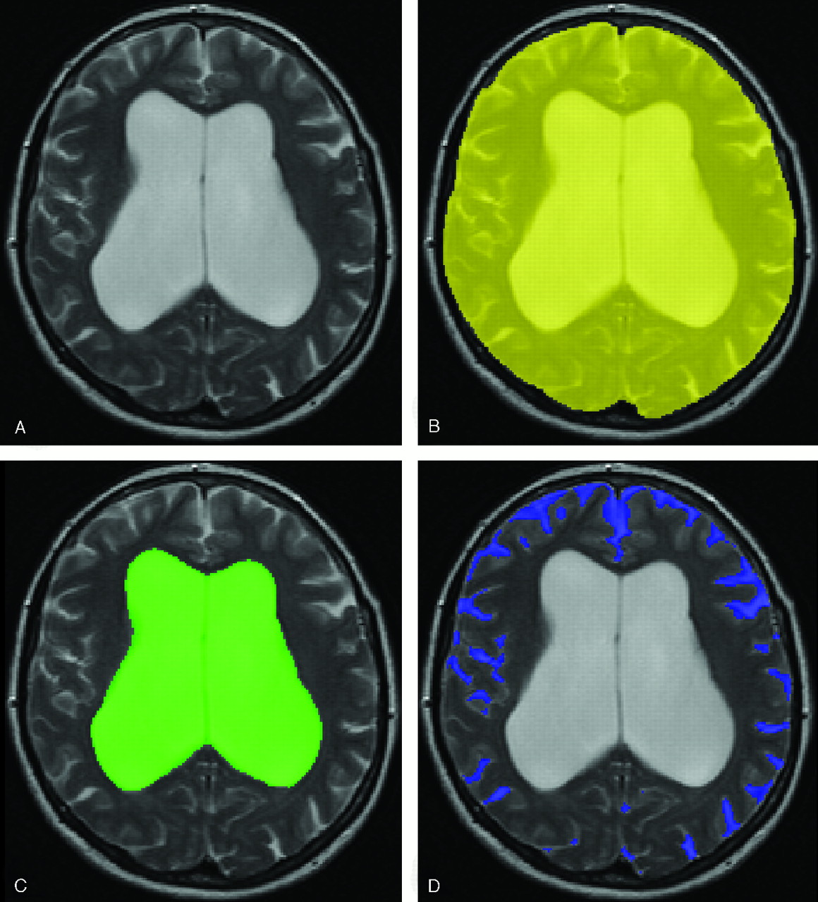
MR brain imaging was performed in all patients before ventriculoperitoneal shunt surgery. Dual spin-echo (proton attenuation and T2-weighted) images were acquired at field strengths of 0.5T (n = 7) or 1.5T (n = 19) (Philips Medical Systems, Best, the Netherlands) with 2500-millisecond repetition time, 27/120-millisecond echo time, a 256 × 256 matrix, and 6-mm section thickness with 0.6-mm intersection gap, covering the whole brain. Locally developed semiautomated segmentation software (SNIPER, Software for Neuro-Image Processing in Experimental Research) that combines knowledge-based fuzzy clustering and region-growing techniques was used to process the images.28 The outer contour of the subarachnoid space was manually delineated on each section by one observer (W.M.P.), which was followed by an automated segmentation procedure that assigned brain tissue and CSF within this region (Fig 1). The volumetric assessment was repeated for 5 of 26 subjects (19%) to analyze the intrarater reliability. The intraclass correlation coefficient was >0.99, which indicates high intrarater reliability. We determined total intracranial volume (TICV, comprising brain parenchyma and CSF), total brain volume, ventricular CSF volume (comprising lateral, third, and fourth ventricles), and extraventricular CSF volume (EVV, reflecting pericerebral CSF volume). Ventricular volume, brain volume, and pericerebral CSF volume were converted into ratios from TICV to normalize for head size. Ventriculomegaly out of proportion to cortical sulcal enlargement, a neuroimaging characteristic of NPH,18,29–31 was quantified by dividing ventricular volume by pericerebral CSF volume. In summary, 4 imaging variables were obtained: ventricular volume ratio, brain volume ratio, pericerebral CSF volume ratio, and a ratio between ventricular and pericerebral CSF volume.

Fig 1.
  • Download figure
  • Open in new tab
  • Download powerpoint
Fig 1.

Intracranial semiautomated segmentation was based on axial T2 (A) and proton attenuation–weighted images. The outer contour of the subarachnoid space was manually delineated on each section (B). This was followed by an automated segmentation procedure that assigned brain tissue and CSF within this region. We determined TICV, ventricular volume (C), brain volume, and pericerebral CSF volume (D).

Ventriculoperitoneal Shunt Surgery

All patients underwent ventriculoperitoneal shunt surgery. The type of shunt was a Codman-Medos nonprogrammable valve system with an opening pressure of 100 mm H2O. The patency of the shunt was judged by a neurosurgeon through clinical examination 2, 6, and 12 months after ventriculoperitoneal shunt surgery. In patients with lack of amelioration or clinical deterioration in combination with lack of decrease of the ventricular volume on CT or MR imaging, a surgical drain revision was carried out. The subjects remained shunted during the entire follow-up period of 1 year after shunt placement.

Statistical Methods

The mean ventricular volume ratio, the mean brain volume ratio, the mean pericerebral CSF volume ratio, and the mean ratio between ventricular and pericerebral CSF volume were compared between the patients who improved after shunt surgery and those who did not. Improvement of gait, cognition, and bladder function were determined by clinical rating (R.W. and H.A.M.M.).27 Mann-Whitney U tests were applied to assess differences between the 4 imaging variables and improvement in gait or cognition or bladder function (SPSS, version 11.5; SPSS, Inc., Chicago, Ill). Adjustments were made for multiple comparisons by using the Bonferroni method. The level of statistical significance was set at P < .05.

Results

All 26 patients had gait impairment, 22 were cognitively impaired, and 22 suffered from urinary incontinence. Clinical follow-up for the assessment of treatment effect took place 1 year after shunt surgery. Three had died in the follow-up period and were excluded from our analysis. After shunt surgery, 19 of the 23 remaining patients (83%) with preoperative gait impairment showed an improved walking pattern. Nine of the 19 remaining patients (47%) with preoperative cognitive impairment showed improved cognition after shunt surgery. Nine of the 20 remaining patients (45%) with preoperative bladder dysfunction reported a decrease in urinary urgency or urinary incontinence after shunt surgery.

Baseline values of TICV, ventricular volume, brain volume, and EVV for all subjects are presented in Table 1. The means and SDs of the 4 imaging variables are presented in Table 2, classified into improvement in each of the symptoms from the NPH clinical triad.

View this table:
  • View inline
  • View popup
Table 1:

Baseline MR imaging values

View this table:
  • View inline
  • View popup
Table 2:

Mean and standard deviation of the four imaging variables, classified into improvement per symptom

The mean of the 4 imaging variables—ventricular volume ratio, brain volume ratio, pericerebral CSF volume ratio, and ratio between ventricular volume and pericerebral CSF volume—did not differ significantly between patients who experienced improvement in gait after shunt surgery and those who did not. No significant differences were found in the mean of the 4 imaging variables between patients who improved in cognition and those who maintained the same level of cognitive impairment. There were also no significant differences in the mean of the 4 imaging variables between patients who did not report an improvement in bladder function and those who did experience less urinary urgency or urinary incontinence after shunt surgery.

Discussion

Our most important finding is that neither the mean ventricular volume ratio, the mean brain volume ratio, the mean pericerebral CSF volume ratio, nor the mean ratio between ventricular and pericerebral CSF volume were different between NPH patients who improved on gait or cognition or bladder function after ventriculoperitoneal shunt surgery and those who did not.

Our results suggest that volumetric assessment of intracranial compartments has no predictive value in differentiating between NPH patients who will respond favorably to ventriculoperitoneal shunt surgery and those who will not. Volumetric assessment has previously been employed to set NPH apart from other conditions and healthy elderly.22,23 The technique has not been used to compare intracranial compartment volumes between NPH patients who improved after ventriculoperitoneal shunt surgery and those who did not. In one study, MR brain images were used to study the preoperative neuroimaging characteristics of NPH in relation to clinical outcome after ventriculoperitoneal shunt surgery.32 In this study, CT and MR brain images were independently evaluated in a qualitative manner by a neuroradiologist and showed an association between preoperative cortical sulci size and postoperative improvement in gait, cognition, and bladder function, whereas ventricular volume was unrelated to postoperative outcome. The finding of an association between preoperative cortical sulci size or pericerebral CSF volume and outcome was not reproduced in our study.

A potential limitation of our study is that all the included subjects were patients eligible for shunt surgery. This resulted in the lack of a control group and a moderate spread of values. Nevertheless, it should be realized that our population represents a consecutive clinical group eligible for shunt surgery, which is the group that benefits most from preoperative risk stratification. In addition, if ventricular volume stays the same after the shunt is placed and the patient does not improve, a shunt function study needs to be done to determine whether the shunt is indeed functioning in light of the fact that the volume of the ventricles has not changed.

Conclusion

Neither the ventricular volume ratio, brain volume ratio, pericerebral CSF volume ratio, nor the ratio between ventricular and pericerebral CSF volume was different between NPH patients who improved on gait or cognition or bladder function and those who did not. Volumetric assessment of intracranial compartments has no predictive value in differentiating between NPH patients who will respond favorably to ventriculoperitoneal shunt surgery and those who will not.

Footnotes

  • This work has been presented in part at the Dutch Radiology Meeting, September 16–17, 2004, and the International Society for Magnetic Resonance in Medicine Workshop on Aging Connections, October 21–23, 2004.

References

  1. ↵
    Adams RD, Fisher CM, Hakim S, et al. Symptomatic occult hydrocephalus with “normal” cerebrospinal-fluid pressure: a treatable syndrome. N Engl J Med 1965;273:117–26
    CrossRefPubMed
  2. ↵
    Vanneste JA. Diagnosis and management of normal-pressure hydrocephalus. J Neurol 2000;247:5–14
    CrossRefPubMed
  3. ↵
    Hebb AO, Cusimano MD. Idiopathic normal pressure hydrocephalus: a systematic review of diagnosis and outcome. Neurosurgery 2001;49:1166–84
    PubMed
  4. ↵
    Esmonde T, Cooke S. Shunting for normal pressure hydrocephalus (NPH). Cochrane Database Syst Rev 2002;CD003157
  5. ↵
    Bret P, Guyotat J, Chazal J. Is normal pressure hydrocephalus a valid concept in 2002? A reappraisal in five questions and proposal for a new designation of the syndrome as “chronic hydrocephalus.” J Neurol Neurosurg Psychiatry 2002;73:9–12
    Abstract/FREE Full Text
  6. ↵
    Sand T, Bovim G, Grimse R, et al. Idiopathic normal pressure hydrocephalus: the CSF tap-test may predict the clinical response to shunting. Acta Neurol Scand 1994;89:311–16
    PubMed
  7. ↵
    Wikkelso C, Andersson H, Blomstrand C, et al. Normal pressure hydrocephalus: predictive value of the cerebrospinal fluid tap-test. Acta Neurol Scand 1986;73:566–73
    PubMed
  8. ↵
    Chen IH, Huang CI, Liu HC, et al. Effectiveness of shunting in patients with normal pressure hydrocephalus predicted by temporary, controlled-resistance, continuous lumbar drainage: a pilot study. J Neurol Neurosurg Psychiatry 1994;57:1430–32
    Abstract/FREE Full Text
  9. Di Lauro L, Mearini M, Bollati A. The predictive value of 5 days CSF diversion for shunting in normal pressure hydrocephalus. J Neurol Neurosurg Psychiatry 1986;49:842–43
    FREE Full Text
  10. ↵
    Haan J, Thomeer RT. Predictive value of temporary external lumbar drainage in normal pressure hydrocephalus. Neurosurgery 1988;22:388–91
    CrossRefPubMed
  11. ↵
    Borgesen SE, Gjerris F. The predictive value of conductance to outflow of CSF in normal pressure hydrocephalus. Brain 1982;105:65–86
    FREE Full Text
  12. ↵
    Bret P, Chazal J, Janny P, et al. [Chronic hydrocephalus in adults]. Neurochirurgie 1990;36(suppl 1):1–159
  13. Crockard HA, Hanlon K, Duda EE, Mullan JF. Hydrocephalus as a cause of dementia: evaluation by computerised tomography and intracranial pressure monitoring. J Neurol Neurosurg Psychiatry 1977;40:736–40
    Abstract/FREE Full Text
  14. Pickard JD, Teasdale G, Matheson M, et al. Intraventricular pressure waves: the best predictive test for shunting in normal pressure hydrocephalus. In: Shulman K, Marmarou A, Miller JD, et al, eds. Intracranial pressure. IV. Berlin: Springer-Verlag;1980;:498–500
  15. ↵
    Symon L, Dorsch NW. Use of long-term intracranial pressure measurement to assess hydrocephalic patients prior to shunt surgery. J Neurosurg 1975;42:258–73
    PubMed
  16. ↵
    Boon AJ, Tans JT, Delwel EJ, et al. Dutch normal-pressure hydrocephalus study: prediction of outcome after shunting by resistance to outflow of cerebrospinal fluid. J Neurosurg 1997;87:687–93
    PubMed
  17. ↵
    Bradley WG Jr, Whittemore AR, Kortman KE, et al. Marked cerebrospinal fluid void: indicator of successful shunt in patients with suspected normal-pressure hydrocephalus. Radiology 1991;178:459–66
    PubMed
  18. ↵
    Bradley WG Jr, Scalzo D, Queralt J, et al. Normal-pressure hydrocephalus: evaluation with cerebrospinal fluid flow measurements at MR imaging. Radiology 1996;198:523–29
    PubMed
  19. ↵
    Condon B, Patterson J, Wyper D, et al. Use of magnetic resonance imaging to measure intracranial cerebrospinal fluid volume. Lancet 1986;1:1355–57
    CrossRefPubMed
  20. Kitagaki H, Mori E, Ishii K, et al. CSF spaces in idiopathic normal pressure hydrocephalus: morphology and volumetry. AJNR Am J Neuroradiol 1998;19:1277–84
    Abstract
  21. Matsumae M, Kikinis R, Morocz I, et al. Intracranial compartment volumes in patients with enlarged ventricles assessed by magnetic resonance-based image processing. J Neurosurg 1996;84:972–81
    PubMed
  22. ↵
    Tsunoda A, Mitsuoka H, Sato K, et al. A quantitative index of intracranial cerebrospinal fluid distribution in normal pressure hydrocephalus using an MRI-based processing technique. Neuroradiology 2000;42:424–29
    CrossRefPubMed
  23. ↵
    Tsunoda A, Mitsuoka H, Bandai H, et al. Intracranial cerebrospinal fluid distribution and its postoperative changes in normal pressure hydrocephalus. Acta Neurochir (Wien) 2001;143:493–99
    PubMed
  24. Tsunoda A, Mitsuoka H, Bandai H, et al. Intracranial cerebrospinal fluid measurement studies in suspected idiopathic normal pressure hydrocephalus, secondary normal pressure hydrocephalus, and brain atrophy. J Neurol Neurosurg Psychiatry 2002;73:552–55
    Abstract/FREE Full Text
  25. ↵
    Yoshihara M, Tsunoda A, Sato K, et al. Differential diagnosis of NPH and brain atrophy assessed by measurement of intracranial and ventricular CSF volume with 3D FASE MRI. Acta Neurochir Suppl 1998;71:371–74
    PubMed
  26. ↵
    Gyldensted C. Measurements of the normal ventricular system and hemispheric sulci of 100 adults with computed tomography. Neuroradiology 1977;14:183–92
    CrossRefPubMed
  27. ↵
    Walchenbach R, Geiger E, Thomeer RT, Vanneste JA. The value of temporary external lumbar CSF drainage in predicting the outcome of shunting on normal pressure hydrocephalus. J Neurol Neurosurg Psychiatry 2002;72:503–506
    Abstract/FREE Full Text
  28. ↵
    Van den Boom R, Lesnik Oberstein SA, Spilt A, et al. Cerebral hemodynamics and white matter hyperintensities in CADASIL. J Cereb Blood Flow Metab 2003;23:599–604
    CrossRefPubMed
  29. ↵
    Bradley WG. Normal pressure hydrocephalus: new concepts on etiology and diagnosis. AJNR Am J Neuroradiol 2000;21:1586–90
    FREE Full Text
  30. Fishman RA, Dillon WP. Normal pressure hydrocephalus: new findings and old questions. AJNR Am J Neuroradiol 2001;22:1640–41
    FREE Full Text
  31. ↵
    Jack CR Jr, Lexa FJ, Trojanowski JQ, et al. Normal aging, dementia, and neurodegenerative disease. In: Atlas SW, ed. Magnetic resonance imaging of the brain and spine. 3rd ed. Philadelphia: Lippincott Williams & Wilkins;2001;1177–1240
  32. ↵
    Poca MA, Mataro M, Del Mar MM, et al. Is the placement of shunts in patients with idiopathic normal-pressure hydrocephalus worth the risk? Results of a study based on continuous monitoring of intracranial pressure. J Neurosurg 2004;100:855–66
    PubMed
  • Received March 29, 2005.
  • Accepted after revision June 6, 2005.
  • Copyright © American Society of Neuroradiology
View Abstract
PreviousNext
Back to top

In this issue

American Journal of Neuroradiology: 27 (1)
American Journal of Neuroradiology
Vol. 27, Issue 1
January, 2006
  • Table of Contents
  • Index by author
Advertisement
Print
Download PDF
Email Article

Thank you for your interest in spreading the word on American Journal of Neuroradiology.

NOTE: We only request your email address so that the person you are recommending the page to knows that you wanted them to see it, and that it is not junk mail. We do not capture any email address.

Enter multiple addresses on separate lines or separate them with commas.
Intracranial Compartment Volumes in Normal Pressure Hydrocephalus: Volumetric Assessment versus Outcome
(Your Name) has sent you a message from American Journal of Neuroradiology
(Your Name) thought you would like to see the American Journal of Neuroradiology web site.
CAPTCHA
This question is for testing whether or not you are a human visitor and to prevent automated spam submissions.
Cite this article
W.M. Palm, R. Walchenbach, B. Bruinsma, F. Admiraal-Behloul, H.A.M. Middelkoop, L.J. Launer, J. van der Grond, M.A. van Buchem
Intracranial Compartment Volumes in Normal Pressure Hydrocephalus: Volumetric Assessment versus Outcome
American Journal of Neuroradiology Jan 2006, 27 (1) 76-79;

Citation Manager Formats

  • BibTeX
  • Bookends
  • EasyBib
  • EndNote (tagged)
  • EndNote 8 (xml)
  • Medlars
  • Mendeley
  • Papers
  • RefWorks Tagged
  • Ref Manager
  • RIS
  • Zotero
0 Responses
Respond to this article
Share
Bookmark this article
Intracranial Compartment Volumes in Normal Pressure Hydrocephalus: Volumetric Assessment versus Outcome
W.M. Palm, R. Walchenbach, B. Bruinsma, F. Admiraal-Behloul, H.A.M. Middelkoop, L.J. Launer, J. van der Grond, M.A. van Buchem
American Journal of Neuroradiology Jan 2006, 27 (1) 76-79;
del.icio.us logo Twitter logo Facebook logo Mendeley logo
  • Tweet Widget
  • Facebook Like
  • Google Plus One

Jump to section

  • Article
    • Abstract
    • Methods
    • Results
    • Discussion
    • Conclusion
    • Footnotes
    • References
  • Figures & Data
  • Info & Metrics
  • Responses
  • References
  • PDF

Related Articles

  • No related articles found.
  • PubMed
  • Google Scholar

Cited By...

  • The First Examination of Diagnostic Performance of Automated Measurement of the Callosal Angle in 1856 Elderly Patients and Volunteers Indicates That 12.4% of Exams Met the Criteria for Possible Normal Pressure Hydrocephalus
  • MR Imaging of Brain Volumes: Evaluation of a Fully Automatic Software
  • Crossref
  • Google Scholar

This article has not yet been cited by articles in journals that are participating in Crossref Cited-by Linking.

More in this TOC Section

  • Fast Contrast-Enhanced 4D MRA and 4D Flow MRI Using Constrained Reconstruction (HYPRFlow): Potential Applications for Brain Arteriovenous Malformations
  • Quiet PROPELLER MRI Techniques Match the Quality of Conventional PROPELLER Brain Imaging Techniques
  • Predictors of Reperfusion in Patients with Acute Ischemic Stroke
Show more Brain

Similar Articles

Advertisement

Indexed Content

  • Current Issue
  • Accepted Manuscripts
  • Article Preview
  • Past Issues
  • Editorials
  • Editors Choice
  • Fellow Journal Club
  • Letters to the Editor

Cases

  • Case Collection
  • Archive - Case of the Week
  • Archive - Case of the Month
  • Archive - Classic Case

Special Collections

  • Special Collections

Resources

  • News and Updates
  • Turn around Times
  • Submit a Manuscript
  • Author Policies
  • Manuscript Submission Guidelines
  • Evidence-Based Medicine Level Guide
  • Publishing Checklists
  • Graphical Abstract Preparation
  • Imaging Protocol Submission
  • Submit a Case
  • Become a Reviewer/Academy of Reviewers
  • Get Peer Review Credit from Publons

Multimedia

  • AJNR Podcast
  • AJNR SCANtastic
  • Video Articles

About Us

  • About AJNR
  • Editorial Board
  • Not an AJNR Subscriber? Join Now
  • Alerts
  • Feedback
  • Advertise with us
  • Librarian Resources
  • Permissions
  • Terms and Conditions

American Society of Neuroradiology

  • Not an ASNR Member? Join Now

© 2025 by the American Society of Neuroradiology All rights, including for text and data mining, AI training, and similar technologies, are reserved.
Print ISSN: 0195-6108 Online ISSN: 1936-959X

Powered by HighWire