Advertisement

Main menu

  • Home
  • Content
    • Current Issue
    • Accepted Manuscripts
    • Article Preview
    • Past Issue Archive
    • AJNR Case Collection
    • Case of the Week Archive
    • Classic Case Archive
    • Case of the Month Archive
  • Special Collections
    • Spinal CSF Leak Articles (Jan 2020-June 2024)
    • 2024 AJNR Journal Awards
    • Most Impactful AJNR Articles
  • Multimedia
    • AJNR Podcast
    • AJNR Scantastics
    • Video Articles
  • For Authors
    • Submit a Manuscript
    • Author Policies
    • Fast publishing of Accepted Manuscripts
    • Graphical Abstract Preparation
    • Manuscript Submission Guidelines
    • Imaging Protocol Submission
    • Submit a Case for the Case Collection
  • About Us
    • About AJNR
    • Editorial Board
  • More
    • Become a Reviewer/Academy of Reviewers
    • Subscribers
    • Permissions
    • Alerts
    • Feedback
    • Advertisers
    • ASNR Home
  • Other Publications
    • ajnr

User menu

  • Alerts
  • Log in

Search

  • Advanced search
American Journal of Neuroradiology

American Journal of Neuroradiology

ASHNR American Society of Functional Neuroradiology ASHNR American Society of Pediatric Neuroradiology ASSR
  • Alerts
  • Log in

Advanced Search

  • Home
  • Content
    • Current Issue
    • Accepted Manuscripts
    • Article Preview
    • Past Issue Archive
    • AJNR Case Collection
    • Case of the Week Archive
    • Classic Case Archive
    • Case of the Month Archive
  • Special Collections
    • Spinal CSF Leak Articles (Jan 2020-June 2024)
    • 2024 AJNR Journal Awards
    • Most Impactful AJNR Articles
  • Multimedia
    • AJNR Podcast
    • AJNR Scantastics
    • Video Articles
  • For Authors
    • Submit a Manuscript
    • Author Policies
    • Fast publishing of Accepted Manuscripts
    • Graphical Abstract Preparation
    • Manuscript Submission Guidelines
    • Imaging Protocol Submission
    • Submit a Case for the Case Collection
  • About Us
    • About AJNR
    • Editorial Board
  • More
    • Become a Reviewer/Academy of Reviewers
    • Subscribers
    • Permissions
    • Alerts
    • Feedback
    • Advertisers
    • ASNR Home
  • Follow AJNR on Twitter
  • Visit AJNR on Facebook
  • Follow AJNR on Instagram
  • Join AJNR on LinkedIn
  • RSS Feeds

Welcome to the new AJNR, Updated Hall of Fame, and more. Read the full announcements.


AJNR is seeking candidates for the position of Associate Section Editor, AJNR Case Collection. Read the full announcement.


OtherBRAIN

Imaging Characteristics of the Subdural Evacuating Port System, a New Bedside Therapy for Subacute/Chronic Subdural Hematoma

S.S. Lollis, M.L. Wolak and A.C. Mamourian
American Journal of Neuroradiology January 2006, 27 (1) 74-75;
S.S. Lollis
  • Find this author on Google Scholar
  • Find this author on PubMed
  • Search for this author on this site
M.L. Wolak
  • Find this author on Google Scholar
  • Find this author on PubMed
  • Search for this author on this site
A.C. Mamourian
  • Find this author on Google Scholar
  • Find this author on PubMed
  • Search for this author on this site
  • Article
  • Figures & Data
  • Info & Metrics
  • Responses
  • References
  • PDF
Loading

Abstract

SUMMARY: The Subdural Evacuating Port System is a new device intended to simplify the treatment of subacute/chronic subdural hematomas. The appearance of the winged canula positioned with its tip in the diploic space overlying the subdural space should allow the radiologist to identify it correctly. Its radiographic features are described here to help the radiologist comment on appropriate placement, and avoid mistaking it for a misplaced subdural drain.

The Subdural Evacuating Port System (SEPS) is a new therapy (Medical Designs, LLC, Sioux Falls, SD) designed to treat subacute or chronic subdural hematomas.1,2 It can be placed at the bedside and permits drainage of fluid during a period of hours to days. The purpose of this report is to describe the imaging characteristics of this device.

Case Report

An 81-year-old man presented to our facility with 4 weeks of progressive gait instability and poor coordination of the left upper extremity. These symptoms began shortly after he fell on an icy pavement, striking his right parieto-occipital region. A head CT scan revealed a right convexity subdural hematoma with mass effect on the adjacent brain. The neurosurgical service was consulted, and they decide to treat the patient with the SEPS.

Description of the Device

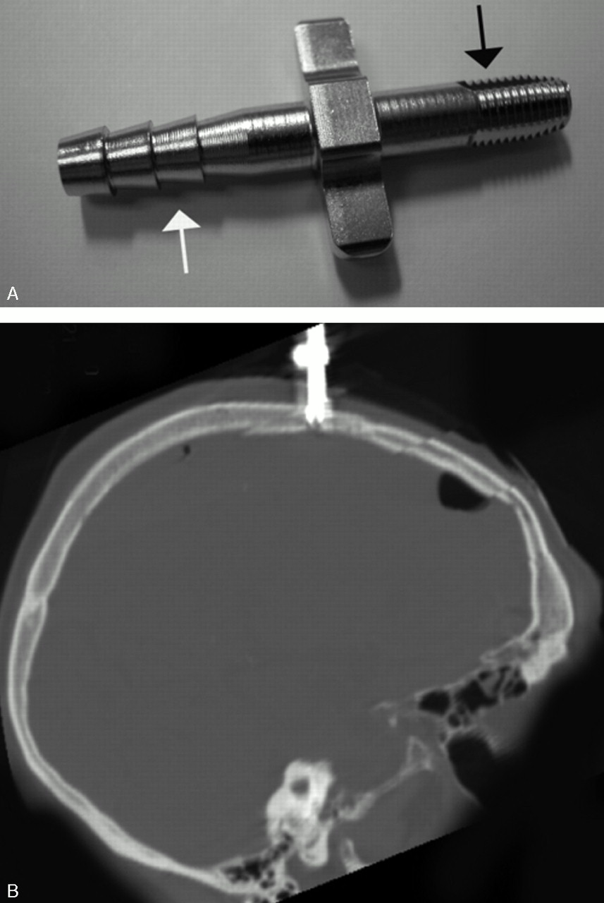
The SEPS consists of a stainless steel evacuating port, radiolucent silicone tubing, and a bulb suction device. It can be placed at the bedside by using a technique similar to that employed for the placement of a subarachnoid bolt used for intracranial pressure monitoring. After localizing the subdural collection by using superficial landmarks and making an appropriate skin incision, the surgeon places a single burr hole in the calvarium using a manual drill with a 5.8-mm drill bit. The dura is then opened and the metal evacuating port is manually screwed into the hole in the skull so that the tip is positioned in the diploic space, not extending beyond the inner table. The external portion of the evacuating port is then connected to the silicone tubing and bulb suction apparatus. Negative pressure (≤2.5 cmHg) is applied by using a bulb for a variable time period, typically 24–48 hours, but until drainage of subdural fluid has ceased. The system is then removed and the skin sutured is closed.

Imaging Characteristics

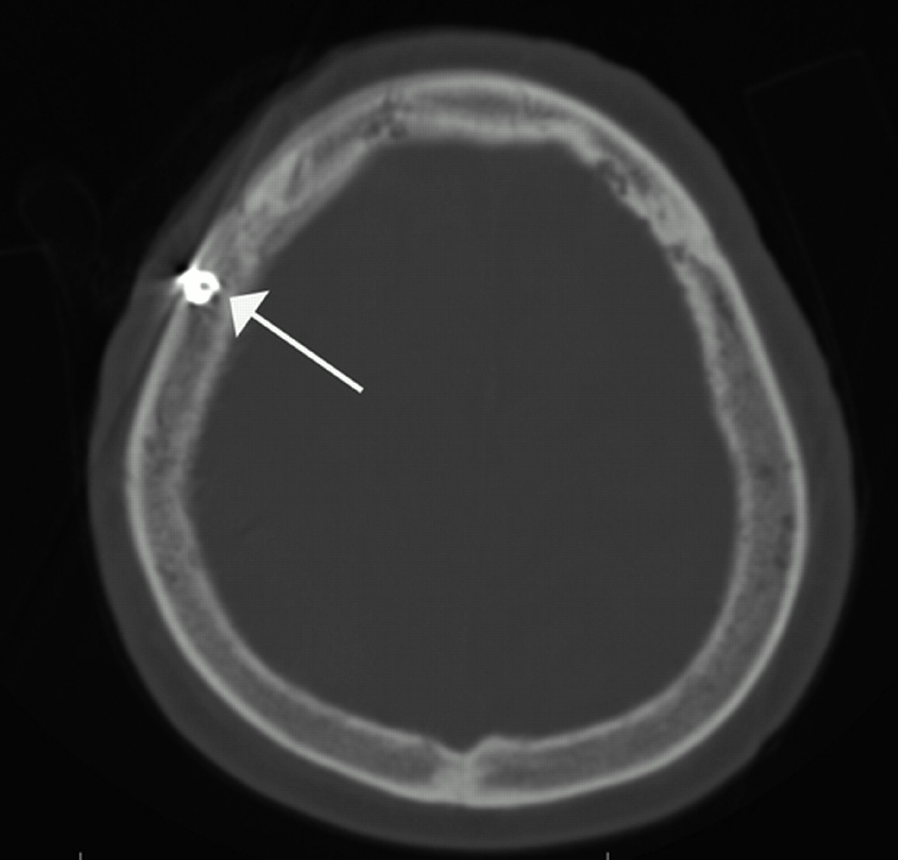
The SEPS can be distinguished radiographically from a typical, bolt-type intracranial pressure monitor. First, the SEPS evacuating port is larger, measuring 4.5 cm in length and 6 mm in tip width (Fig 1). It has metal wings, giving it the appearance of a cross on reformatted images (Fig 2). Second, the evacuating port consists entirely of stainless steel; typical intracranial pressure monitors consist of a metal central core, with radiolucent wings to assist in placement. The SEPS is also easily distinguished from subdural catheters. The evacuating port is anchored into the diploic space and represents the most medial end of the device (Figs 3 and 4)—ie, there is no subdural catheter extending along the surface of the brain.

Fig 1.
  • Download figure
  • Open in new tab
  • Download powerpoint
Fig 1.

A, This image shows the metal device that is screwed into the burr hole. The threaded end (black arrow) is self-tapping. The notched tubing (white arrow) is then attached to the silicone tubing.

B, This reformat of the CT data shows the metal insertion portion of the device orientation perpendicular to the skull.

Fig 2.
  • Download figure
  • Open in new tab
  • Download powerpoint
Fig 2.

Oblique (A) and A-P (B) views, reformatted from CT data on a 3D workstation (Vitrea), demonstrate the winged canula and its relationship to the skull.

Fig 3.
  • Download figure
  • Open in new tab
  • Download powerpoint
Fig 3.

The bone image from that same study shows the tip of the device positioned in the diploic space of the right calvaria (arrow).

Fig 4.
  • Download figure
  • Open in new tab
  • Download powerpoint
Fig 4.

The reformatted 2D image demonstrates the relationships of the subdural hematoma (short arrows) and the device positioned in the skull (long arrow).

Discussion

Although several surgical options exist for treatment of subacute and chronic subdural hematomas, recent studies suggest that optimal management involves twist-drill catheter placement and drainage via a closed external drainage system.3 This procedure can be performed either in the operative theater or at the bedside. The drainage system typically consists of a radiopaque catheter placed in the subdural compartment, which is connected to a Jackson-Pratt-type suction reservoir via a radiolucent connector tube. The SEPS is a new approach to treatment of subdural hematomas. The decision to use the SEPS may be based on several potential advantages over twist-drill catheter placement and drainage. The SEPS introduces mild, uniform negative pressure throughout the subdural space without direct contact with the brain, cortical or bridging vessels, or vascularized hematoma membranes. The risk of catheter-related parenchymal injury and iatrogenic hemorrhage are theoretically reduced. It is likely that radiologists practicing at neurosurgical centers will see an increasing number of CT scans after placement of this system. The system has unique imaging characteristics that distinguish it from bolt-type intracranial pressure monitors, as well as subdural catheters. It is important to be able to recognize the SEPS and comment on its appropriate placement.

References

  1. ↵
    Asfora WT, Schwebach L. A modified technique to treat chronic and subacute subdural hematoma: techincal note. Surg Neurol 2003;59:329–32
    PubMed
  2. ↵
    Asfora WT, Schwebach L, Louw D. A modified technique to treat subdural hematomas: the Subdural Evacuating Port System. S D J Med 2001;54:495–98
    PubMed
  3. ↵
    Williams GR, Baskaya MK, Menendez J, et al. Burr-hole versus twist drill drainage for the evacuation of chronic subdural hematoma: a comparison of clinical results. J Clin Neurosci 2001;8:551–54
    PubMed
  • Received June 7, 2005.
  • Accepted after revision July 20, 2005.
  • Copyright © American Society of Neuroradiology
View Abstract
PreviousNext
Back to top

In this issue

American Journal of Neuroradiology: 27 (1)
American Journal of Neuroradiology
Vol. 27, Issue 1
January, 2006
  • Table of Contents
  • Index by author
Advertisement
Print
Download PDF
Email Article

Thank you for your interest in spreading the word on American Journal of Neuroradiology.

NOTE: We only request your email address so that the person you are recommending the page to knows that you wanted them to see it, and that it is not junk mail. We do not capture any email address.

Enter multiple addresses on separate lines or separate them with commas.
Imaging Characteristics of the Subdural Evacuating Port System, a New Bedside Therapy for Subacute/Chronic Subdural Hematoma
(Your Name) has sent you a message from American Journal of Neuroradiology
(Your Name) thought you would like to see the American Journal of Neuroradiology web site.
CAPTCHA
This question is for testing whether or not you are a human visitor and to prevent automated spam submissions.
Cite this article
S.S. Lollis, M.L. Wolak, A.C. Mamourian
Imaging Characteristics of the Subdural Evacuating Port System, a New Bedside Therapy for Subacute/Chronic Subdural Hematoma
American Journal of Neuroradiology Jan 2006, 27 (1) 74-75;

Citation Manager Formats

  • BibTeX
  • Bookends
  • EasyBib
  • EndNote (tagged)
  • EndNote 8 (xml)
  • Medlars
  • Mendeley
  • Papers
  • RefWorks Tagged
  • Ref Manager
  • RIS
  • Zotero
0 Responses
Respond to this article
Share
Bookmark this article
Imaging Characteristics of the Subdural Evacuating Port System, a New Bedside Therapy for Subacute/Chronic Subdural Hematoma
S.S. Lollis, M.L. Wolak, A.C. Mamourian
American Journal of Neuroradiology Jan 2006, 27 (1) 74-75;
del.icio.us logo Twitter logo Facebook logo Mendeley logo
  • Tweet Widget
  • Facebook Like
  • Google Plus One

Jump to section

  • Article
    • Abstract
    • Case Report
    • Discussion
    • References
  • Figures & Data
  • Info & Metrics
  • Responses
  • References
  • PDF

Related Articles

  • No related articles found.
  • PubMed
  • Google Scholar

Cited By...

  • No citing articles found.
  • Crossref
  • Google Scholar

This article has not yet been cited by articles in journals that are participating in Crossref Cited-by Linking.

More in this TOC Section

  • Progression of Microstructural Damage in Spinocerebellar Ataxia Type 2: A Longitudinal DTI Study
  • Usefulness of Quantitative Susceptibility Mapping for the Diagnosis of Parkinson Disease
  • Evaluating the Effects of White Matter Multiple Sclerosis Lesions on the Volume Estimation of 6 Brain Tissue Segmentation Methods
Show more Brain

Similar Articles

Advertisement

Indexed Content

  • Current Issue
  • Accepted Manuscripts
  • Article Preview
  • Past Issues
  • Editorials
  • Editors Choice
  • Fellow Journal Club
  • Letters to the Editor

Cases

  • Case Collection
  • Archive - Case of the Week
  • Archive - Case of the Month
  • Archive - Classic Case

Special Collections

  • Special Collections

Resources

  • News and Updates
  • Turn around Times
  • Submit a Manuscript
  • Author Policies
  • Manuscript Submission Guidelines
  • Evidence-Based Medicine Level Guide
  • Publishing Checklists
  • Graphical Abstract Preparation
  • Imaging Protocol Submission
  • Submit a Case
  • Become a Reviewer/Academy of Reviewers
  • Get Peer Review Credit from Publons

Multimedia

  • AJNR Podcast
  • AJNR SCANtastic
  • Video Articles

About Us

  • About AJNR
  • Editorial Board
  • Not an AJNR Subscriber? Join Now
  • Alerts
  • Feedback
  • Advertise with us
  • Librarian Resources
  • Permissions
  • Terms and Conditions

American Society of Neuroradiology

  • Not an ASNR Member? Join Now

© 2025 by the American Society of Neuroradiology All rights, including for text and data mining, AI training, and similar technologies, are reserved.
Print ISSN: 0195-6108 Online ISSN: 1936-959X

Powered by HighWire