
Article Figures & Data
Figures
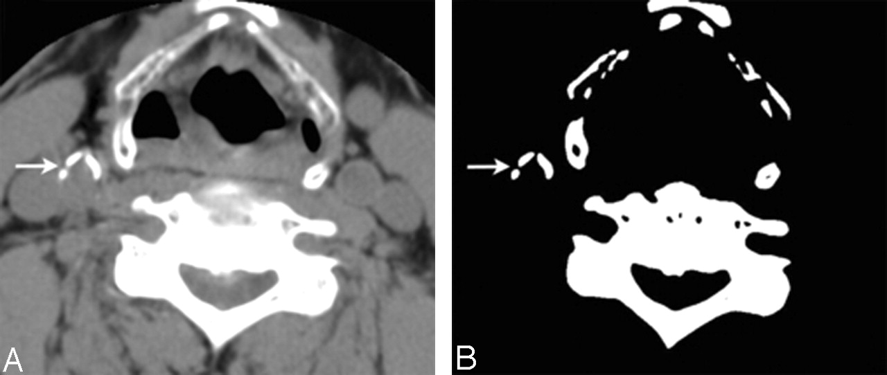
- Fig 1.
Axial CT at level of thyroid cartilage shown on soft tissue window (width, 340 HU; center, 43 HU) in panel A and narrow window (width, 1 HU; center, 130 HU) in panel B. Three calcific plaques are seen at the right distal common carotid artery (arrow).
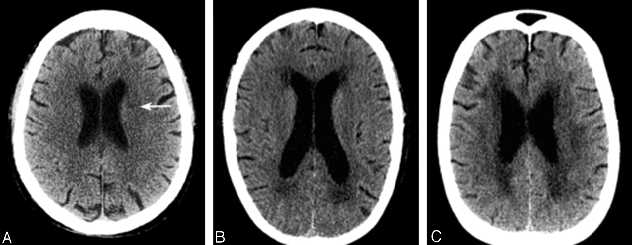
- Fig 2.
Examples of the white matter rating scores by using the European Task Force on Age-Related White Matter Changes, from 3 separate study cases. Score 1 (A): focal ill-defined hypoattenuation in the left corona radiate (arrow); score 2 (B): beginning confluence of lesions; score 3 (C): diffuse involvement of the frontal and parieto-occipital region seen in this example.


Tables
White Matter Score Number of Hemispheres Median Age (y) 25th Percentile of Age (y) 75th Percentile of Age (y) 0 214 50 42.0 57.0 1 87 69 57.3 80.5 2 90 73 65.0 82.0 3 27 83 82.0 89.0 Carotid Artery Calcium Score Number of Carotids Median Age (y) 25th Percentile of Age (y) 75th Percentile of Age (y) 0 257 52 45.0 61.0 1–100 59 72 58.3 78.8 101–400 49 77 63.0 84.0 400+ 53 82 77.3 85.0 Test Pearson Correlation Pearson Correlation with Age Adjustment White matter grade and age 0.67 (P < .001) Log10 total carotid calcium score and age 0.60 (P < .001) White matter grade and log10 total carotid calcium score 0.45 (P < .001) 0.07 (P = .14) White matter grade and log10 CCA calcium score 0.44 (P < .001) 0.07 (P = .13) White matter grade and log10 ICA calcium score 0.32 (P < .001) 0.06 (P = .23) Note.— ICA indicates internal carotid artery; CCA, common carotid artery.
- Table 4:
Prospective population-based cohort studies of white matter-lesion severity and incident stroke
Study Group Clinical Entry Criteria Total Number of Patients Follow-up (y) Definition of WM Outcome Variable % of Incident Strokes During Follow-up by WM Outcome Variable Cumulative Stroke Incidence Multivariable Adjusted Relative Risk (95% CI) Subtype of Stroke Subtype of Ischemic Stroke Dutch TIA Trial Group7 TIA or non-disabling ischemic stroke (Rankin ≤3) 3017 Not clearly defined WMH vs No WMH 15% vs 8% 19% vs 10% (3 y) 1.6 (1.2–2.2) Ischemic 90% vs 89%; hemorrhage 6% vs 9%; unknown 4% vs 2% Large artery 47% vs 45%; lacunar 33% vs 29%; vertebrobasilar 10% vs 15% NASCET Group5 Symptomatic carotid artery disease without WMH on entry CT 596 6.0 (median) Development of WMH vs no WMH 36% vs 23.5% Not defined 1.5 (1.0–2.4) Not defined Large artery 66% vs 73%; lacunar 29% vs 21%; cardioembolic 5% vs 6% ARIC Group10 No history of stroke of TIA 1684 4.7 (median) Grade ≥3 vs grade <3* 6.5% vs 1.3% 6.8% vs 1.4% (5 y) 3.4 (1.5–7.7) Not subdivided by WM status: ischemic 78%; hemorrhage 16%; combined 6% Not defined Rotterdam Scan Study Group9 No history of stroke or TIA 1077 4.2 (mean) Third tertile grade vs first tertile grade Not defined Not defined 4.7 (2.0–11.2) (periventricular lesions); 3.6 (1.4–9.2) (subcortical lesions) Not subdivided by WM status: ischemic 74%; hemorrhage 10%; unspecified 16% Not defined CHS Group8 No history of stroke or TIA 3293 7 (median) Grade ≥5 vs grade ≤1† 27.6% vs 6.0% 2.8% vs 0.6% (annual risk) 3.0 (1.9–4.7) Ischemic (81%) Lacunar (15%); cardioembolic (27%); atherosclerotic (5%); unknown (47%); mixed (6%) Note.— TIA indicates transient ischemic attack; WMH, white matter hypodensity; NASCET, North American Symptomatic Carotid Endarterectomy Trial Group; ARIC, Atherosclerosis Risk in Communities Study; CHS, Cardiovascular Health Study Collaborative Research Group; CI, confidence interval.
* WM grade 3 defined as thicker continuous periventricular rim with scattered patches of subcortical WM lesions.
† Grade 1 defined as discontinuous periventricular rim or minimal “dot”of subcortical disease; grade 5 defined as mild periventricular confluence surrounding the frontal and occipital horn.
In this issue


Jump to section
Related Articles
Cited By...
- Correlation between carotid bifurcation calcium burden on non-enhanced CT and percentage stenosis, as confirmed by digital subtraction angiography
- Calcification in Major Vessel Beds Relates to Vascular Brain Disease
- The Relation of Carotid Calcium Volume with Carotid Artery Stenosis in Symptomatic Patients
- Carotid Artery Wall Thickness and Leukoaraiosis: Preliminary Results Using Multidetector Row CT Angiography
- Authors' response