Advertisement

Main menu

  • Home
  • Content
    • Current Issue
    • Accepted Manuscripts
    • Article Preview
    • Past Issue Archive
    • AJNR Case Collection
    • Case of the Week Archive
    • Classic Case Archive
    • Case of the Month Archive
  • Special Collections
    • Spinal CSF Leak Articles (Jan 2020-June 2024)
    • 2024 AJNR Journal Awards
    • Most Impactful AJNR Articles
  • Multimedia
    • AJNR Podcast
    • AJNR Scantastics
    • Video Articles
  • For Authors
    • Submit a Manuscript
    • Author Policies
    • Fast publishing of Accepted Manuscripts
    • Graphical Abstract Preparation
    • Manuscript Submission Guidelines
    • Imaging Protocol Submission
    • Submit a Case for the Case Collection
  • About Us
    • About AJNR
    • Editorial Board
  • More
    • Become a Reviewer/Academy of Reviewers
    • Subscribers
    • Permissions
    • Alerts
    • Feedback
    • Advertisers
    • ASNR Home
  • Other Publications
    • ajnr

User menu

  • Alerts
  • Log in

Search

  • Advanced search
American Journal of Neuroradiology

American Journal of Neuroradiology

ASHNR American Society of Functional Neuroradiology ASHNR American Society of Pediatric Neuroradiology ASSR
  • Alerts
  • Log in

Advanced Search

  • Home
  • Content
    • Current Issue
    • Accepted Manuscripts
    • Article Preview
    • Past Issue Archive
    • AJNR Case Collection
    • Case of the Week Archive
    • Classic Case Archive
    • Case of the Month Archive
  • Special Collections
    • Spinal CSF Leak Articles (Jan 2020-June 2024)
    • 2024 AJNR Journal Awards
    • Most Impactful AJNR Articles
  • Multimedia
    • AJNR Podcast
    • AJNR Scantastics
    • Video Articles
  • For Authors
    • Submit a Manuscript
    • Author Policies
    • Fast publishing of Accepted Manuscripts
    • Graphical Abstract Preparation
    • Manuscript Submission Guidelines
    • Imaging Protocol Submission
    • Submit a Case for the Case Collection
  • About Us
    • About AJNR
    • Editorial Board
  • More
    • Become a Reviewer/Academy of Reviewers
    • Subscribers
    • Permissions
    • Alerts
    • Feedback
    • Advertisers
    • ASNR Home
  • Follow AJNR on Twitter
  • Visit AJNR on Facebook
  • Follow AJNR on Instagram
  • Join AJNR on LinkedIn
  • RSS Feeds

Welcome to the new AJNR, Updated Hall of Fame, and more. Read the full announcements.


AJNR is seeking candidates for the position of Associate Section Editor, AJNR Case Collection. Read the full announcement.


Research ArticleBRAIN

Comparison of Microvascular Permeability Measurements, Ktrans, Determined with Conventional Steady-State T1-Weighted and First-Pass T2*-Weighted MR Imaging Methods in Gliomas and Meningiomas

S. Cha, L. Yang, G. Johnson, A. Lai, M.-H. Chen, T. Tihan, M. Wendland and W.P. Dillon
American Journal of Neuroradiology February 2006, 27 (2) 409-417;
S. Cha
  • Find this author on Google Scholar
  • Find this author on PubMed
  • Search for this author on this site
L. Yang
  • Find this author on Google Scholar
  • Find this author on PubMed
  • Search for this author on this site
G. Johnson
  • Find this author on Google Scholar
  • Find this author on PubMed
  • Search for this author on this site
A. Lai
  • Find this author on Google Scholar
  • Find this author on PubMed
  • Search for this author on this site
M.-H. Chen
  • Find this author on Google Scholar
  • Find this author on PubMed
  • Search for this author on this site
T. Tihan
  • Find this author on Google Scholar
  • Find this author on PubMed
  • Search for this author on this site
M. Wendland
  • Find this author on Google Scholar
  • Find this author on PubMed
  • Search for this author on this site
W.P. Dillon
  • Find this author on Google Scholar
  • Find this author on PubMed
  • Search for this author on this site
  • Article
  • Figures & Data
  • Info & Metrics
  • Responses
  • References
  • PDF
Loading

Article Figures & Data

Figures

  • Tables
  • Fig 1.
    • Download figure
    • Open in new tab
    • Download powerpoint
    Fig 1.

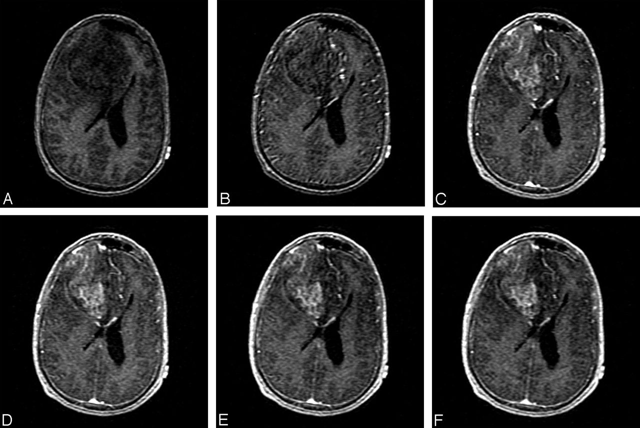
    Right frontal anaplastic oligoastrocytoma (WHO grade III) in a 43-year-old man. A dynamic series of ssT1 SPGR images through one anatomic location before, during, and after the administration of intravenous Gd-DTPA demonstrate earlier enhancement of normal vessels followed by delayed and persistent enhancement of the right frontal high-grade glioma.

  • Fig 2.
    • Download figure
    • Open in new tab
    • Download powerpoint
    Fig 2.

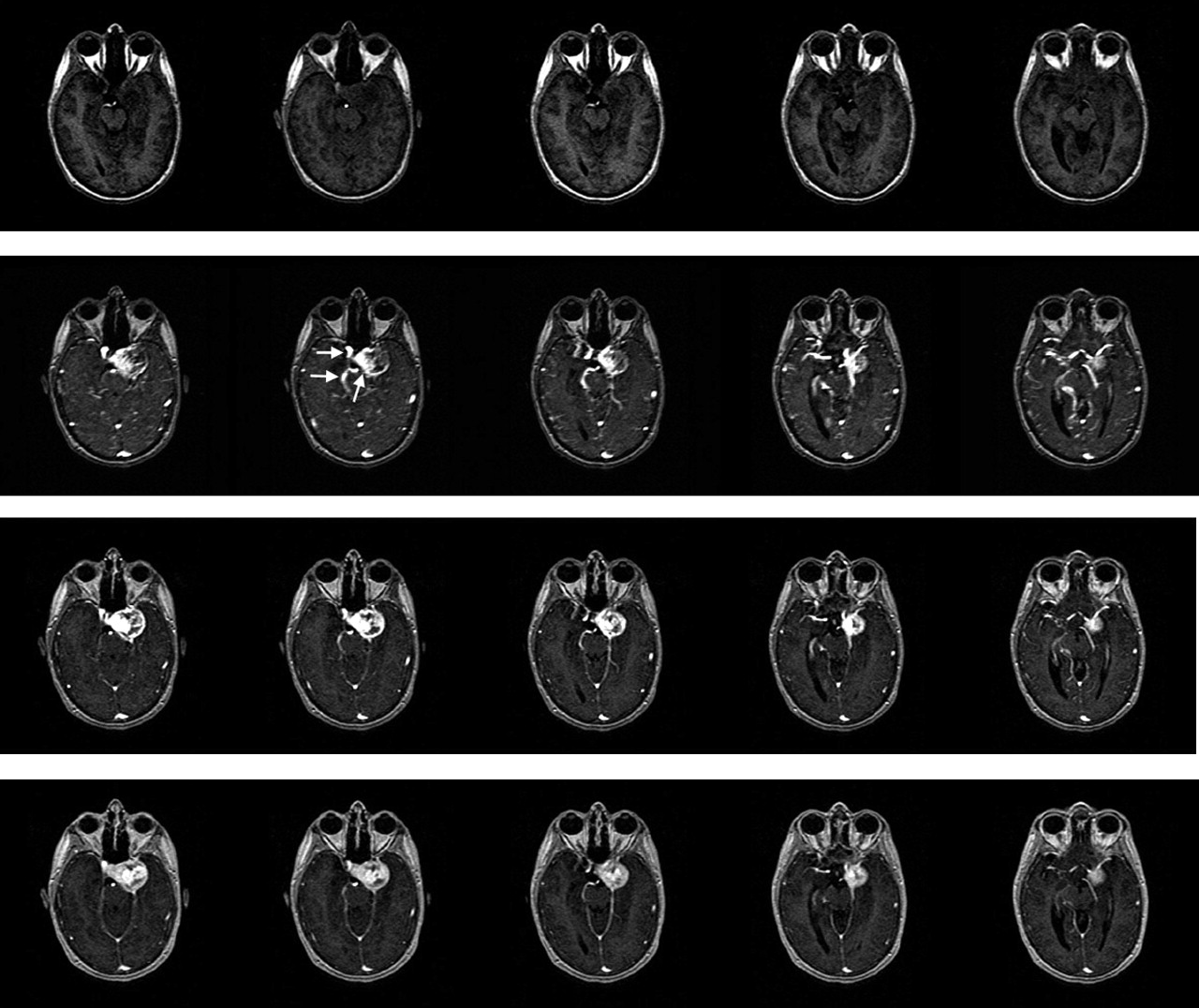
    Left cavernous sinus hemangiopericytoma in a 30-year-old woman. A dynamic series of ssT1 SPGR images through multiple anatomic locations before (top row), during (middle 2 rows), and after (bottom row) the administration of intravenous Gd-DTPA demonstrate simultaneous contrast agent arrival within the normal vessels (second row, horizontal arrows) and within this highly vascular extra-axial brain tumor (slanted arrow).

  • Fig 3.
    • Download figure
    • Open in new tab
    • Download powerpoint
    Fig 3.

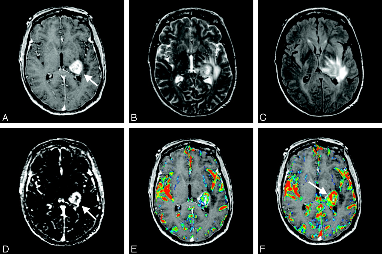
    Left thalamic/posterior frontal GBM (WHO grade IV) in a 63-year-old man. Upper panel, Left, Transaxial contrast-enhanced SPGR image demonstrates an enhancing left dorsolateral thalamic and posterior frontal lobe tumor (arrow). Middle and Right, Transaxial T2-weighted image (middle) and FLAIR (right) show moderate degree of surrounding edema (arrowheads). Lower panel, Left, Transaxial ssT1 Ktrans map demonstrates a rim of increased permeability (arrow). Middle, Transaxial fpT2* Ktrans color map overlayed onto SPGR image also shows a rim of increased permeability. Right, Transaxial fpT2* rCBV color map overlayed onto SPGR image demonstrates similar rim shape of increased blood volume but more focused on the medial aspect of the tumor (arrow).
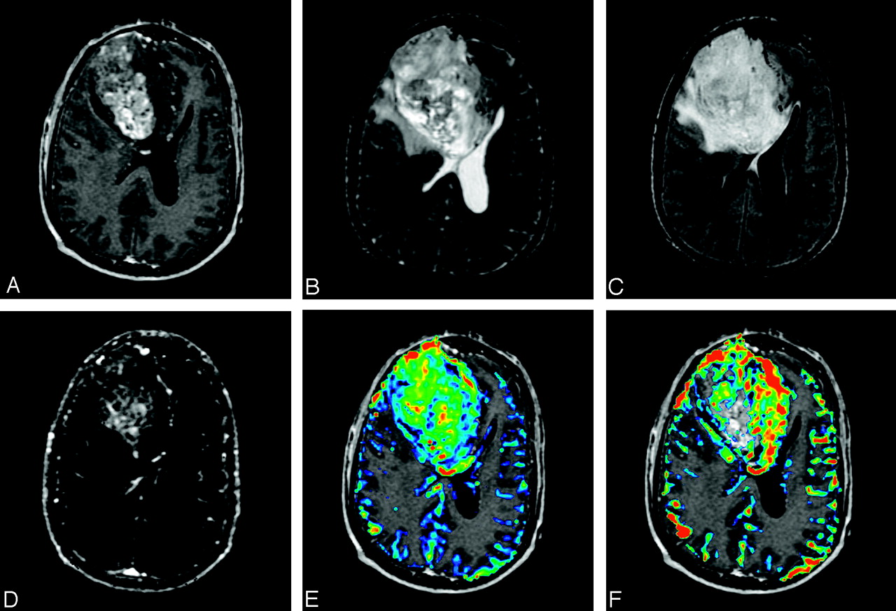
  • Fig 4.
    • Download figure
    • Open in new tab
    • Download powerpoint
    Fig 4.

    Right frontal anaplastic oligoastrocytoma (WHO grade III) in a 43-year-old man. Upper panel, Left, Transaxial contrast-enhanced SPGR image demonstrates a large heterogeneously right frontal lobe tumor. Middle and Right, Transaxial T2-weighted image (middle) and FLAIR (right) show moderate degree of surrounding edema. Lower panel, Left, Transaxial ssT1 Ktrans map demonstrates a large central area of increased permeability. Middle, Transaxial fpT2* Ktrans color map overlayed onto SPGR image also shows global increase in permeability throughout the tumor. Right, Transaxial fpT2* rCBV color map overlayed onto SPGR image demonstrates increased blood volume mostly involving the medial aspect of the tumor.
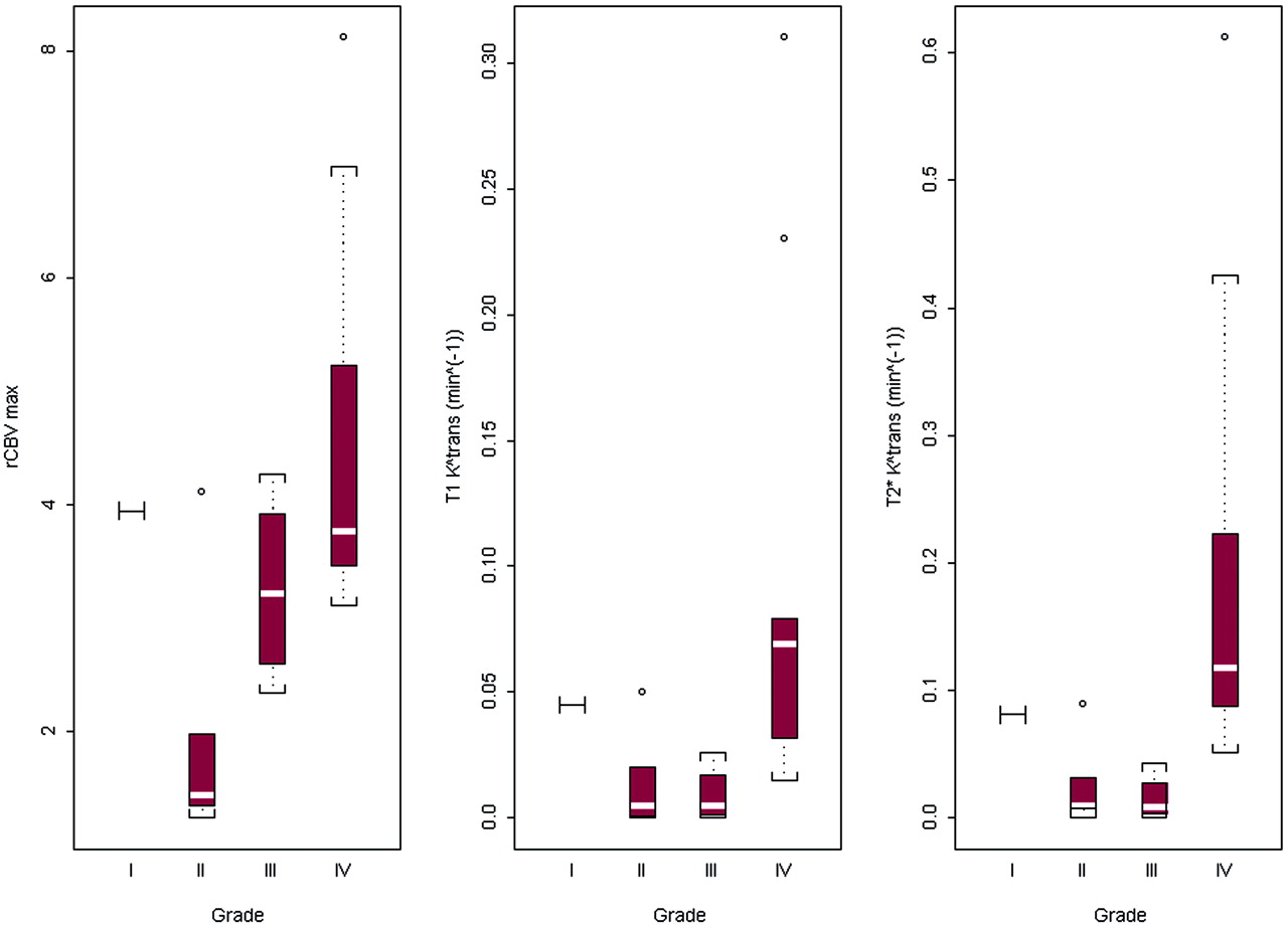
  • Fig 5.
    • Download figure
    • Open in new tab
    • Download powerpoint
    Fig 5.

    Box plots of fpT2* rCBV maximum, ssT1- and fpT2*-derived Ktrans values (in minutes−1) for grades I, II, III, and IV gliomas. Box plots of fpT2* rCBV maximum, ssT1- and fpT2*-derived Ktrans values (in minutes−1) for grades I, II, III, and IV gliomas. The red box extends from the first quartile to the third quartile of the data, with the white line marking the median. The black lines with end brackets represent the most extreme observations in the data that are not more than 1.5 times the height of the box beyond either quartile. All points outside this range are presented by a circle and are considered to be outliers. There was only one patient with grade I gliomas (pilocytic astrocytoma), and the value was presented as horizontal [I] bar in the plots.

  • Fig 6.
    • Download figure
    • Open in new tab
    • Download powerpoint
    Fig 6.

    Scatter plots and fitted regression line of ssT1-derived Ktrans on fpT2*-derived Ktrans. The Pearson correlation coefficient for gliomas is high and estimated to be 0.95 (95% CI [0.89, 0.98]); however, no linear correlation exists between fpT2* and ssT1 Ktrans values for meningiomas, with the Pearson correlation coefficient estimated to be 0.16 (95% CI [−0.68, 0.81]).

Tables

  • Figures
    • View popup
    Table 1:

    Comparison between the ssT1 and fpT2* methods: theoretical and practical considerations

    ssT1 MethodfpT2* Method
    Theoretical considerations
        Shape of contrast agent concentration time curveBi-exponential decayGamma-variate
        Concentration of intravascular contrast agentLowerHigher
        Ktrans for normal brain tissueZeroNegligible or very small, but not zero
        Rate of contrast agent movement from intravascular to extravascular space within a single voxel of tissueSlowerFaster
    Practical considerations
        Spatial resolutionHigherLower
        Subjectivity to susceptibility artifactNoYes
        Imaging timeLonger (>6 min)Shorter (<1.5 min)
        Postprocessing algorithm complexityHigherLower
    • View popup
    Table 2:

    P values from the Kruskal-Wallis test of overall equality of all three grades for rCBV max, ssT1-, and fpT2*-derived Ktrans

    rCBV maxssT1-Derived KtransfpT2*-Derived Ktrans
    Kruskal-Wallis test.03*.006*.003*
    Grade II vs III.11>.5>0.5
    Grade II vs IV.019.017.005†
    Grade III vs IV.19.009†.002†
    • * For significant Kruskal-Wallis test, P values from 3 Wilcoxon rank sum tests for 2 sample data were listed with Bonferroni adjustment for multiple comparisons.

    • † Difference was statistically significant after Bonferroni adjustment for multiple comparisons

PreviousNext
Back to top

In this issue

American Journal of Neuroradiology: 27 (2)
American Journal of Neuroradiology
Vol. 27, Issue 2
February, 2006
  • Table of Contents
  • Index by author
Advertisement
Print
Download PDF
Email Article

Thank you for your interest in spreading the word on American Journal of Neuroradiology.

NOTE: We only request your email address so that the person you are recommending the page to knows that you wanted them to see it, and that it is not junk mail. We do not capture any email address.

Enter multiple addresses on separate lines or separate them with commas.
Comparison of Microvascular Permeability Measurements, Ktrans, Determined with Conventional Steady-State T1-Weighted and First-Pass T2*-Weighted MR Imaging Methods in Gliomas and Meningiomas
(Your Name) has sent you a message from American Journal of Neuroradiology
(Your Name) thought you would like to see the American Journal of Neuroradiology web site.
CAPTCHA
This question is for testing whether or not you are a human visitor and to prevent automated spam submissions.
Cite this article
S. Cha, L. Yang, G. Johnson, A. Lai, M.-H. Chen, T. Tihan, M. Wendland, W.P. Dillon
Comparison of Microvascular Permeability Measurements, Ktrans, Determined with Conventional Steady-State T1-Weighted and First-Pass T2*-Weighted MR Imaging Methods in Gliomas and Meningiomas
American Journal of Neuroradiology Feb 2006, 27 (2) 409-417;

Citation Manager Formats

  • BibTeX
  • Bookends
  • EasyBib
  • EndNote (tagged)
  • EndNote 8 (xml)
  • Medlars
  • Mendeley
  • Papers
  • RefWorks Tagged
  • Ref Manager
  • RIS
  • Zotero
0 Responses
Respond to this article
Share
Bookmark this article
Comparison of Microvascular Permeability Measurements, Ktrans, Determined with Conventional Steady-State T1-Weighted and First-Pass T2*-Weighted MR Imaging Methods in Gliomas and Meningiomas
S. Cha, L. Yang, G. Johnson, A. Lai, M.-H. Chen, T. Tihan, M. Wendland, W.P. Dillon
American Journal of Neuroradiology Feb 2006, 27 (2) 409-417;
del.icio.us logo Twitter logo Facebook logo Mendeley logo
  • Tweet Widget
  • Facebook Like
  • Google Plus One

Jump to section

  • Article
    • Abstract
    • Methods
    • Results
    • Discussion
    • Conclusion
    • Acknowledgments
    • Footnotes
    • References
  • Figures & Data
  • Info & Metrics
  • Responses
  • References
  • PDF

Related Articles

  • No related articles found.
  • PubMed
  • Google Scholar

Cited By...

  • Advances in endovascular neuro-oncology: endovascular selective intra-arterial (ESIA) infusion of targeted biologic therapy for brain tumors
  • Local Glioma Cells Are Associated with Vascular Dysregulation
  • Differentiation between Treatment-Induced Necrosis and Recurrent Tumors in Patients with Metastatic Brain Tumors: Comparison among 11C-Methionine-PET, FDG-PET, MR Permeability Imaging, and MRI-ADC--Preliminary Results
  • Quantifying Intracranial Plaque Permeability with Dynamic Contrast-Enhanced MRI: A Pilot Study
  • On the Use of DSC-MRI for Measuring Vascular Permeability
  • Quantifying Intracranial Aneurysm Wall Permeability for Risk Assessment Using Dynamic Contrast-Enhanced MRI: A Pilot Study
  • Evaluation of Microvascular Permeability with Dynamic Contrast-Enhanced MRI for the Differentiation of Primary CNS Lymphoma and Glioblastoma: Radiologic-Pathologic Correlation
  • Effects of Microvascular Permeability Changes on Contrast-Enhanced T1 and Pharmacokinetic MR Imagings After Ischemia
  • MR Imaging of Neoplastic Central Nervous System Lesions: Review and Recommendations for Current Practice
  • Increased Blood-Brain Barrier Permeability on Perfusion CT Might Predict Malignant Middle Cerebral Artery Infarction
  • Enhancing Fraction in Glioma and Its Relationship to the Tumoral Vascular Microenvironment: A Dynamic Contrast-Enhanced MR Imaging Study
  • Patterns and Predictors of Blood-Brain Barrier Permeability Derangements in Acute Ischemic Stroke
  • Crossref
  • Google Scholar

This article has not yet been cited by articles in journals that are participating in Crossref Cited-by Linking.

More in this TOC Section

  • Fast Contrast-Enhanced 4D MRA and 4D Flow MRI Using Constrained Reconstruction (HYPRFlow): Potential Applications for Brain Arteriovenous Malformations
  • Quiet PROPELLER MRI Techniques Match the Quality of Conventional PROPELLER Brain Imaging Techniques
  • Predictors of Reperfusion in Patients with Acute Ischemic Stroke
Show more Brain

Similar Articles

Advertisement

Indexed Content

  • Current Issue
  • Accepted Manuscripts
  • Article Preview
  • Past Issues
  • Editorials
  • Editors Choice
  • Fellow Journal Club
  • Letters to the Editor

Cases

  • Case Collection
  • Archive - Case of the Week
  • Archive - Case of the Month
  • Archive - Classic Case

Special Collections

  • Special Collections

Resources

  • News and Updates
  • Turn around Times
  • Submit a Manuscript
  • Author Policies
  • Manuscript Submission Guidelines
  • Evidence-Based Medicine Level Guide
  • Publishing Checklists
  • Graphical Abstract Preparation
  • Imaging Protocol Submission
  • Submit a Case
  • Become a Reviewer/Academy of Reviewers
  • Get Peer Review Credit from Publons

Multimedia

  • AJNR Podcast
  • AJNR SCANtastic
  • Video Articles

About Us

  • About AJNR
  • Editorial Board
  • Not an AJNR Subscriber? Join Now
  • Alerts
  • Feedback
  • Advertise with us
  • Librarian Resources
  • Permissions
  • Terms and Conditions

American Society of Neuroradiology

  • Not an ASNR Member? Join Now

© 2025 by the American Society of Neuroradiology All rights, including for text and data mining, AI training, and similar technologies, are reserved.
Print ISSN: 0195-6108 Online ISSN: 1936-959X

Powered by HighWire