
Article Figures & Data
Figures
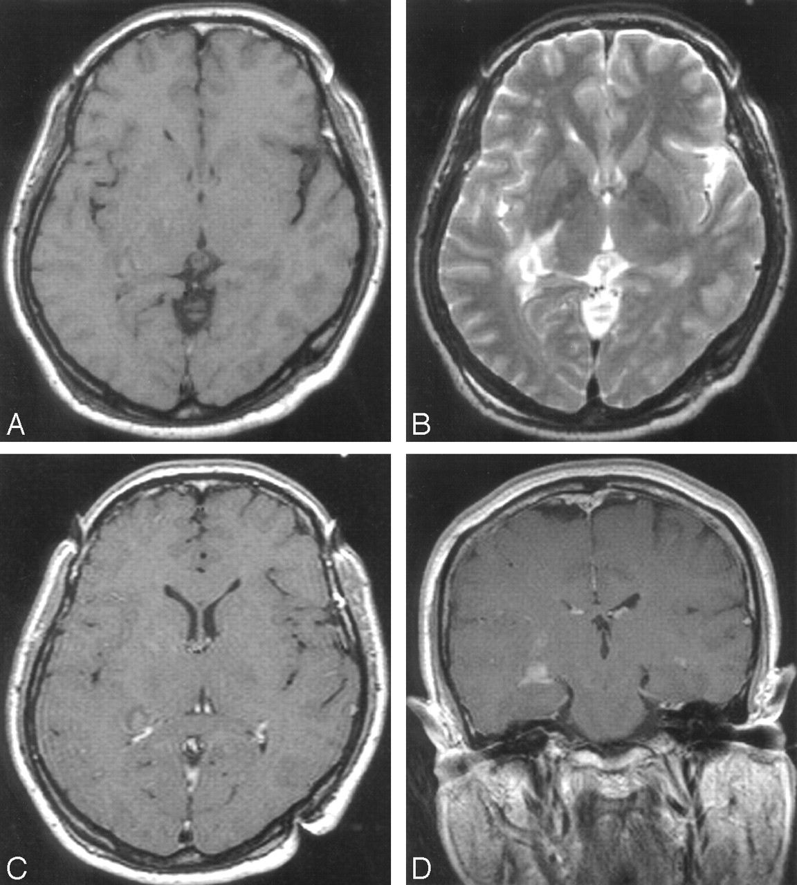
- Fig 1.
Initial MR imaging before stereotactic biopsy. Right temporal periventricular mass lesion is seen on T1-weighted (A), T2-weighted (B), and contrast-enhanced T1-weighted images (C and D).
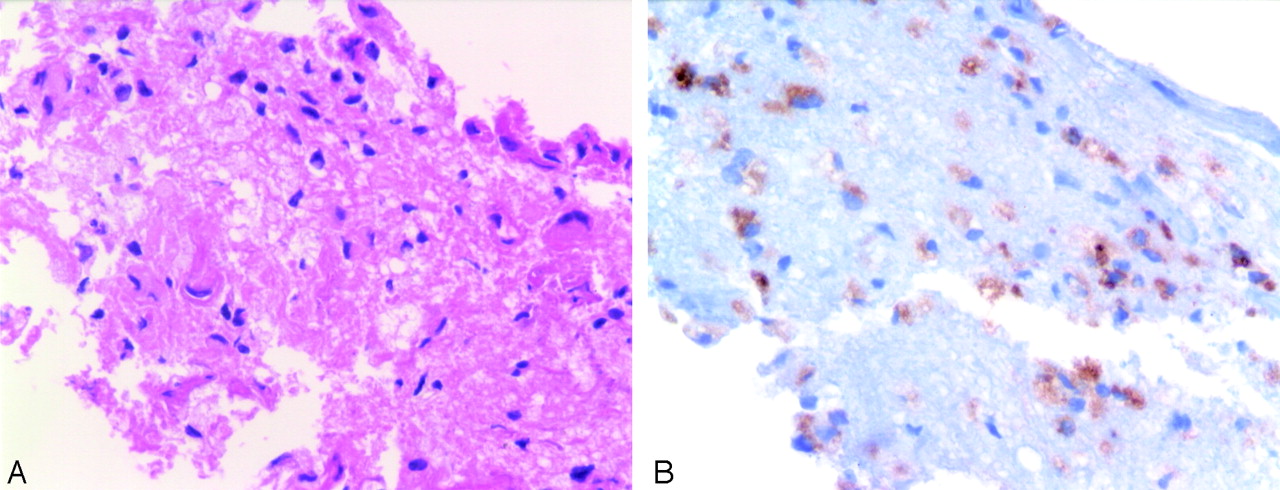
- Fig 2.
Histologic findings. A, Abundant foamy histiocytes infiltrated in glial parenchyma (H&E, ×400). B, Most infiltrating cells show immunoreactivity for CD68 with intense cytoplasmic pattern (×400).
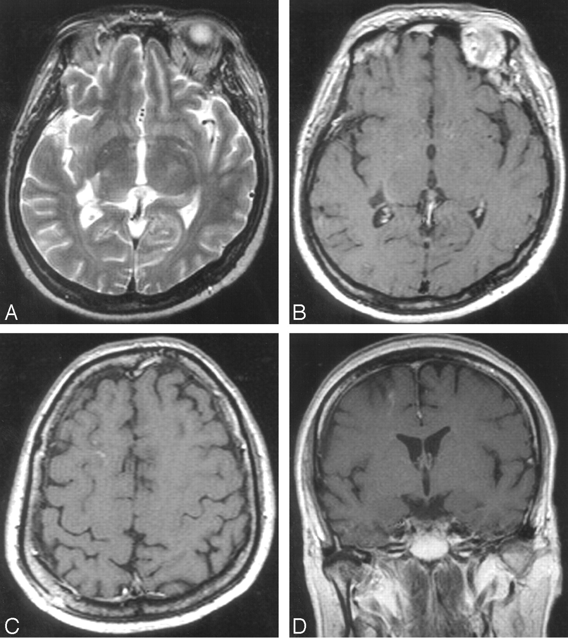
- Fig 3.
One-month follow-up MR imaging after treatment. T2-weighted (A) and contrast-enhanced T1-weighted (B) images show focal malacic cavity. Contrast-enhanced T1-weighted images (C and D) show newly developed lesions in right frontal lobe.
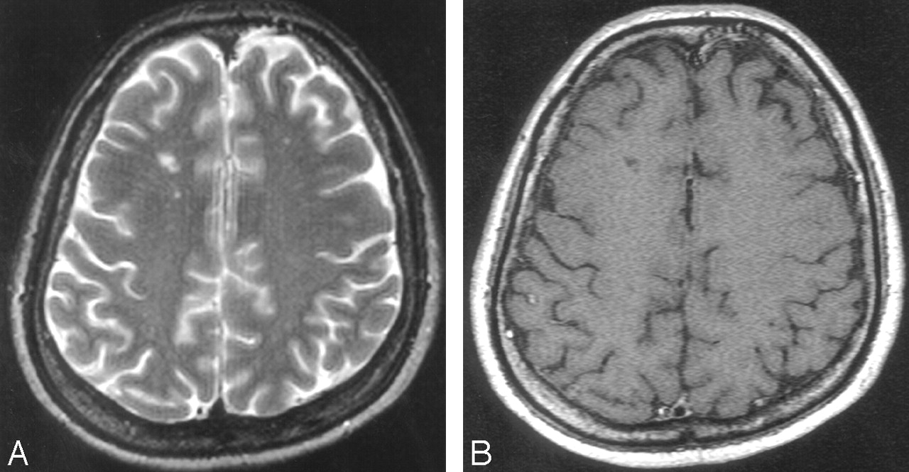
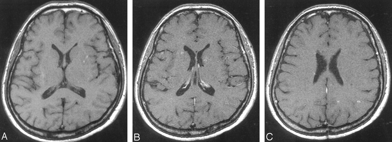
- Fig 4.
Four-month follow-up MR imaging. Focal malacic cavity is seen on T2-weighted (A) and contrast-enhanced T1-weighted (B) images.
- Fig 5.
One-year follow-up MR imaging. Focal high-signal-intensity spots are seen in left basal ganglia on T1-weighted image (A). Contrast-enhanced T1-weighted images show lesions in both basal ganglia (B) and in left parietal lobe (C).
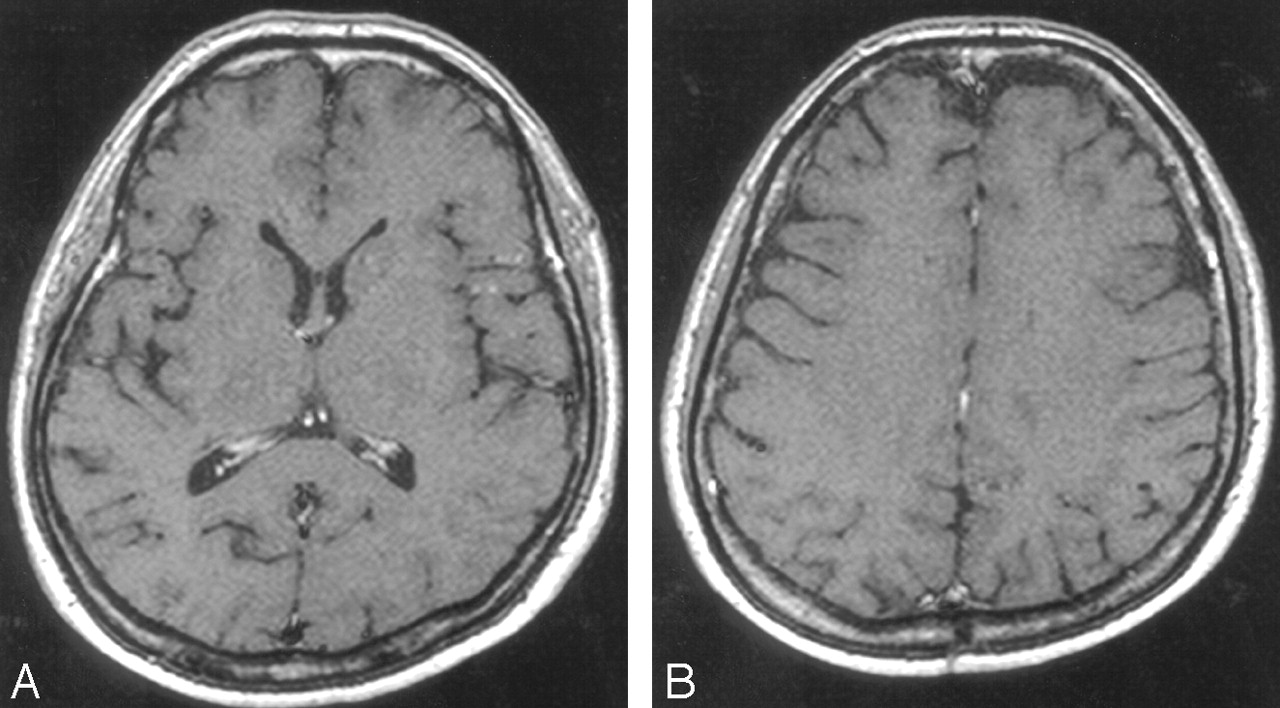
- Fig 6.
Fifteen-month follow-up MR imaging. Perivascular space widening with subtle enhancement is noted in both basal ganglia (A) and left parietal lobe (B) on contrast-enhanced T1-weighted images.


In this issue


Jump to section
Related Articles
Cited By...
- No citing articles found.