Advertisement

Main menu

  • Home
  • Content
    • Current Issue
    • Accepted Manuscripts
    • Article Preview
    • Past Issue Archive
    • AJNR Case Collection
    • Case of the Week Archive
    • Classic Case Archive
    • Case of the Month Archive
  • Special Collections
    • Spinal CSF Leak Articles (Jan 2020-June 2024)
    • 2024 AJNR Journal Awards
    • Most Impactful AJNR Articles
  • Multimedia
    • AJNR Podcast
    • AJNR Scantastics
    • Video Articles
  • For Authors
    • Submit a Manuscript
    • Author Policies
    • Fast publishing of Accepted Manuscripts
    • Graphical Abstract Preparation
    • Manuscript Submission Guidelines
    • Imaging Protocol Submission
    • Submit a Case for the Case Collection
  • About Us
    • About AJNR
    • Editorial Board
  • More
    • Become a Reviewer/Academy of Reviewers
    • Subscribers
    • Permissions
    • Alerts
    • Feedback
    • Advertisers
    • ASNR Home
  • Other Publications
    • ajnr

User menu

  • Alerts
  • Log in

Search

  • Advanced search
American Journal of Neuroradiology

American Journal of Neuroradiology

ASHNR American Society of Functional Neuroradiology ASHNR American Society of Pediatric Neuroradiology ASSR
  • Alerts
  • Log in

Advanced Search

  • Home
  • Content
    • Current Issue
    • Accepted Manuscripts
    • Article Preview
    • Past Issue Archive
    • AJNR Case Collection
    • Case of the Week Archive
    • Classic Case Archive
    • Case of the Month Archive
  • Special Collections
    • Spinal CSF Leak Articles (Jan 2020-June 2024)
    • 2024 AJNR Journal Awards
    • Most Impactful AJNR Articles
  • Multimedia
    • AJNR Podcast
    • AJNR Scantastics
    • Video Articles
  • For Authors
    • Submit a Manuscript
    • Author Policies
    • Fast publishing of Accepted Manuscripts
    • Graphical Abstract Preparation
    • Manuscript Submission Guidelines
    • Imaging Protocol Submission
    • Submit a Case for the Case Collection
  • About Us
    • About AJNR
    • Editorial Board
  • More
    • Become a Reviewer/Academy of Reviewers
    • Subscribers
    • Permissions
    • Alerts
    • Feedback
    • Advertisers
    • ASNR Home
  • Follow AJNR on Twitter
  • Visit AJNR on Facebook
  • Follow AJNR on Instagram
  • Join AJNR on LinkedIn
  • RSS Feeds

Welcome to the new AJNR, Updated Hall of Fame, and more. Read the full announcements.


AJNR is seeking candidates for the position of Associate Section Editor, AJNR Case Collection. Read the full announcement.


Research ArticleBRAIN

Quantitative MR Imaging R2 Relaxometry in Elderly Participants Reporting Memory Loss

M.J. House, T.G. St. Pierre, J.K. Foster, R.N. Martins and R. Clarnette
American Journal of Neuroradiology February 2006, 27 (2) 430-439;
M.J. House
  • Find this author on Google Scholar
  • Find this author on PubMed
  • Search for this author on this site
T.G. St. Pierre
  • Find this author on Google Scholar
  • Find this author on PubMed
  • Search for this author on this site
J.K. Foster
  • Find this author on Google Scholar
  • Find this author on PubMed
  • Search for this author on this site
R.N. Martins
  • Find this author on Google Scholar
  • Find this author on PubMed
  • Search for this author on this site
R. Clarnette
  • Find this author on Google Scholar
  • Find this author on PubMed
  • Search for this author on this site
  • Article
  • Figures & Data
  • Info & Metrics
  • Responses
  • References
  • PDF
Loading

Article Figures & Data

Figures

  • Tables
  • Fig 1.
    • Download figure
    • Open in new tab
    • Download powerpoint
    Fig 1.

    Representative spin-echo images (TE = 6 ms) of the anterior (A) and posterior (B) coronal sections of a control subject. The 14 gray and white matter regions for which R2 data were acquired are shown as representative regions of interest. Lateral frontal cortex (1), lateral temporal cortex (2), corpus callosum (body) (3), caudate nucleus (4), internal capsule (anterior limb) (5), subcortical frontal white matter (6), frontal white matter (7), putamen (8), globus pallidus (9), temporal white matter (10), hippocampus (11), red nucleus (12), thalamus (13), and substantia nigra (14), The 3 circular objects surrounding the head are MnCl2-doped water bags that were used as part of another experiment not described here. The red nucleus is not well defined on this image and was typically delineated on longer echo-time images.
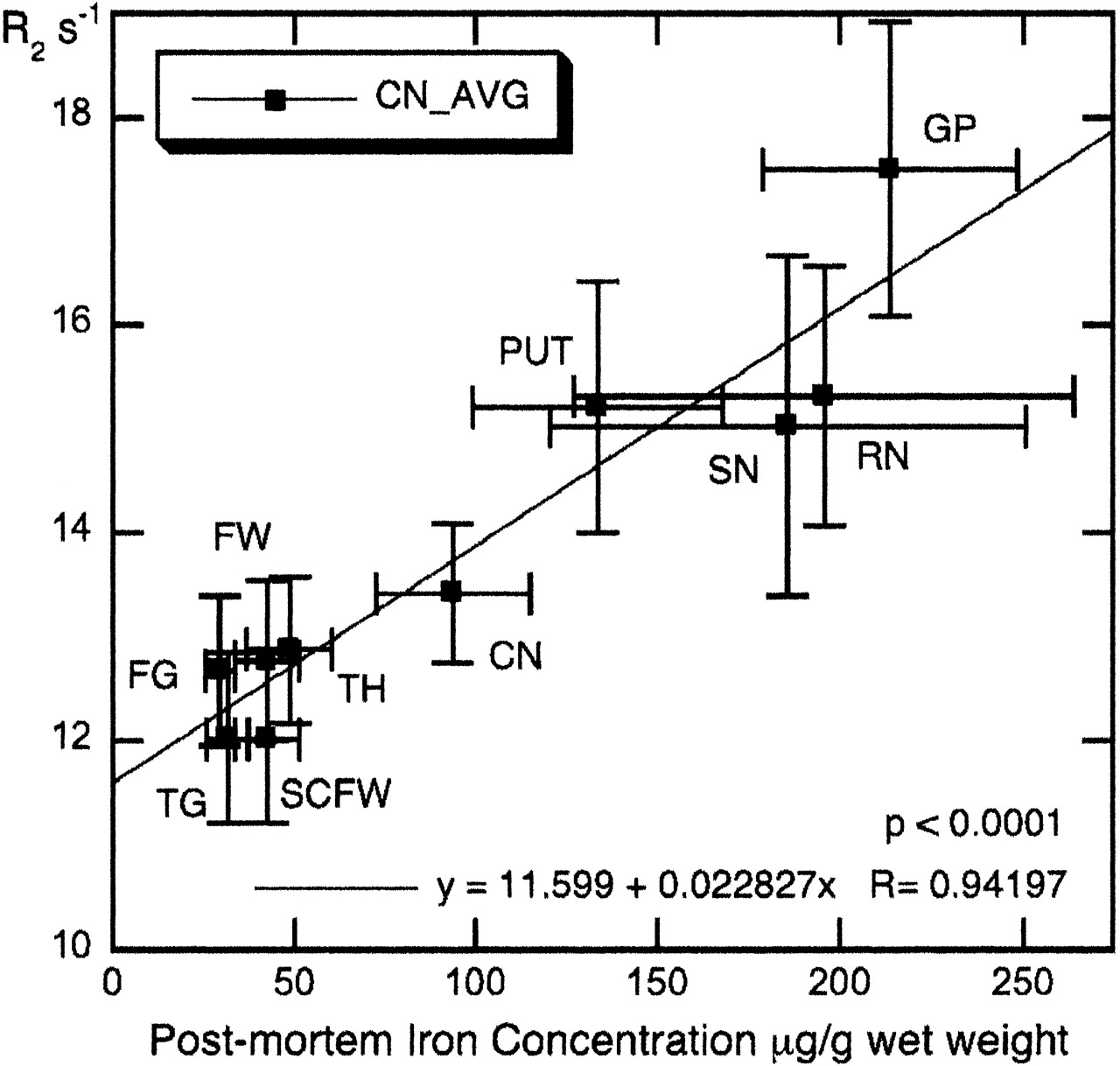
  • Fig 2.
    • Download figure
    • Open in new tab
    • Download powerpoint
    Fig 2.

    Mean R2 values of the control participants for 10 brain regions versus postmortem iron content of normal individuals. Error bars represent SD. Iron concentrations from Hallgren and Sourander.21 Note that frontal (FW) and subcortical frontal white matter (SCFW) regions from this study have been assigned the same iron concentration, because Hallgren and Sourander21 provide iron data for frontal white matter only. CN = caudate nucleus; FG = frontal cortex gray; FW = frontal cortex white; GP = globus pallidus; PUT = putamen; RN = red nucleus; SCFW = subcortical frontal white; SN = substantia nigra; TG = temporal cortex gray; TH = thalamus.
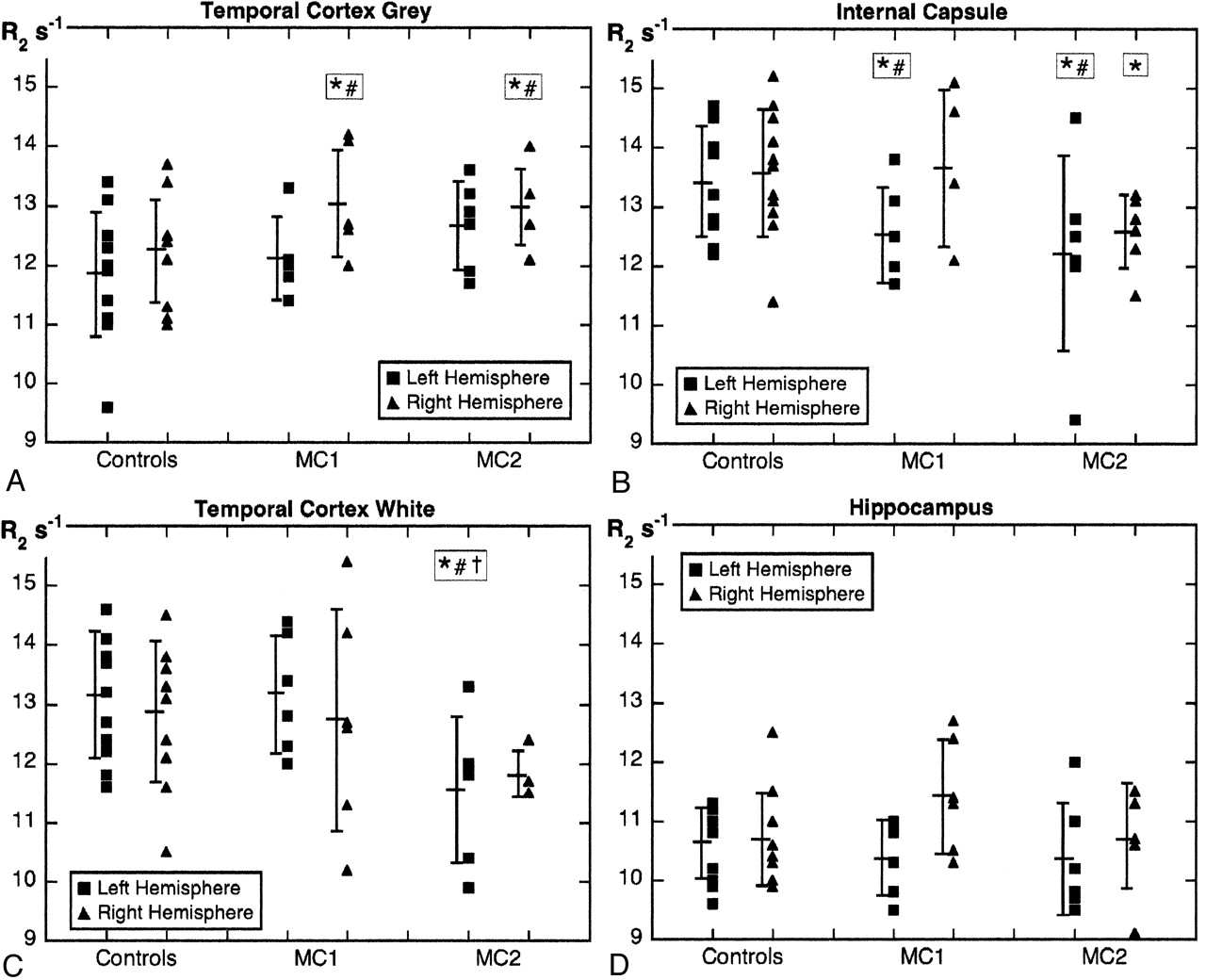
  • Fig 3.
    • Download figure
    • Open in new tab
    • Download powerpoint
    Fig 3.

    R2 values for temporal cortex gray matter (A), the internal capsule (B), temporal cortex white matter (C), and the hippocampus (D) for the control participants and memory-complaint subgroups MC1 and MC2. Error bars around the mean are SD. *Significant difference between controls and MC1 or MC2 (P < .05, one-tailed t test). #Significant difference between controls and MC1 or MC2 (P < .05, after ANCOVA adjustment for age where applicable). †Significant difference between MC1 and MC2 (P < .05, one-tailed t test).
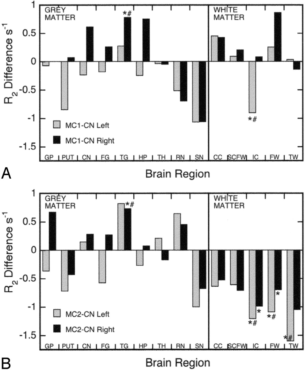
  • Fig 4.
    • Download figure
    • Open in new tab
    • Download powerpoint
    Fig 4.

    Plot of R2 differences between the MC1 subgroup and controls (A), and the MC2 subgroup and controls (B), for the left and right hemispheres. CC = corpus callosum; CN = caudate nucleus; FG = frontal cortex gray; FW = frontal cortex white; GP = globus pallidus; HP = hippocampus; IC = internal capsule; PUT = putamen; RN = red nucleus; SCFW = subcortical frontal white; SN = substantia nigra; TG = temporal cortex gray; TH = thalamus; TW = temporal cortex white. *Significant difference between controls and MC1 or MC2 (P < .05, one-tailed t test). #Significant difference between controls and MC1 or MC2 (P < .05, after ANCOVA adjustment for age where applicable).

Tables

  • Figures
    • View popup
    Table 1:

    Demographic details and neuropsychological test scores of the participants

    ControlsMC1MC2
    No. of individuals1166
    Age (y)70.7 (6.9)69.2 (6.8)75.5 (8.8)
    Gender % (no.) of men27 (3)33 (2)17 (1)
    Education (y)11.6 (2.5)10.3 (3.7)11.2 (2.6)
    MMSE28.0 (1.7)25.5 (1.8)*18.5 (4.3)*
    NART110.7 (6.5)104.9 (4.2)113.5 (6.0)
    CVLT-IFR52.46 (9.1)43.17 (8.4)18.67 (8.6)*
    CVLT-SDFR10.00 (2.76)8.67 (1.97)0.50 (0.84)*
    CVLT-LDFR10.64 (2.98)9.00 (2.97)0.17 (0.41)*
    • Note.— Data are expressed as means (±SD), except for gender. All neuropsychological data are presented here as raw scores. CVLT indicates California Verbal Learning Test; IFR, immediate free recall; LDFR, long delay free recall; MMSE, Mini Mental State Examination; NART, National Adult Reading Test; SDFR, short delay free recall.

    • * Significant difference between controls and MC1 or MC2 subgroups (P < .05, two-tailed t-test).

    • View popup
    Table 2:

    Correlation coefficient signs between R2 values and neuropsychological test scores for the 12 subjects with memory complaints

    RegionHemisphereMMSECVLT IFRCVLT SDFRCVLT LDFR
    Gray matter
        Globus pallidusL++−−
    R+−−−
        PutamenL−−−−
    R−−−−
        Caudate nucleusL−−−−
    R−−−−
        Frontal cortexL+−−−
    R−−−+
        Temporal cortexL+−−−
    R−−−−
        HippocampusL+++−
    R−−−−
        ThalamusL−−−−
    R−−−−
        Red nucleusL−−−−
    R−−−−
        Substantia nigraL−−−−
    R−−−−
    White matter
        Corpus callosumL++++
    R+++−
        Subcortical frontal whiteL++++
    R++++
        Internal capsuleL−+−−
    R−−−+
        Frontal whiteL++++
    R++++
        Temporal whiteL++++
    R−−−−
    • Note.— CVLT indicates California Verbal Learning Test; IFR, immediate free recall; LDFR, long delay free recall; MMSE, Mini Mental State Examination; SDFR, short delay free recall. Data are displayed as the revelant Spearman’s rank-order partial correlation coefficient sign (+, positive; −, negative) for each brain region and test score. Note the overall frequency of negative correlation coefficients for gray matter regions and positive correlation coefficients for white matter regions.

PreviousNext
Back to top

In this issue

American Journal of Neuroradiology: 27 (2)
American Journal of Neuroradiology
Vol. 27, Issue 2
February, 2006
  • Table of Contents
  • Index by author
Advertisement
Print
Download PDF
Email Article

Thank you for your interest in spreading the word on American Journal of Neuroradiology.

NOTE: We only request your email address so that the person you are recommending the page to knows that you wanted them to see it, and that it is not junk mail. We do not capture any email address.

Enter multiple addresses on separate lines or separate them with commas.
Quantitative MR Imaging R2 Relaxometry in Elderly Participants Reporting Memory Loss
(Your Name) has sent you a message from American Journal of Neuroradiology
(Your Name) thought you would like to see the American Journal of Neuroradiology web site.
CAPTCHA
This question is for testing whether or not you are a human visitor and to prevent automated spam submissions.
Cite this article
M.J. House, T.G. St. Pierre, J.K. Foster, R.N. Martins, R. Clarnette
Quantitative MR Imaging R2 Relaxometry in Elderly Participants Reporting Memory Loss
American Journal of Neuroradiology Feb 2006, 27 (2) 430-439;

Citation Manager Formats

  • BibTeX
  • Bookends
  • EasyBib
  • EndNote (tagged)
  • EndNote 8 (xml)
  • Medlars
  • Mendeley
  • Papers
  • RefWorks Tagged
  • Ref Manager
  • RIS
  • Zotero
0 Responses
Respond to this article
Share
Bookmark this article
Quantitative MR Imaging R2 Relaxometry in Elderly Participants Reporting Memory Loss
M.J. House, T.G. St. Pierre, J.K. Foster, R.N. Martins, R. Clarnette
American Journal of Neuroradiology Feb 2006, 27 (2) 430-439;
del.icio.us logo Twitter logo Facebook logo Mendeley logo
  • Tweet Widget
  • Facebook Like
  • Google Plus One

Jump to section

  • Article
    • Abstract
    • Methods
    • Results
    • Discussion
    • Conclusion
    • Acknowledgments
    • References
  • Figures & Data
  • Info & Metrics
  • Responses
  • References
  • PDF

Related Articles

  • No related articles found.
  • PubMed
  • Google Scholar

Cited By...

  • High-Resolution 3D Ultra-Short Echo Time MRI with Rosette k-Space Pattern for Brain Iron Content Mapping
  • Measuring brain integrity using MRI: a novel biomarker for Alzheimers disease using T2 relaxometry
  • Sodium MR Imaging Detection of Mild Alzheimer Disease: Preliminary Study
  • Crossref
  • Google Scholar

This article has not yet been cited by articles in journals that are participating in Crossref Cited-by Linking.

More in this TOC Section

  • Fast Contrast-Enhanced 4D MRA and 4D Flow MRI Using Constrained Reconstruction (HYPRFlow): Potential Applications for Brain Arteriovenous Malformations
  • Quiet PROPELLER MRI Techniques Match the Quality of Conventional PROPELLER Brain Imaging Techniques
  • Predictors of Reperfusion in Patients with Acute Ischemic Stroke
Show more Brain

Similar Articles

Advertisement

Indexed Content

  • Current Issue
  • Accepted Manuscripts
  • Article Preview
  • Past Issues
  • Editorials
  • Editors Choice
  • Fellow Journal Club
  • Letters to the Editor

Cases

  • Case Collection
  • Archive - Case of the Week
  • Archive - Case of the Month
  • Archive - Classic Case

Special Collections

  • Special Collections

Resources

  • News and Updates
  • Turn around Times
  • Submit a Manuscript
  • Author Policies
  • Manuscript Submission Guidelines
  • Evidence-Based Medicine Level Guide
  • Publishing Checklists
  • Graphical Abstract Preparation
  • Imaging Protocol Submission
  • Submit a Case
  • Become a Reviewer/Academy of Reviewers
  • Get Peer Review Credit from Publons

Multimedia

  • AJNR Podcast
  • AJNR SCANtastic
  • Video Articles

About Us

  • About AJNR
  • Editorial Board
  • Not an AJNR Subscriber? Join Now
  • Alerts
  • Feedback
  • Advertise with us
  • Librarian Resources
  • Permissions
  • Terms and Conditions

American Society of Neuroradiology

  • Not an ASNR Member? Join Now

© 2025 by the American Society of Neuroradiology All rights, including for text and data mining, AI training, and similar technologies, are reserved.
Print ISSN: 0195-6108 Online ISSN: 1936-959X

Powered by HighWire