Advertisement

Main menu

  • Home
  • Content
    • Current Issue
    • Accepted Manuscripts
    • Article Preview
    • Past Issue Archive
    • AJNR Case Collection
    • Case of the Week Archive
    • Classic Case Archive
    • Case of the Month Archive
  • Special Collections
    • Spinal CSF Leak Articles (Jan 2020-June 2024)
    • 2024 AJNR Journal Awards
    • Most Impactful AJNR Articles
  • Multimedia
    • AJNR Podcast
    • AJNR Scantastics
    • Video Articles
  • For Authors
    • Submit a Manuscript
    • Author Policies
    • Fast publishing of Accepted Manuscripts
    • Graphical Abstract Preparation
    • Manuscript Submission Guidelines
    • Imaging Protocol Submission
    • Submit a Case for the Case Collection
  • About Us
    • About AJNR
    • Editorial Board
  • More
    • Become a Reviewer/Academy of Reviewers
    • Subscribers
    • Permissions
    • Alerts
    • Feedback
    • Advertisers
    • ASNR Home
  • Other Publications
    • ajnr

User menu

  • Alerts
  • Log in

Search

  • Advanced search
American Journal of Neuroradiology

American Journal of Neuroradiology

ASHNR American Society of Functional Neuroradiology ASHNR American Society of Pediatric Neuroradiology ASSR
  • Alerts
  • Log in

Advanced Search

  • Home
  • Content
    • Current Issue
    • Accepted Manuscripts
    • Article Preview
    • Past Issue Archive
    • AJNR Case Collection
    • Case of the Week Archive
    • Classic Case Archive
    • Case of the Month Archive
  • Special Collections
    • Spinal CSF Leak Articles (Jan 2020-June 2024)
    • 2024 AJNR Journal Awards
    • Most Impactful AJNR Articles
  • Multimedia
    • AJNR Podcast
    • AJNR Scantastics
    • Video Articles
  • For Authors
    • Submit a Manuscript
    • Author Policies
    • Fast publishing of Accepted Manuscripts
    • Graphical Abstract Preparation
    • Manuscript Submission Guidelines
    • Imaging Protocol Submission
    • Submit a Case for the Case Collection
  • About Us
    • About AJNR
    • Editorial Board
  • More
    • Become a Reviewer/Academy of Reviewers
    • Subscribers
    • Permissions
    • Alerts
    • Feedback
    • Advertisers
    • ASNR Home
  • Follow AJNR on Twitter
  • Visit AJNR on Facebook
  • Follow AJNR on Instagram
  • Join AJNR on LinkedIn
  • RSS Feeds

Welcome to the new AJNR, Updated Hall of Fame, and more. Read the full announcements.


AJNR is seeking candidates for the position of Associate Section Editor, AJNR Case Collection. Read the full announcement.


  • Getting new auth cookie, if you see this message a lot, tell someone!
  • Getting new auth cookie, if you see this message a lot, tell someone!
Research ArticleBRAIN

Correlation of Carotid Stenosis Diameter and Cross-Sectional Areas with CT Angiography

E.S. Bartlett, S.P. Symons and A.J. Fox
American Journal of Neuroradiology March 2006, 27 (3) 638-642;
E.S. Bartlett
  • Find this author on Google Scholar
  • Find this author on PubMed
  • Search for this author on this site
S.P. Symons
  • Find this author on Google Scholar
  • Find this author on PubMed
  • Search for this author on this site
A.J. Fox
  • Find this author on Google Scholar
  • Find this author on PubMed
  • Search for this author on this site
  • Article
  • Figures & Data
  • Info & Metrics
  • Responses
  • References
  • PDF
Loading

Article Figures & Data

Figures

  • Fig 1.
    • Download figure
    • Open in new tab
    • Download powerpoint
    Fig 1.

    Quantification of a symmetric right carotid bulb stenosis. A, Axial CTA source image showing symmetric stenosis of the right carotid bulb (white arrow). B, Axial magnification of the symmetric right carotid bulb stenosis with measurement calipers (calipers marked [A], showing measurement of 0.21 cm [2.1 mm]). C, Axial magnification of the symmetric right carotid bulb stenosis with the AGFA Impax 4.5 VT measuring the cross-sectional 2D area of 0.02 cm2 (2.0 mm2).
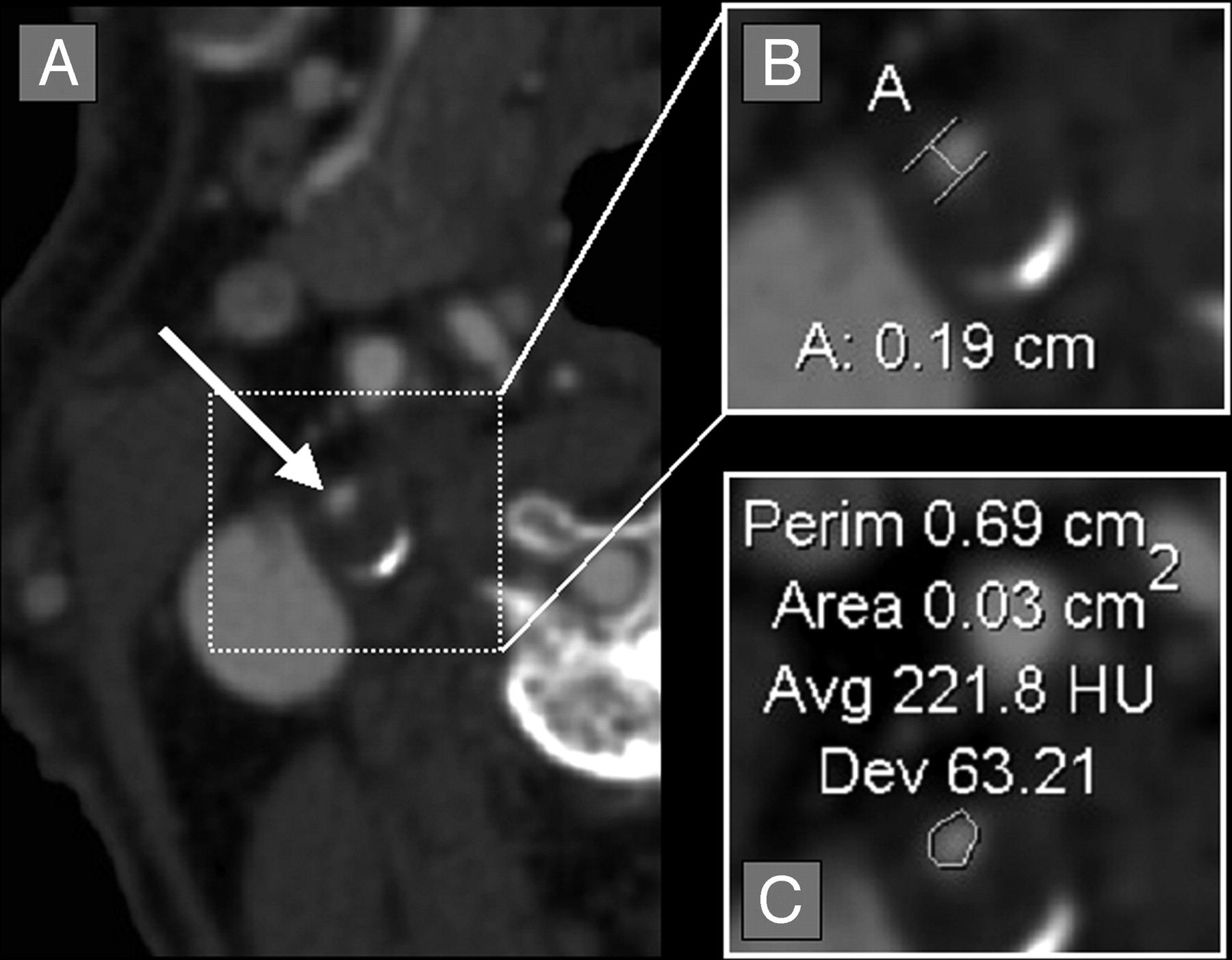
  • Fig 2.
    • Download figure
    • Open in new tab
    • Download powerpoint
    Fig 2.

    Quantification of an asymmetric right carotid bulb stenosis. A, Axial CTA source image showing asymmetric stenosis of the right carotid bulb (white arrow), with partially calcified posterior carotid bulb wall. B, Axial magnification of the asymmetric right carotid bulb stenosis with measurement calipers placed at the region of narrowest stenosis (calipers marked [A], showing measurement of 0.19 cm [1.9 mm]). C, Axial magnification of the asymmetric right carotid bulb stenosis with the AGFA Impax 4.5 VT measuring the cross-sectional 2D area of 0.03 cm2 (3.0 mm2). Note that the narrowest diameter is slightly smaller than on Fig 1, though the area is slightly larger than on Fig 1.
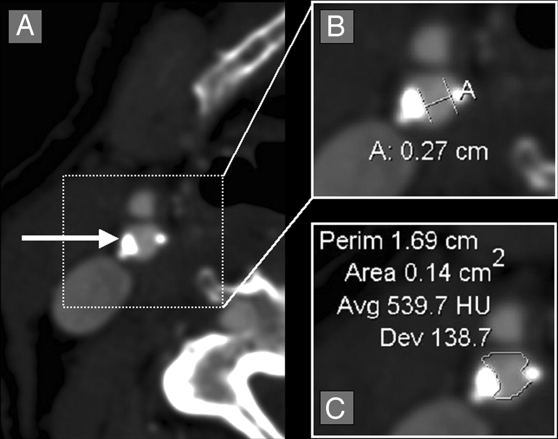
  • Fig 3.
    • Download figure
    • Open in new tab
    • Download powerpoint
    Fig 3.

    Quantification of an irregular, asymmetric carotid bulb stenosis with calcification. A, Axial CTA source image showing an asymmetric stenosis (white arrow). B, Axial magnification of the asymmetric carotid bulb stenosis with measurement calipers placed at the region of narrowest stenosis (calipers marked [A], showing measurement of 0.27 cm [2.7 mm]). C, Axial magnification of the asymmetric carotid bulb stenosis with the AGFA Impax 4.5 VT measuring the cross-sectional 2D area of 0.14 cm2 (14.0 mm2).
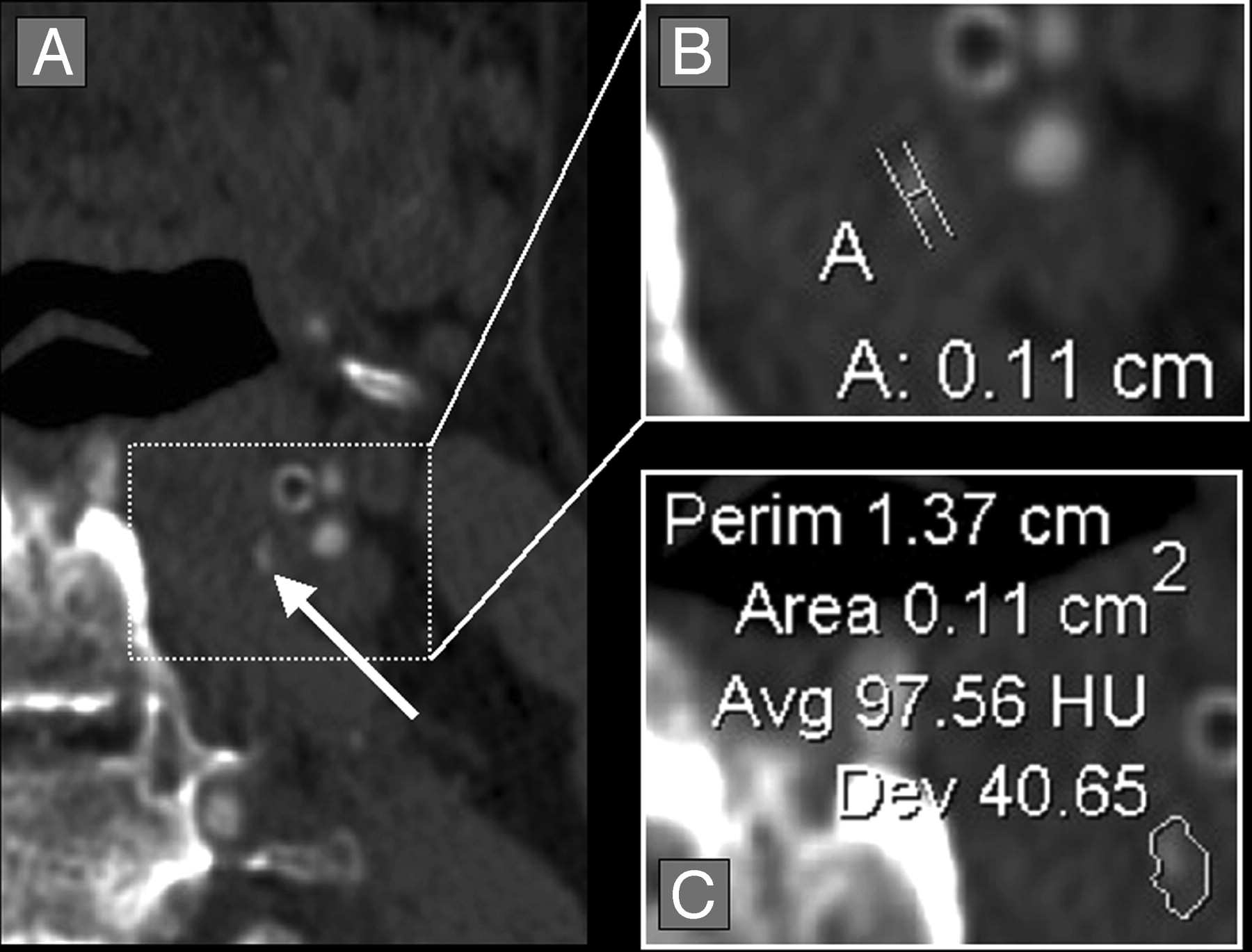
  • Fig 4.
    • Download figure
    • Open in new tab
    • Download powerpoint
    Fig 4.

    Quantification of a diminutive residual carotid bulb lumen. A, Axial CTA source image showing a diminutive left residual carotid bulb lumen with poor contrast filling (white arrow). B, Axial magnification of the residual carotid bulb lumen with measurement calipers placed at the region of narrowest stenosis (calipers marked [A], showing measurement of 0.11 cm [1.1 mm]). C, Axial magnification of the residual carotid bulb lumen with the AGFA Impax 4.5 VT. The VT could not accurately measure the cross-sectional 2D area of the lumen because there was very little contrast-filling of the diminutive carotid bulb. The difference in the HUs between the contrast-filled lumen and the surrounding tissues was not great enough for the VT to accurately measure the lumen.

  • Fig 5.
    • Download figure
    • Open in new tab
    • Download powerpoint
    Fig 5.

    Quantification of a diminutive residual carotid bulb lumen. A, Axial CTA source image showing a diminutive right residual carotid bulb lumen (white arrow). B, Axial magnification of the residual carotid bulb lumen with measurement calipers placed at the region of narrowest stenosis (calipers marked [A], showing measurement of 0.10 cm [1.0 mm]). C, Axial magnification of the residual carotid bulb lumen with the AGFA Impax 4.5 VT, showing the cross-sectional 2D area of 0.02 cm2 (2.0 mm2). The HUs between the contrast-filled lumen and the surrounding tissues was great enough for the VT to accurately measure the lumen, despite the small lumen size.
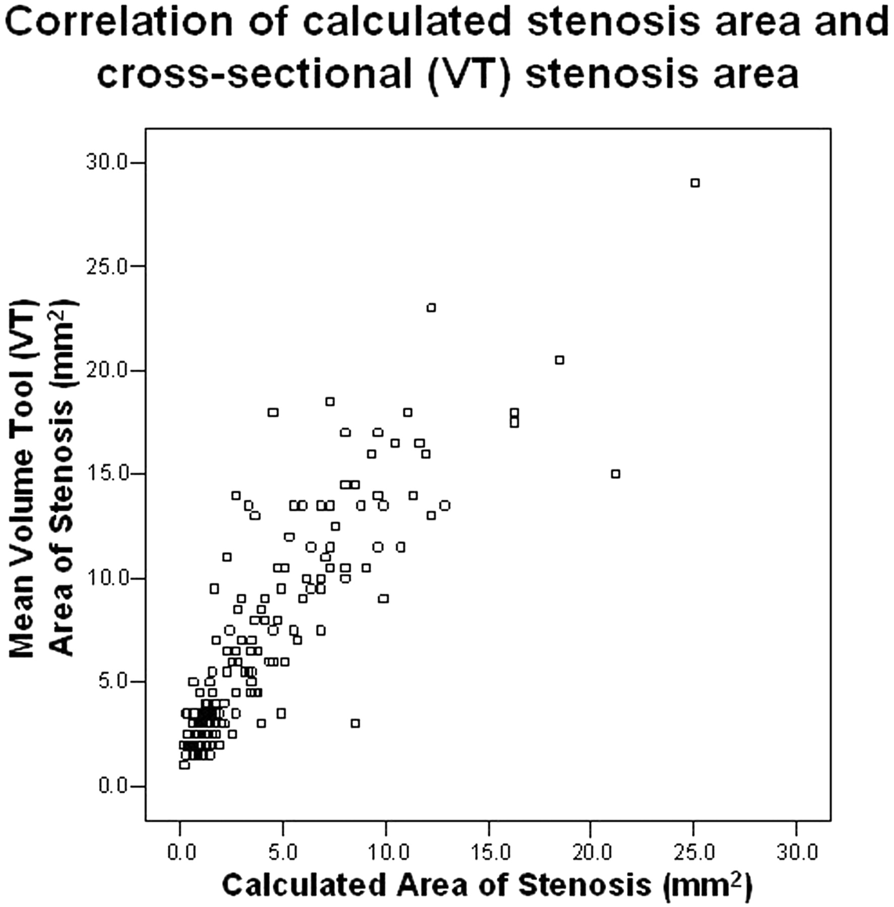
  • Fig 6.
    • Download figure
    • Open in new tab
    • Download powerpoint
    Fig 6.

    Pearson correlation between the calculated stenosis area (based upon the narrowest diameter) and the measured cross-sectional VT area of stenosis (mm2). The calculated area showed a trend of underestimating the area in comparison to the measured area. This trend is not surprising, because the calculated area is based upon the narrowest stenosis, which does not account for noncircular stenoses. Nonetheless, there was excellent correlation between these 2 methods of area quantification (correlation coefficient = 0.87; n = 176).
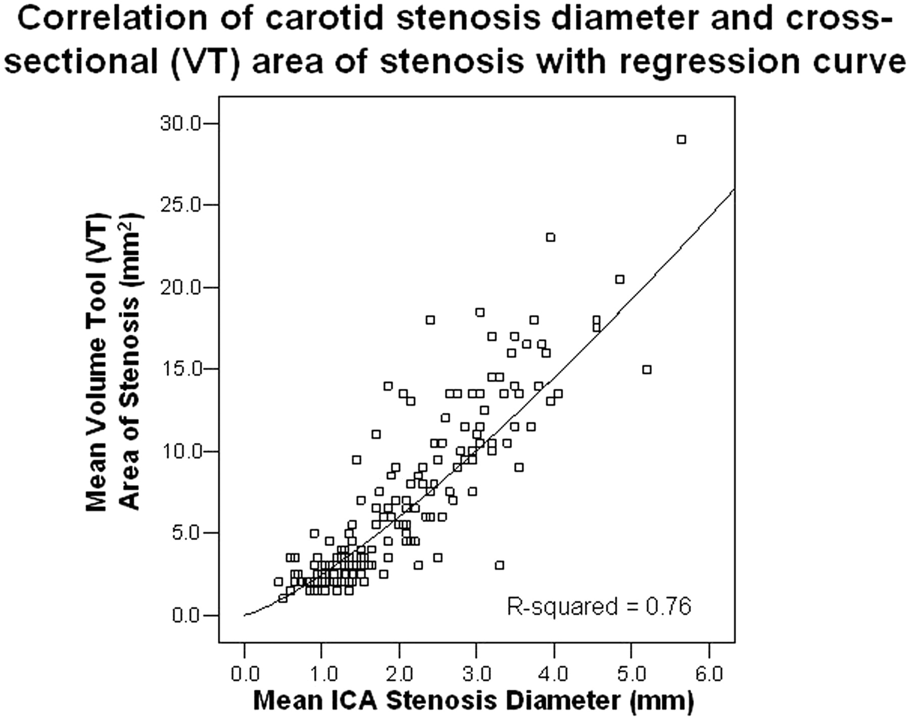
  • Fig 7.
    • Download figure
    • Open in new tab
    • Download powerpoint
    Fig 7.

    Pearson correlation between the mean carotid stenosis narrowest diameter (mm) and the mean cross-sectional VT stenosis area (mm2). There was excellent correlation between the 2 stenosis quantification measures at 0.88 (n = 176). This supports the use of the narrowest diameter measurement to quantify carotid bulb stenosis, in lieu of the more precise cross-sectional area measurement. A regression curve was plotted over the data with an R2 value of 0.76, which indicates that the narrowest diameter has an excellent predictive ability to estimate the cross-sectional area (as defined by the VT).

  • Fig 8.
    • Download figure
    • Open in new tab
    • Download powerpoint
    Fig 8.

    Pearson correlation between the mean carotid stenosis narrowest diameter (mm) and the mean calculated stenosis area (mm2). As anticipated, there was perfect correlation between the 2 methods of stenosis quantification at 1.0 (n = 176). A regression curve was plotted over the data, showing that the ability to predict the calculated area from the stenosis diameter is perfect with an R2 value of 1.0. This is expected, because the calculated area is based on the diameter measurements. The data provide a graphic example of the nonlinear relationship between the narrowest diameter and the area, which demonstrates that minimal changes in the diameter of the carotid lumen cause more dramatic changes in the area of the lumen.

PreviousNext
Back to top

In this issue

American Journal of Neuroradiology: 27 (3)
American Journal of Neuroradiology
Vol. 27, Issue 3
March, 2006
  • Table of Contents
  • Index by author
Advertisement
Print
Download PDF
Email Article

Thank you for your interest in spreading the word on American Journal of Neuroradiology.

NOTE: We only request your email address so that the person you are recommending the page to knows that you wanted them to see it, and that it is not junk mail. We do not capture any email address.

Enter multiple addresses on separate lines or separate them with commas.
Correlation of Carotid Stenosis Diameter and Cross-Sectional Areas with CT Angiography
(Your Name) has sent you a message from American Journal of Neuroradiology
(Your Name) thought you would like to see the American Journal of Neuroradiology web site.
CAPTCHA
This question is for testing whether or not you are a human visitor and to prevent automated spam submissions.
Cite this article
E.S. Bartlett, S.P. Symons, A.J. Fox
Correlation of Carotid Stenosis Diameter and Cross-Sectional Areas with CT Angiography
American Journal of Neuroradiology Mar 2006, 27 (3) 638-642;

Citation Manager Formats

  • BibTeX
  • Bookends
  • EasyBib
  • EndNote (tagged)
  • EndNote 8 (xml)
  • Medlars
  • Mendeley
  • Papers
  • RefWorks Tagged
  • Ref Manager
  • RIS
  • Zotero
0 Responses
Respond to this article
Share
Bookmark this article
Correlation of Carotid Stenosis Diameter and Cross-Sectional Areas with CT Angiography
E.S. Bartlett, S.P. Symons, A.J. Fox
American Journal of Neuroradiology Mar 2006, 27 (3) 638-642;
del.icio.us logo Twitter logo Facebook logo Mendeley logo
  • Tweet Widget
  • Facebook Like
  • Google Plus One

Jump to section

  • Article
    • Abstract
    • Methods
    • Results
    • Discussion
    • Conclusion
    • References
  • Figures & Data
  • Info & Metrics
  • Responses
  • References
  • PDF

Related Articles

  • No related articles found.
  • PubMed
  • Google Scholar

Cited By...

  • Reporting standards for angioplasty and stent-assisted angioplasty for intracranial atherosclerosis
  • Should Modeling Methodology Suppress Anatomic Excellence?
  • Reporting Standards for Angioplasty and Stent-Assisted Angioplasty for Intracranial Atherosclerosis
  • Response to Letter by Bladin et al
  • Simplification of the Residual Lumen Geometry in Measuring Carotid Stenosis
  • Composition of the Stable Carotid Plaque: Insights From a Multidetector Computed Tomography Study of Plaque Volume
  • Carotid Stenosis Index Revisited With Direct CT Angiography Measurement of Carotid Arteries to Quantify Carotid Stenosis
  • Crossref
  • Google Scholar

This article has not yet been cited by articles in journals that are participating in Crossref Cited-by Linking.

More in this TOC Section

  • Fast Contrast-Enhanced 4D MRA and 4D Flow MRI Using Constrained Reconstruction (HYPRFlow): Potential Applications for Brain Arteriovenous Malformations
  • Quiet PROPELLER MRI Techniques Match the Quality of Conventional PROPELLER Brain Imaging Techniques
  • Predictors of Reperfusion in Patients with Acute Ischemic Stroke
Show more Brain

Similar Articles

Advertisement

Indexed Content

  • Current Issue
  • Accepted Manuscripts
  • Article Preview
  • Past Issues
  • Editorials
  • Editors Choice
  • Fellow Journal Club
  • Letters to the Editor

Cases

  • Case Collection
  • Archive - Case of the Week
  • Archive - Case of the Month
  • Archive - Classic Case

Special Collections

  • Special Collections

Resources

  • News and Updates
  • Turn around Times
  • Submit a Manuscript
  • Author Policies
  • Manuscript Submission Guidelines
  • Evidence-Based Medicine Level Guide
  • Publishing Checklists
  • Graphical Abstract Preparation
  • Imaging Protocol Submission
  • Submit a Case
  • Become a Reviewer/Academy of Reviewers
  • Get Peer Review Credit from Publons

Multimedia

  • AJNR Podcast
  • AJNR SCANtastic
  • Video Articles

About Us

  • About AJNR
  • Editorial Board
  • Not an AJNR Subscriber? Join Now
  • Alerts
  • Feedback
  • Advertise with us
  • Librarian Resources
  • Permissions
  • Terms and Conditions

American Society of Neuroradiology

  • Not an ASNR Member? Join Now

© 2025 by the American Society of Neuroradiology All rights, including for text and data mining, AI training, and similar technologies, are reserved.
Print ISSN: 0195-6108 Online ISSN: 1936-959X

Powered by HighWire