Advertisement

Main menu

  • Home
  • Content
    • Current Issue
    • Accepted Manuscripts
    • Article Preview
    • Past Issue Archive
    • AJNR Case Collection
    • Case of the Week Archive
    • Classic Case Archive
    • Case of the Month Archive
  • Special Collections
    • Spinal CSF Leak Articles (Jan 2020-June 2024)
    • 2024 AJNR Journal Awards
    • Most Impactful AJNR Articles
  • Multimedia
    • AJNR Podcast
    • AJNR Scantastics
    • Video Articles
  • For Authors
    • Submit a Manuscript
    • Author Policies
    • Fast publishing of Accepted Manuscripts
    • Graphical Abstract Preparation
    • Manuscript Submission Guidelines
    • Imaging Protocol Submission
    • Submit a Case for the Case Collection
  • About Us
    • About AJNR
    • Editorial Board
  • More
    • Become a Reviewer/Academy of Reviewers
    • Subscribers
    • Permissions
    • Alerts
    • Feedback
    • Advertisers
    • ASNR Home
  • Other Publications
    • ajnr

User menu

  • Alerts
  • Log in

Search

  • Advanced search
American Journal of Neuroradiology

American Journal of Neuroradiology

ASHNR American Society of Functional Neuroradiology ASHNR American Society of Pediatric Neuroradiology ASSR
  • Alerts
  • Log in

Advanced Search

  • Home
  • Content
    • Current Issue
    • Accepted Manuscripts
    • Article Preview
    • Past Issue Archive
    • AJNR Case Collection
    • Case of the Week Archive
    • Classic Case Archive
    • Case of the Month Archive
  • Special Collections
    • Spinal CSF Leak Articles (Jan 2020-June 2024)
    • 2024 AJNR Journal Awards
    • Most Impactful AJNR Articles
  • Multimedia
    • AJNR Podcast
    • AJNR Scantastics
    • Video Articles
  • For Authors
    • Submit a Manuscript
    • Author Policies
    • Fast publishing of Accepted Manuscripts
    • Graphical Abstract Preparation
    • Manuscript Submission Guidelines
    • Imaging Protocol Submission
    • Submit a Case for the Case Collection
  • About Us
    • About AJNR
    • Editorial Board
  • More
    • Become a Reviewer/Academy of Reviewers
    • Subscribers
    • Permissions
    • Alerts
    • Feedback
    • Advertisers
    • ASNR Home
  • Follow AJNR on Twitter
  • Visit AJNR on Facebook
  • Follow AJNR on Instagram
  • Join AJNR on LinkedIn
  • RSS Feeds

Welcome to the new AJNR, Updated Hall of Fame, and more. Read the full announcements.


AJNR is seeking candidates for the position of Associate Section Editor, AJNR Case Collection. Read the full announcement.


Research ArticleHEAD AND NECK

The Many Faces of Facial Nerve Schwannoma

R.H. Wiggins, H.R. Harnsberger, K.L. Salzman, C. Shelton, T.R. Kertesz and C.M. Glastonbury
American Journal of Neuroradiology March 2006, 27 (3) 694-699;
R.H. Wiggins III
  • Find this author on Google Scholar
  • Find this author on PubMed
  • Search for this author on this site
H.R. Harnsberger
  • Find this author on Google Scholar
  • Find this author on PubMed
  • Search for this author on this site
K.L. Salzman
  • Find this author on Google Scholar
  • Find this author on PubMed
  • Search for this author on this site
C. Shelton
  • Find this author on Google Scholar
  • Find this author on PubMed
  • Search for this author on this site
T.R. Kertesz
  • Find this author on Google Scholar
  • Find this author on PubMed
  • Search for this author on this site
C.M. Glastonbury
  • Find this author on Google Scholar
  • Find this author on PubMed
  • Search for this author on this site
  • Article
  • Figures & Data
  • Info & Metrics
  • Responses
  • References
  • PDF
Loading

Article Figures & Data

Figures

  • Fig 1.
    • Download figure
    • Open in new tab
    • Download powerpoint
    Fig 1.

    Axial T2-weighted MR imaging at the level of the internal auditory canals, demonstrating a large, homogeneous mass filling the right internal auditory canal and extending into the cerebellopontine angle (between white arrows). From Wiggins and Harnsberger (2001). Used with permission.
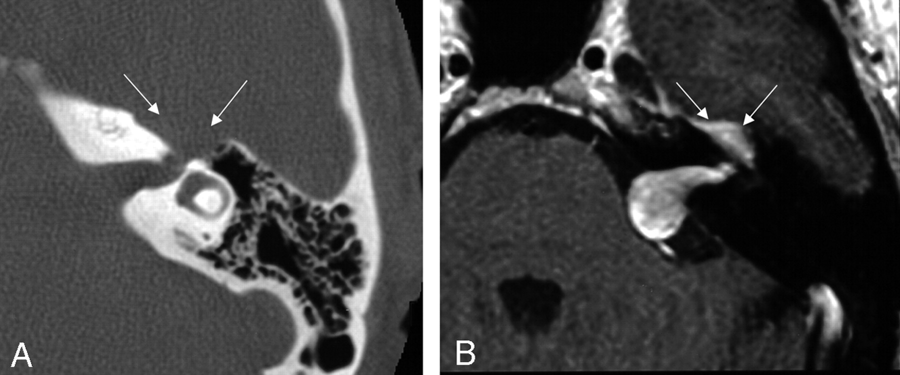
  • Fig 2.
    • Download figure
    • Open in new tab
    • Download powerpoint
    Fig 2.

    Axial images demonstrating focal enlargement of the labyrinthine segment of the facial nerve on the axial bone algorithm CT (left), and a homogeneously enhancing mass filling the internal auditory canal with extension into the cerebellopontine angle and labyrinthine segment on the axial T1-weighted postcontrast-enhanced MR image from a similar level (right). These images are from the same case as in Fig 1, and the patient was misdiagnosed preoperatively because of failure to note the enhancement and enlargement along the labyrinthine segment into the geniculate fossa (arrows). From Wiggins and Harnsberger (2001). Used with permission.
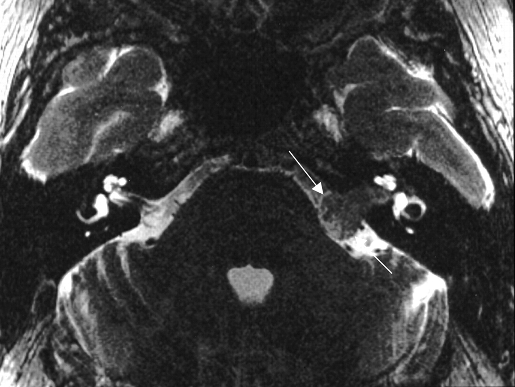
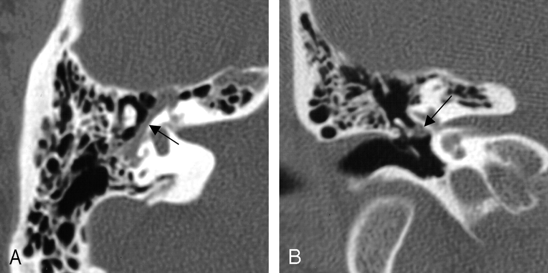
  • Fig 3.
    • Download figure
    • Open in new tab
    • Download powerpoint
    Fig 3.

    Axial bone algorithm CT image demonstrating focal enlargement of the labyrinthine segment of the facial nerve canal from a facial nerve schwannoma (between arrows). From Wiggins and Harnsberger (2001). Used with permission.
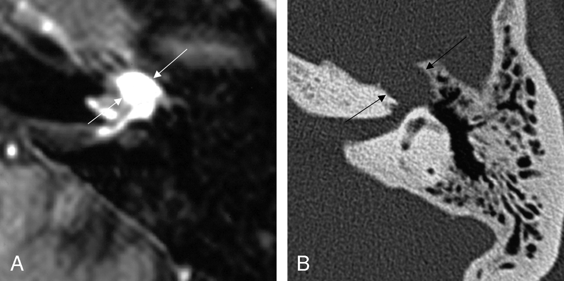
  • Fig 4.
    • Download figure
    • Open in new tab
    • Download powerpoint
    Fig 4.

    Axial images from the same case of a facial nerve schwannoma, demonstrating homogeneous enhancement of the labyrinthine segment and geniculate fossa on the axial T1-weighted postcontrast-enhanced MR imaging (left), and focal enlargement of the corresponding geniculate fossa and the labyrinthine segment of the facial nerve on the bone algorithm CT (right), at a similar level (between arrows). From Wiggins and Harnsberger (2001). Used with permission.

  • Fig 5.
    • Download figure
    • Open in new tab
    • Download powerpoint
    Fig 5.

    MR images demonstrating a large left middle cranial fossa mass. The axial T2-weighted image (left), and sagittal T1-weighted postcontrast image (right) show an extra-axial lesion, with a visible CSF/vascular cleft and associated buckling of the gray/white junction. The right image demonstrates a focal bulbous portion of the large mass extending to the geniculate fossa (between arrows), which was the origin of this FNS. From Wiggins and Harnsberger (2001). Used with permission.
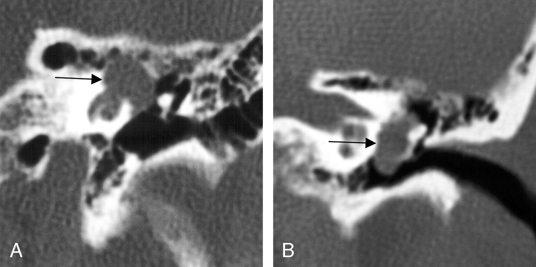
  • Fig 6.
    • Download figure
    • Open in new tab
    • Download powerpoint
    Fig 6.

    Bone algorithm CT images from the same case, demonstrating focal enlargement of the right tympanic segment, in the axial (left) and coronal (right) planes (at arrows). From Wiggins and Harnsberger (2001). Used with permission.

  • Fig 7.
    • Download figure
    • Open in new tab
    • Download powerpoint
    Fig 7.

    Two different cases of FNS, both coronal plane bone algorithm CT images show significant enlargement of the tympanic segment of the facial nerve with the case on the left demonstrating extension of the lesion primarily superiorly and medially, as well as extension into the cochlea, whereas the lesion on the right shows extension primarily inferiorly and laterally, displacing the ossicles, explaining this patient’s presentation with conductive hearing loss and a middle ear mass on clinical examination (at arrows). From Wiggins and Harnsberger (2001). Used with permission.
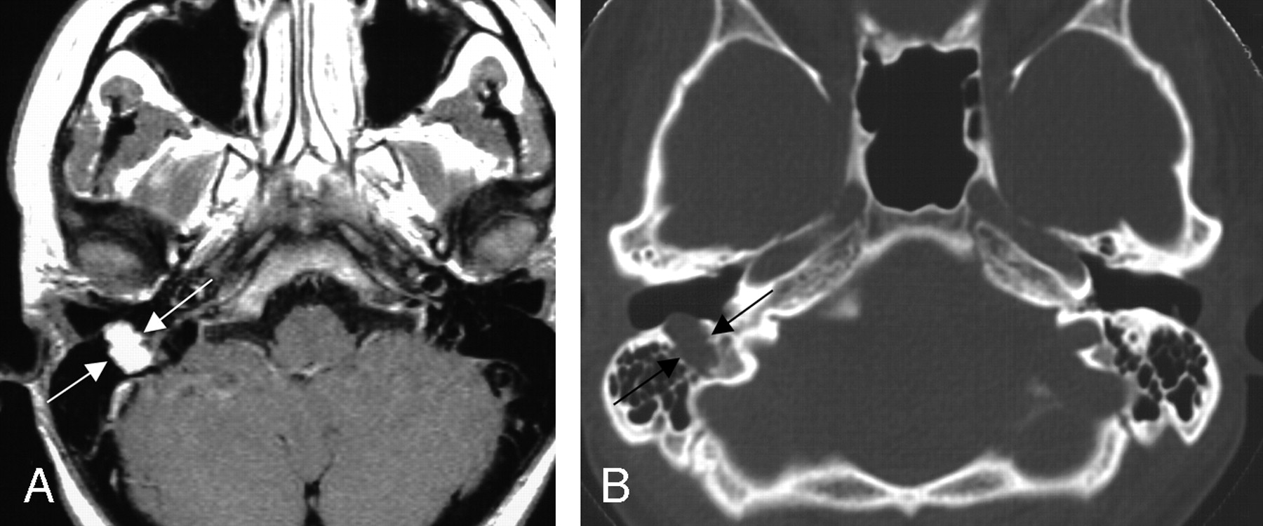
  • Fig 8.
    • Download figure
    • Open in new tab
    • Download powerpoint
    Fig 8.

    Two axial images from the same case of a facial nerve schwannoma involving the right mastoid segment of the facial nerve canal. The axial T1-weighted postcontrast MR image (left) shows homogeneous enhancement of the mass (between arrows). The bone algorithm CT (right) at the same level shows focal enlargement of the descending segment with extension toward the external auditory canal (between arrows). From Wiggins and Harnsberger (2001). Used with permission.
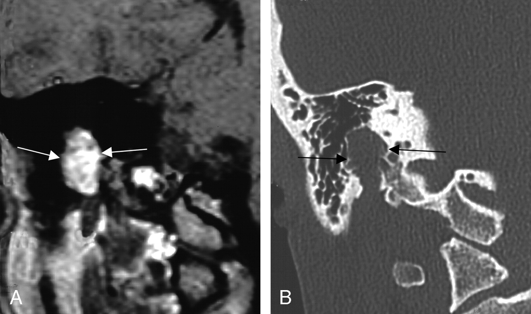
  • Fig 9.
    • Download figure
    • Open in new tab
    • Download powerpoint
    Fig 9.

    Two coronal views of the same case, demonstrating the aggressive appearing imaging features of mastoid segment FNSs. The left coronal T1-weighted postcontrast-enhanced image shows homogeneous enhancement. The right coronal bone algorithm CT study demonstrated an aggressive appearing lesion, as the FNS erupts into the surrounding thin-walled mastoid air cells of the same lesion (between arrows). From Wiggins and Harnsberger (2001). Used with permission.

PreviousNext
Back to top

In this issue

American Journal of Neuroradiology: 27 (3)
American Journal of Neuroradiology
Vol. 27, Issue 3
March, 2006
  • Table of Contents
  • Index by author
Advertisement
Print
Download PDF
Email Article

Thank you for your interest in spreading the word on American Journal of Neuroradiology.

NOTE: We only request your email address so that the person you are recommending the page to knows that you wanted them to see it, and that it is not junk mail. We do not capture any email address.

Enter multiple addresses on separate lines or separate them with commas.
The Many Faces of Facial Nerve Schwannoma
(Your Name) has sent you a message from American Journal of Neuroradiology
(Your Name) thought you would like to see the American Journal of Neuroradiology web site.
CAPTCHA
This question is for testing whether or not you are a human visitor and to prevent automated spam submissions.
Cite this article
R.H. Wiggins, H.R. Harnsberger, K.L. Salzman, C. Shelton, T.R. Kertesz, C.M. Glastonbury
The Many Faces of Facial Nerve Schwannoma
American Journal of Neuroradiology Mar 2006, 27 (3) 694-699;

Citation Manager Formats

  • BibTeX
  • Bookends
  • EasyBib
  • EndNote (tagged)
  • EndNote 8 (xml)
  • Medlars
  • Mendeley
  • Papers
  • RefWorks Tagged
  • Ref Manager
  • RIS
  • Zotero
0 Responses
Respond to this article
Share
Bookmark this article
The Many Faces of Facial Nerve Schwannoma
R.H. Wiggins, H.R. Harnsberger, K.L. Salzman, C. Shelton, T.R. Kertesz, C.M. Glastonbury
American Journal of Neuroradiology Mar 2006, 27 (3) 694-699;
del.icio.us logo Twitter logo Facebook logo Mendeley logo
  • Tweet Widget
  • Facebook Like
  • Google Plus One

Jump to section

  • Article
    • Abstract
    • Materials and Methods
    • Results
    • Discussion
    • Conclusion
    • References
  • Figures & Data
  • Info & Metrics
  • Responses
  • References
  • PDF

Related Articles

  • No related articles found.
  • PubMed
  • Google Scholar

Cited By...

  • Extratemporal intraparotid facial nerve schwannoma
  • Crossref
  • Google Scholar

This article has not yet been cited by articles in journals that are participating in Crossref Cited-by Linking.

More in this TOC Section

  • Correlation of Apparent Diffusion Coefficient at 3T with Prognostic Parameters of Retinoblastoma
  • Parathyroid Lesions: Characterization with Dual-Phase Arterial and Venous Enhanced CT of the Neck
  • MR Diagnosis of Facial Neuritis: Diagnostic Performance of Contrast-Enhanced 3D-FLAIR Technique Compared with Contrast-Enhanced 3D-T1-Fast-Field Echo with Fat Suppression
Show more Head and Neck

Similar Articles

Advertisement

Indexed Content

  • Current Issue
  • Accepted Manuscripts
  • Article Preview
  • Past Issues
  • Editorials
  • Editors Choice
  • Fellow Journal Club
  • Letters to the Editor

Cases

  • Case Collection
  • Archive - Case of the Week
  • Archive - Case of the Month
  • Archive - Classic Case

Special Collections

  • Special Collections

Resources

  • News and Updates
  • Turn around Times
  • Submit a Manuscript
  • Author Policies
  • Manuscript Submission Guidelines
  • Evidence-Based Medicine Level Guide
  • Publishing Checklists
  • Graphical Abstract Preparation
  • Imaging Protocol Submission
  • Submit a Case
  • Become a Reviewer/Academy of Reviewers
  • Get Peer Review Credit from Publons

Multimedia

  • AJNR Podcast
  • AJNR SCANtastic
  • Video Articles

About Us

  • About AJNR
  • Editorial Board
  • Not an AJNR Subscriber? Join Now
  • Alerts
  • Feedback
  • Advertise with us
  • Librarian Resources
  • Permissions
  • Terms and Conditions

American Society of Neuroradiology

  • Not an ASNR Member? Join Now

© 2025 by the American Society of Neuroradiology All rights, including for text and data mining, AI training, and similar technologies, are reserved.
Print ISSN: 0195-6108 Online ISSN: 1936-959X

Powered by HighWire