
Article Figures & Data
Figures
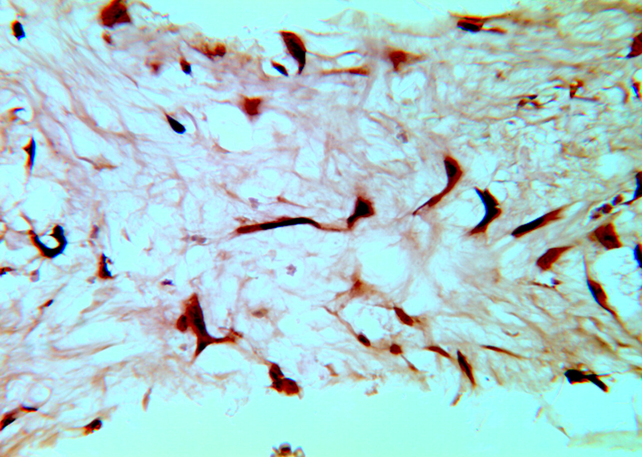
- Fig 1.
Photomicrographs of rabbit aneurysms embolized with platinum coils show that spindle cells within the aneurysm dome at 4 weeks (A) are positive for SMA and negative for SMA at 16 weeks (B) (immunohistochemistry, antibody to SMA, original magnification ×200).
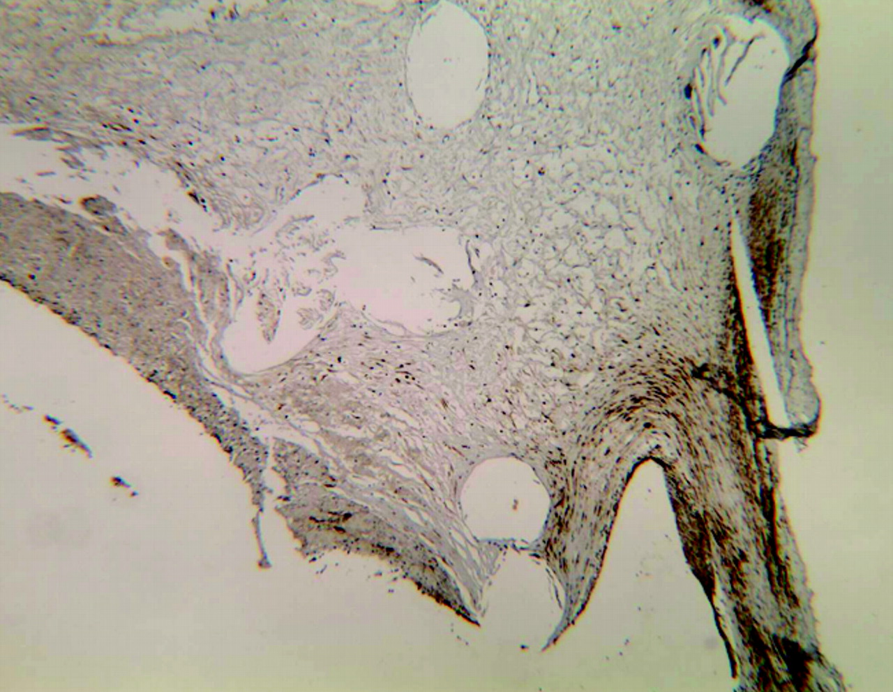
- Fig 2.
Photomicrograph of a rabbit aneurysm 10 weeks after embolization with platinum coils shows sparse cells within the dome that are positive for SMA staining (arrow); no positive cells were observed at the neck (dashed arrow). The cells at the side edge of neck (the transition zone of aneurysm wall to aneurysm neck) are positive for SMA (dotted arrow) (immunohistochemistry, antibody to SMA, original magnification ×60).
- Fig 3.
Photomicrograph of TUNEL staining for a 4-week rabbit aneurysm embolized with platinum coils. The attenuated brown or dark brown signal intensity is localized within the nuclei of the spindle cells, indicating positive staining (TUNEL, original magnification ×200).


Tables
Time (wk) Total No. of Animals No. of Animals Used for H&E, Masson Trichrome, and Immunohistochemistry No. of Animals Used for TUNEL 2 5 5 0 4 6 6 4 10 5 5 3 16 6 6 1 24 6 6 5 Note:—TUNEL indicates terminal deoxynucleotidyl transferase mediated nick and end- labeling.
Group H&E Masson Trichrome Immunostains TUNEL 2 Weeks (n = 5) Dome/lumen Unorganized thrombus (n = 5) No collagen deposition (n = 5) • Positive SMA (n = 5); mean number = 5 ± 3 • Weak positive myosin heavy chain (n = 5) • Weak positive vimentin (n = 5) No TUNEL staining performed in this group due to lack of nucleated cells Neck Unorganized thrombus, no endothelial cell lining (n = 2) Indication Myofibroblasts (n = 5) Unorganized thrombus, partial endothelial cell lining (n = 3) 4 Weeks (n = 6) Dome/lumen Loose connective tissue (n = 3) Disorganized collagen fibers (n = 1) • Positive SMA (n = 6); mean number = 45 ± 9 • Mildly positive myosin heavy chain (n = 6) • Mildly positive vimentin (n = 6) • Mildly positive desmin (n = 6) Sample size (n = 4) TUNEL positive (n = 4) Unorganized thrombus (n = 3) No collagen deposition (n = 5) Indication Myofibroblastic immuno-phenotype (n = 6) Neck Unorganized fibrin and/or laminated thrombus (n = 5) 10 Weeks (n = 5) Dome/lumen Unorganized thrombus (n = 2) No collagen deposition (n = 5) • Sparse SMA (n = 5); mean number = 10 ± 5 • Sparse vimentin (n = 5) • Negative myosin heavy chain (n = 5) • Negative desmin (n = 5) Sample size (n = 5) Sparse TUNEL positive cells (n = 5) Vascularized, loose, hypocellular tissue (n = 3) Indication Myofibroblastic immuno-phenotype (n = 5) Neck Thin layers of fibrinous/fibrous tissue (n = 5) 16 Weeks (n = 6) Dome/lumen Neovascular, loose, hypocellular tissue (n = 6) No collagen deposition (n = 6) • Negative SMA (n = 6); mean number = 0 ± 0 • Negative myosin heavy chain (n = 6) • Negative vimentin (n = 6) • Negative desmin (n = 6) Sample size (n = 1) No TUNEL positive cells (n = 1) Neck Thin layers of fibrinous/fibrous tissue (n = 5) 24 Weeks (n = 6) Dome/lumen Loose, hypocellular tissue (n = 6) No collagen deposition (n = 6) • Negative SMA (n = 6); mean number = 0 ± 0 • Negative myosin heavy chain (n = 6) • Negative vimentin (n = 6) • Negative desmin (n = 6) Sample size (n = 5) No TUNEL positive cells (n = 5) Neck Thin layers of fibrous tissue (n = 5) Unorganized fibrin (n = 1) Note:—TUNEL indicates terminal deoxynucleotidyl transferase mediated nick and end-labeling; SMA, smooth muscle actin.
In this issue


Jump to section
Related Articles
Cited By...
- Rabbit Elastase Aneurysm Model Mimics the Recurrence Rate of Human Intracranial Aneurysms following Platinum Coil Embolization
- Histologic and Biomolecular Similarities in Healing between Aneurysms and Cutaneous Skin Wounds
- Autologous adipose-derived mesenchymal stem cells improve healing of coiled experimental saccular aneurysms: an angiographic and histopathological study
- From bench to bedside: utility of the rabbit elastase aneurysm model in preclinical studies of intracranial aneurysm treatment
- Characterizing patterns of endothelialization following coil embolization: a whole-mount, dual immunostaining approach
- Mechanisms of Healing in Coiled Intracranial Aneurysms: A Review of the Literature
- A Large and Giant Bifurcation Aneurysm Model in Canines: Proof of Feasibility
- In Vivo Experimental Intracranial Aneurysm Models: A Systematic Review
- Angiographic and Histologic Comparison of Experimental Aneurysms Embolized with Hydrogel Filaments
- Endovascular Histologic Effects of Ultrathin Gold- or Vitronectin-Coated Platinum Aneurysm Coils in a Rodent Arterial Occlusion Model: A Preliminary Investigation
- Molecular Indices of Apoptosis Activation in Elastase-Induced Aneurysms After Embolization With Platinum Coils