
Article Figures & Data
Figures
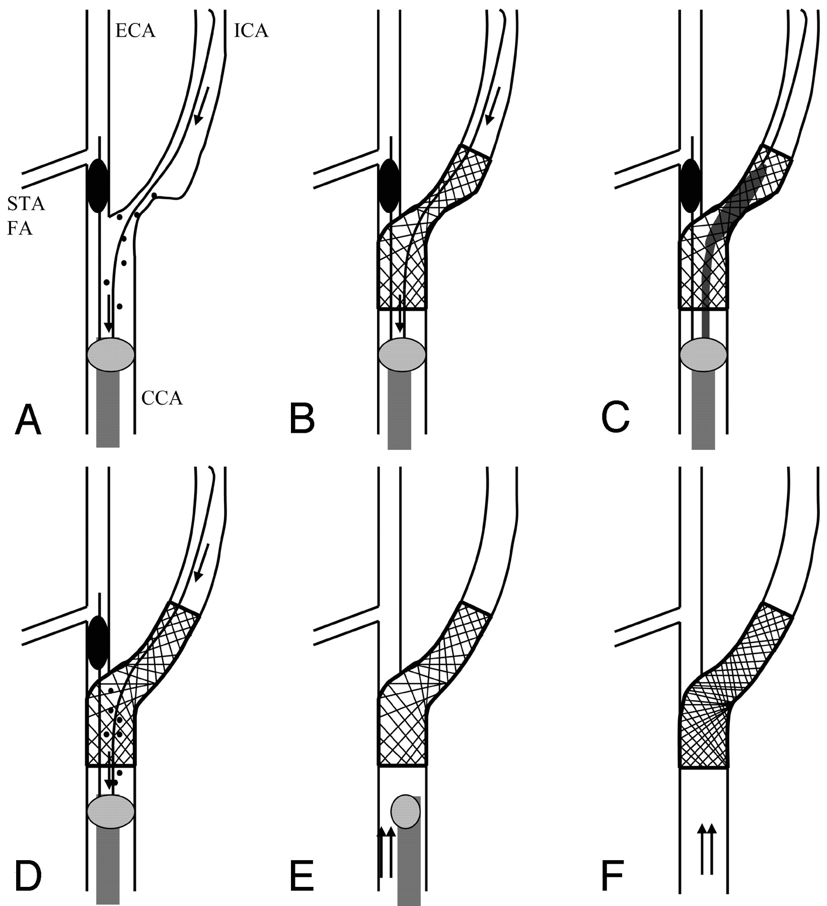
- Fig 1.
Schema of the procedure by using the Parodi Anti-Emboli System. STA, superior thyroid artery; FA, facial artery; arrow, reversed blood flow; double arrow, normal blood flow; spot, debris
A, Flow reversal from the intracranial ICA by blockade of both the proximal CCA and ECA.
B, Placement of stent.
C, Postdilation.
D, Irrigation of debris by flow reversal.
E, Return to normal flow.
F, Final
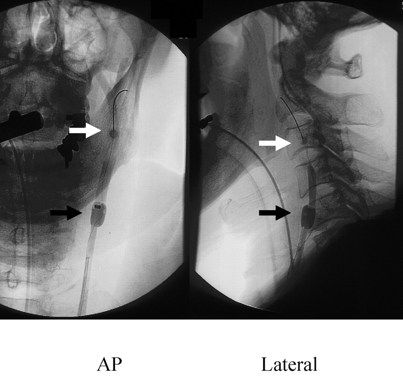
- Fig 2.
Carotid angioplasty with stent by Parodi Anti-Emboli System (left carotid angiogram, anteroposterior and lateral view). White arrow, blocking balloon in the external carotid artery; black arrow, proximal blocking balloon in the common carotid artery.
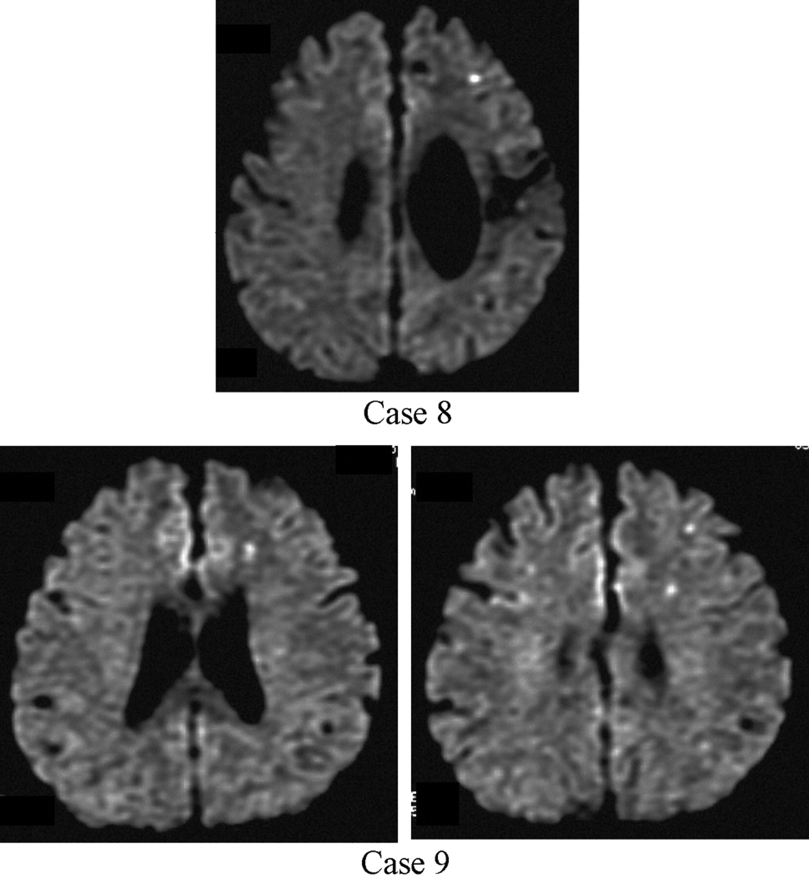
- Fig 3.
Ischemic lesions after carotid angioplasty with stent by Parodi Anti-Emboli System (diffusion-weighted MR image).
Top, Case 8 (after left carotid stent placement).
Bottom, Case 9 (after left carotid stent placement).


Tables
- Table 1:
Patients undergoing carotid angioplasty with stenting using the Parodi Anti-Emboli System in Mie University Hospital from 2003 to 2004
Patient No./Age (y)/Sex Side Symptom before Treatment Systemic Complication Stenosis Ratio (%) Postprocedural Stenosis (%) Occlusion Time during Procedure (s) Control ACT (s) ACT during Procedure (s) Prolonged ACT times Predilation Stent MRI-DWI Findings after Procedure (High-Signal Spot) 1/71/M R Asymptomatic HTN 90 10 418 148 222 1.50 (−) SMARTeR (−) 2/88/M L TIA HTN 90 30 660 150 318 2.12 (+) SMARTeR (−) 3/69/M R Asymptomatic DM, HL 90 15 964 161 304 1.89 (+) SMARTeR (−) 4/75/M R TIA (Amaurosis) DM 90 10 683 151 389 2.58 (+) SMARTeR (−) 5/62/M R Minor (−) 80 15 600 138 286 2.07 (+) SMARTeR (−) 6/69/M L Minor HL 90 5 420 133 250 1.88 (−) SMARTeR (−) 7/61/M R Asymptomatic DM, HTN 90 0 830 167 523 3.13 (+) SMARTeR (−) 8/63/M L Major DVT 70 5 624 167 344 2.06 (+) SMARTeR (+) 9/65/M L Asymptomatic HTN, HL 70 10 517 137 337 2.46 (−) SMARTeR (+) 10/77/M L Minor CRF, OMI 90 5 1354 144 428 2.97 (+) SMARTeR (−) 11/66/M R Minor DM 95 5 931 110 254 2.31 (+) Protege (−) Note:—ACT indicates activated clotting time; MRI-DWI, magnetic resonance imaging–diffusion weighted imaging; TIA, transient ischemia attack; HTN, hypertension; DM, diabetes mellitus; HL, hyperlipidemia; DVT, deep venous thrombosis; CRF, chronic renal failure; OMI, old myocardial infarction.
Averages are as follows: age, 69.7 ± 7.8 years; stenosis ratio, 85.4 ± 8.5%; occlusion time, 722.5 ± 255.2 seconds; ACT (control), 146.1 ± 15.2 seconds; ACT (during procedures, 329.5 ± 80.7 seconds.
- Table 2:
MRI-DWI assessment after DSA in Mie University Hospital from 2002 to 2004 (control group)
Patient No./Age (y)/Sex Diagnosis DSA Study (No. of vessels) MRI-DWI Study after DSA High-Intensity Spot on MRI-DWI 1/71/M Bilateral IC stenosis 4 1 day after (−) 2/46/F Unruptured aneurysm 4 1 day after (+) (1 spot, 2mm) 3/29/F Dural AVF 6 Same day (−) 4/31/F Brain tumor 3 6 days after (−) 5/76/M Right IC stenosis 4 10 days after (−) 6/39/M Brain tumor 4 1 day after (−) 7/59/F Brain tumor 6 1 day after (+) (1 spot, 2mm) 8/56/F Brain tumor 4 Same day (−) 9/77/F Unruptured aneurysm 4 1 day after (−) 10/37/M Brain tumor 3 5 days after (−) 11/64/M Right IC stenosis 4 3 days after (−) 12/76/F Right IC stenosis 4 8 days after (−) 13/77/M Left IC occlusion 4 Same day (−) 14/49/M Left MC occlusion 4 Same day (−) 15/59/F Left IC occlusion 4 1 day after (−) 16/76/M Right IC stenosis 4 1 day after (−) 17/43/F Unruptured aneurysm 4 4 days after (+) (2 spots, 2mm) 18/58/M Left IC stenosis 4 10 days after (−) 19/53/F Right VA dissection 4 1 day after (−) 20/69/M Right IC stenosis 4 1 day after (−) 21/48/M Unruptured aneurysm 4 1 day after (−) 22/71/M Right IC stenosis 4 1 day after (−) 23/74/M Left IC stenosis, Right IC occlusion 4 1 day after (−) 24/74/F Unruptured aneurysm 4 1 day after (−) 25/59/M Left VA occlusion 4 1 day after (−) 26/44/M Unconsciousness due to hypnotics 4 Same day (−) Note:—MRI-DWI indicates magnetic resonance imaging–diffusion weighted imaging; DSA, digital subtraction angiography; within the DSA study column, 4 vessels, bilateral common carotid arteries and bilateral vertebral arteries, and 6 vessels, bilateral internal and external carotid arteries and bilateral vertebral arteries; IC, internal carotid artery; MC, middle cerebral artery; VA, vertebral artery; AVF, arteriovenous fistula. Average age was 58.3 ± 15.3 years.
- Table 3:
Newly appearing high-intensity spots after carotid angioplasty with stent on MRI-DWI
Patient No. Side Findings on MRI-DWI Position No. of Spots Size of Spots 8 Left Ipsilateral frontal lobe 1 2 mm 9 Left Ipsilateral frontal lobe 3 2 mm Note:— MRI-DWI indicates magnetic resonance imaging–diffusion weighted imaging.


