
Article Figures & Data
Figures
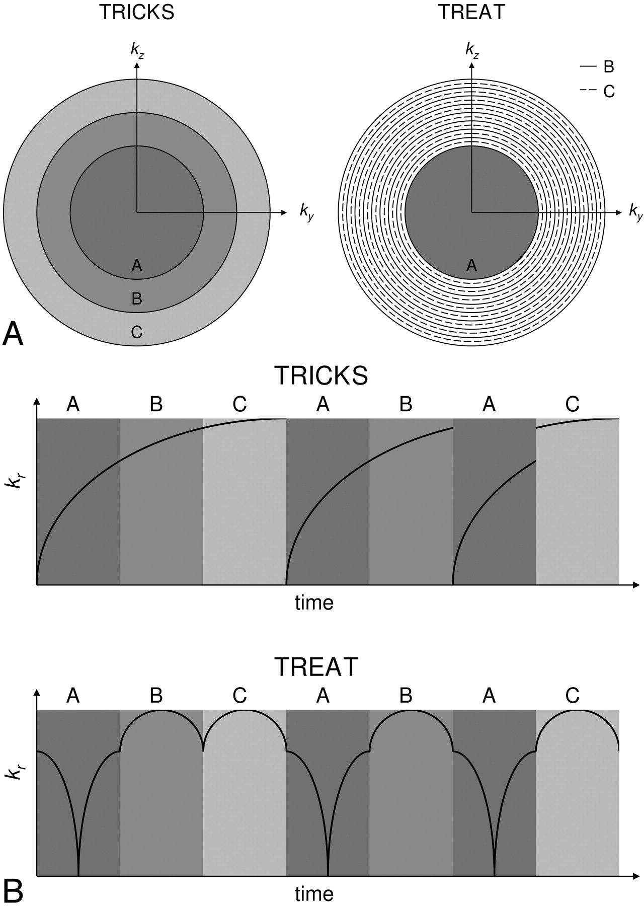
- Fig 1.
Comparison of the TRICKS and TREAT acquisition schemes. The segmentation methods are shown in panel A. In panel B, the distance of each sample from the center of k-space (kr) is plotted as a function of time to demonstrate the different ordering methods. Notice that the TREAT scheme acquires data without discontinuities in kr.
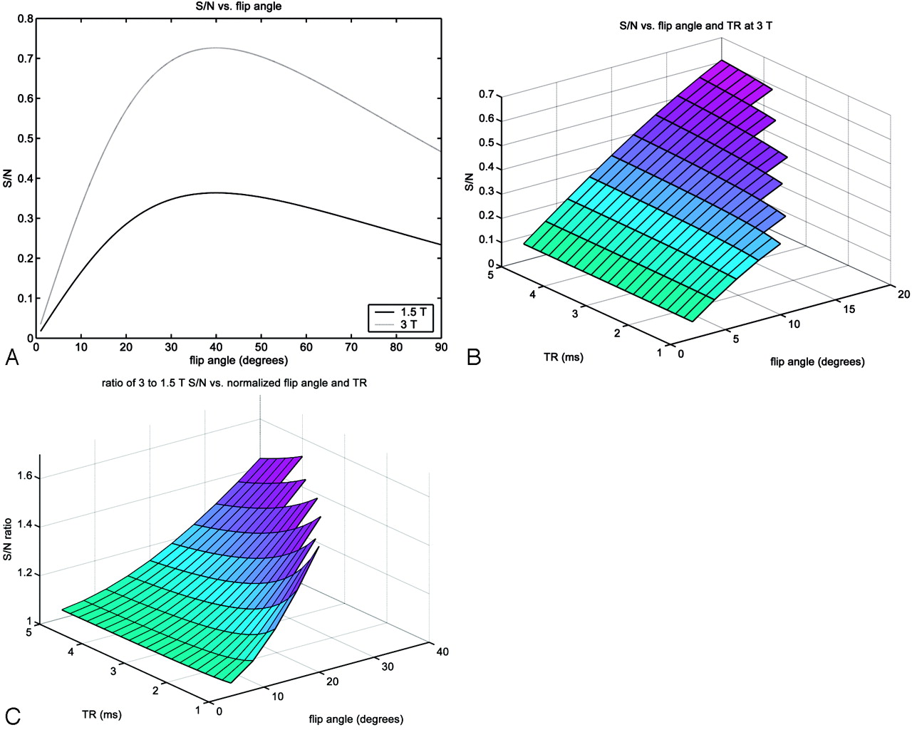
- Fig 2.
For enhanced intra-arterial blood: signal intensity versus flip angle at 1.5T and 3T (A), signal intensity versus flip angle and TR at 3T (B), and the ratio of 3T to 1.5T signals versus normalized flip angle and TR (C). The normalized flip angle was that which yielded an equivalent SAR at 1.5T. Missing values at high flip angles and low TRs were experimentally determined to exceed the SAR threshold.
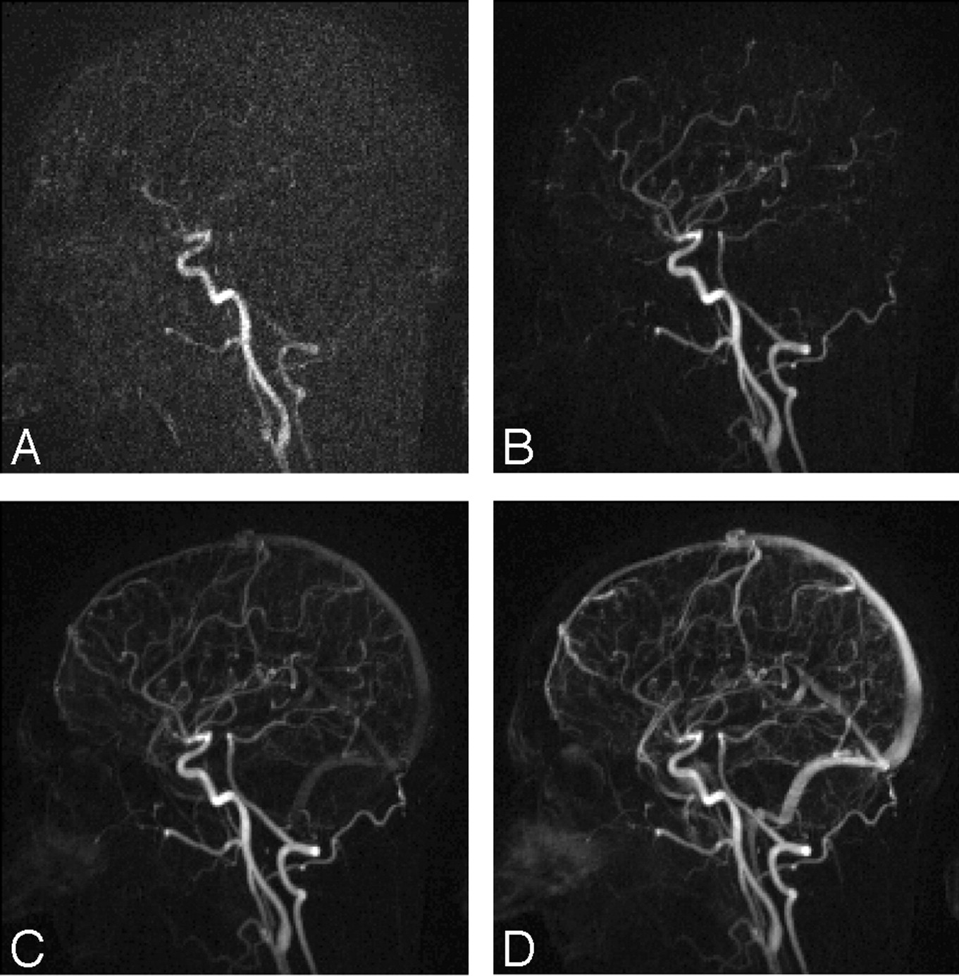
- Fig 3.
A qualitative comparison of S/N at 1.5T (A and C) and 3T (B and D) with timed coronal acquisitions is shown in these arterial phase mask subtraction MIP images. Magnified views (C and D) demonstrate better depiction of small distal middle cerebral arterial branches (circled) at 3T because of increased S/N.
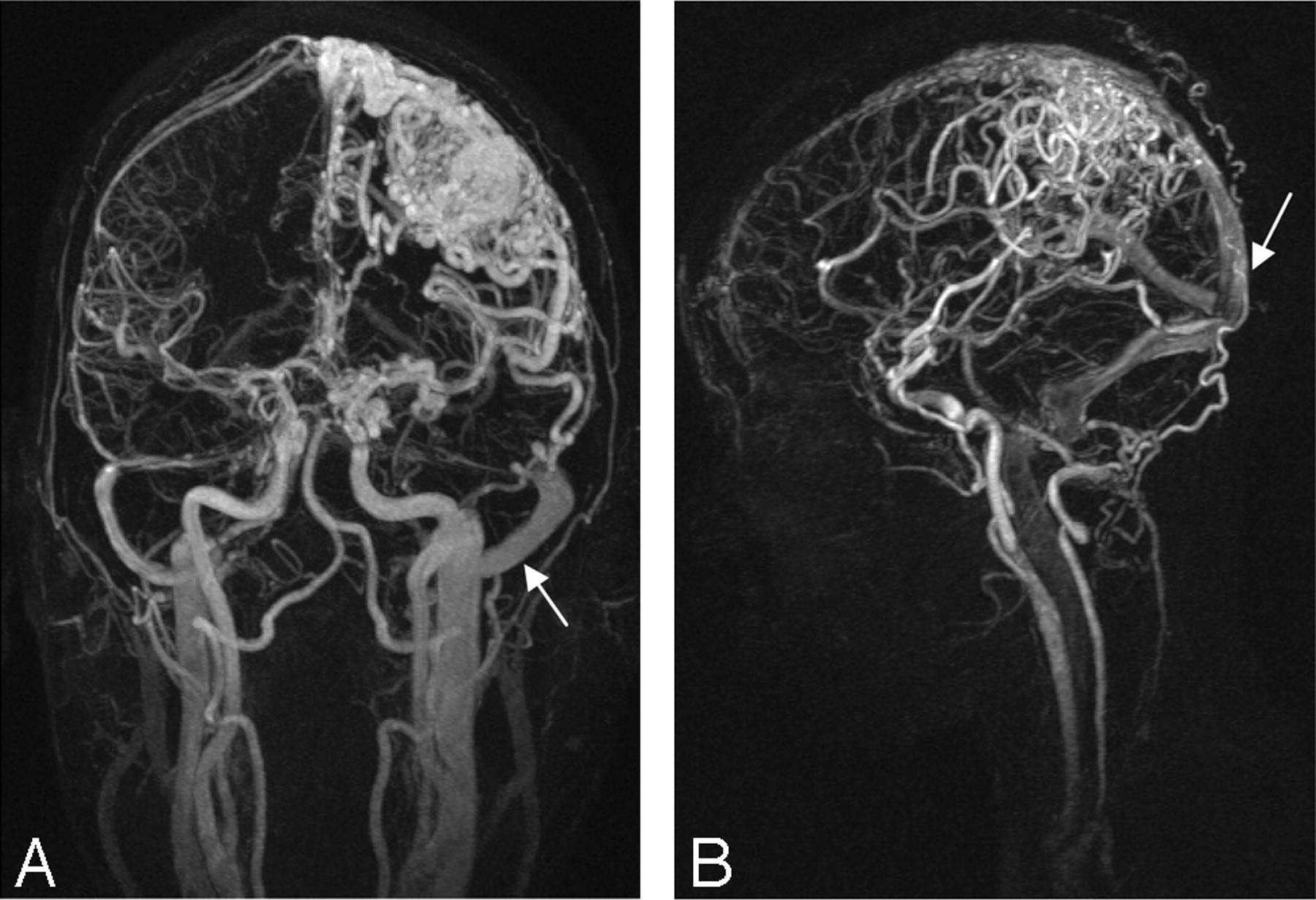
- Fig 4.
Timed coronal (A) and sagittal (B) MRAs at 3T of different AVM patients are shown in these subtracted MIP images. Despite high S/N and spatial resolution, low temporal resolution makes it difficult to determine the order in which vessels fill and the direction of flow. Early venous enhancement (arrows) due to arteriovenous shunting is present and “degrades” the arterial phase.
- Fig 5.
Time-resolved sagittal MRA at 3T of healthy volunteer. Consecutive timeframes are shown with temporal resolution of 2.5 seconds/frame and spatial resolution of 1.0 × 1.0 × 2.5 mm.
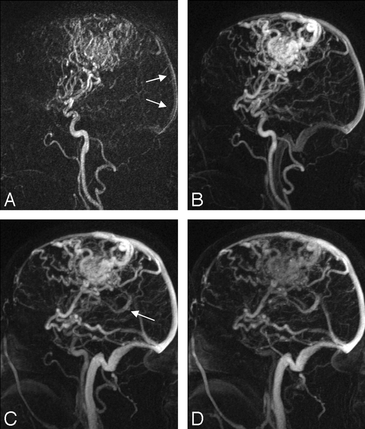
- Fig 6.
Time-resolved sagittal MRA at 3T of an AVM patient. Consecutive MIP timeframes are shown with temporal resolution of 2.5 seconds/frame and spatial resolution of 1.0 × 1.0 × 2.5 mm. Notice subtle early venous enhancement in the sagittal sinus during the early arterial phase (A, arrows) before enhancement of the basal vein of Rosenthal (C, arrow).
- Fig 7.
Consecutive timeframes of a conventional radiographic angiogram (A–D) and a 3D MIP MR angiogram (E and F) of the same patient with an arteriovenous malformation. The MR imaging is displayed with inverse grayscale contrast for better comparison. Conventional radiographic images are temporally blurred (G and H) for comparison with the MR images. For the radiographic and MR angiograms, respectively, the spatial resolutions are 0.1 × 0.1 and 1.0 × 1.0 × 2.5 mm and the temporal resolutions are 0.3 seconds/frame and 2.5 seconds/frame, respectively.


Tables
- Table 1:
T1 values (ms) at 1.5T and 3T for relevant intracranial contrast-enhanced MR angiography tissues
Nonenhanced Blood Cortical Gray Matter 1.5T 1381 ± 95 931 ± 21 3T 1872 ± 125 1232 ± 62 % Increase 35.5 32.3 - Table 2:
Qualitative comparison of 1.5T and 3T times intracranial contrast-enhanced MR angiography
M2 Segment M4 Segment Image Quality 1.5T 2.8 ± 0.45 1.6 ± 0.55 2.8 ± 0.45 3T 3.4 ± 0.55 2.4 ± 0.55 3.4 ± 0.55 P value <.05 <.05 <.05
In this issue


Jump to section
Related Articles
Cited By...
- Intracranial Arteriovenous Shunting: Detection with Arterial Spin-Labeling and Susceptibility-Weighted Imaging Combined
- Contrast-Enhanced Time-Resolved MRA for Follow-Up of Intracranial Aneurysms Treated with the Pipeline Embolization Device
- Noninvasive Evaluation of Cerebral Arteriovenous Malformations by 4D-MRA for Preoperative Planning and Postoperative Follow-Up in 56 Patients: Comparison with DSA and Intraoperative Findings
- Postcontrast Susceptibility-Weighted Imaging: A Novel Technique for the Detection of Arteriovenous Shunting in Vascular Malformations of the Brain
- Accuracy of Susceptibility-Weighted Imaging for the Detection of Arteriovenous Shunting in Vascular Malformations of the Brain
- Prediction of Response to Chemoradiation Therapy in Squamous Cell Carcinomas of the Head and Neck Using Dynamic Contrast-Enhanced MR Imaging
- A Compartment-Based Approach for the Imaging Evaluation of Tinnitus
- Cranial Dural Arteriovenous Fistula: Diagnosis and Classification with Time-Resolved MR Angiography at 3T
- 4D Radial Acquisition Contrast-Enhanced MR Angiography and Intracranial Arteriovenous Malformations: Quickly Approaching Digital Subtraction Angiography
- High-Resolution 3T MR Angiography of the Carotid Arteries: Comparison of Manual and Semiautomated Quantification of Stenosis
- Diagnostic Value of Multidetector-Row CT Angiography in the Evaluation of Thrombosis of the Cerebral Venous Sinuses