
Article Figures & Data
Figures
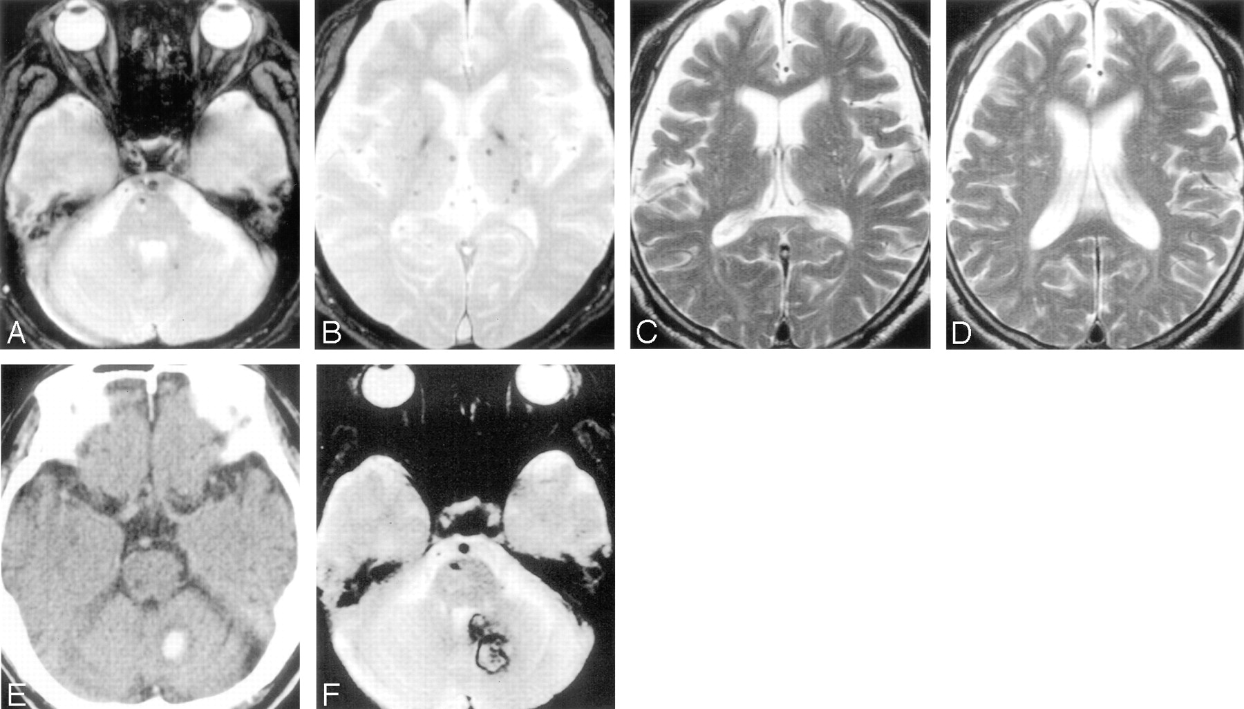
- Fig 1.
MR and CT images obtained from a patient (70-year-old man) with intracerebral hemorrhage in the cerebellum, who had been treated with aspirin after the occurrence of lacunar infarction. A and B, Initial T2*-weighted gradient-echo images (TR/TE, 800/26; flip angle, 20°) show multiple microbleeds in the brain stem, cerebellum, basal ganglia, and cerebral hemispheres. C and D, T2-weighted spin-echo images (TR/TE, 4500/112) do not show advanced white matter hyperintensity. E and F, CT image (E) and T2*-weighted gradient-echo image (F) obtained 9 months after the lacunar infarction show occurrence of cerebellar hemorrhage.
- Fig 2.
MR images obtained from a patient (85-year-old woman) with lacunar infarction in the right internal capsule after the occurrence of lacunar infarction in the right corona radiata. A and B, Initial T2*-weighted gradient-echo images (TR/TE, 800/26; flip angle, 20°) show no microbleeds. C, T2-weighted spin-echo image (TR/TE, 4500/112) shows advanced white matter hyperintensity. D, Diffusion-weighted image (single-shot echo-planar spin-echo sequence; TR/TE, 5300/135; b = 1000 mm2/s) obtained 23 months after the lacunar infarction shows a hyperintense lesion in the right internal capsule, consistent with acute infarction.


Tables
Group A Group B Group C Group D P Patients, n (M/F) 39 (22/17) 52 (34/18) 133 (85/48) 42 (26/16) .8219 Age, y (SD) 74.7 (9.0) 70.5 (10.1) 64.3 (11.4) 65.4 (11.3) .0001 Stroke types, n (ischemic/hemorrhagic) 39 (33/6) 52 (33/19) 133 (97/36) 42 (20.22) .0018 Antiplatelet therapy, n (%) 32 (82.1) 31 (59.6) 96 (72.2) 16 (38.1) .0001 Hypertension, n (%) 26 (66.7) 44 (84.6) 81 (60.9) 36 (85.7) .0013 Diabetes mellitus, n (%) 10 (25.6) 10 (19.2) 42 (31.6) 8 (19.0) .2214 Hypercholesterolemia, n (%) 17 (43.6) 14 (26.9) 31 (23.3) 9 (21.4) .0698 Note:— Group A, patients with advanced white matter hyperintensity (WMH) but without microbleeds; group B, patients with coexistence of microbleeds and advanced WMH; group C, patients without either mcribleeds or advanced WMH; group D, patients with microbleeds but without advanced WMH.
Recurrent Stroke, n (%) Recurrence Rate by Kaplan-Meier Method 1 y 2 y Group A (n = 39) 6 (15.4) 10.5 17.4 Group B (n = 52) 6 (11.5) 9.6 14.9 Group C (n = 133) 6 (4.5) 1.5 5.8 Group D (n = 42) 8 (19.0) 14.3 21.2 Note:— Group A, patients with advanced white matter hyperintensity (WMH) but without microbleeds; group B, patients with coexistence of microbleeds and advanced WMH; group C, patients without either microbleeds or advanced WMH; group D, patients with microbleeds but without advanced WMH.
Recurrent Stroke, n (%) Recurrence Rate by Kaplan-Meier Method (%) 1 y 2 y Group A (n = 39) 0 (0.0) 0 0 Group B (n = 52) 1 (1.9) 0 5.9 Group C (n = 133) 1 (0.8) 0 1.5 Group D (n = 42) 8 (19.0) 14.3 21.2 Note:— Group A, patients with advanced white matter hyperintensity (WMH) but without microbleeds; group B, patients with coexistence of microbleeds and advanced WMH; group C, patients without either microbleeds or advanced WMH; group D, patients with microbleeds but without advanced WMH.
Recurrent Stroke, n (%) Recurrence Rate by Kaplan-Meier Method (%) 1 y 2 y Group A (n = 39) 6 (15.4) 10.5 17.4 Group B (n = 52) 5 (9.6) 9.6 9.6 Group C (n = 133) 5 (3.8) 1.5 4.4 Group D (n = 42) 0 (0.0) 0 0 Note:— Group A, patients with advanced white matter hyperintensity (WMH) but without microbleeds; group B, patients with coexistence of microbleeds and advanced WMH; group C, patients without either microbleeds or advanced WMH; group D, patients with microbleeds but without advanced WMH.
Group/Age (y)/Sex Previous stroke Microbleeds, n WMH, Grade Antiplatelet Therapy Hyper-tension Diabetes Mellitus Hypercho-lesterolemia Recurrent Stroke A/84/F Lacunar infarction 0 2 Cilostazol (+) (−) (−) Lacunar infarction A/71/M Lacunar infarction 0 2 Ticlopidine (+) (−) (+) Lacunar infarction A/78/F Atherothrombotic infarction 0 2 Ticlopidine (−) (−) (+) Atherothrombotic infarction A/87/F Lacunar infarction 0 2 Aspirin (+) (−) (−) Lacunar infarction A/74/F Lacunar infarction 0 2 Aspirin (−) (−) (−) Lacunar infarction A/85/F Lacunar infarction 0 3 Cilostrazol (+) (−) (−) Lacunar infarction B/70/M Atherothrombotic infarction 3 3 Ticlopidine (+) (−) (+) Atherothrombotic infarction B/57/M Intracerebral hemorrhage 19 2 (−) (+) (−) (−) Lacunar infarction B/76/F Lacunar infarction 2 2 Cilostazol (+) (−) (+) Lacunar infarction B/66/M Lacunar infarction 1 2 Aspirin (+) (+) (+) Lacunar infarction B/55/M Lacunar infarction 2 2 Cilostazol (−) (−) (−) Lacunar infarction B/69/M Lacunar infarction 13 3 Aspirin (+) (−) (−) Intracerebral hemorrhage C/54/M Lacunar infarction 0 1 Cilostazol (+) (+) (−) Lacunar infarction C/61/F Intracerebral hemorrhage 0 1 Aspirin + ticlopidine (+) (+) (−) Lacunar infarction C/61/F Atherothrombotic infarction 0 1 Aspirin + ticlopidine (+) (−) (−) Lacunar infarction C/57/F Lacunar infarction 0 0 Aspirin (−) (+) (−) Atherothrombotic infarction C/54/M Lacunar infarction 0 1 Aspirin (−) (+) (−) Lacunar infarction C/74/M Lacunar infarction 0 1 Aspirin (−) (+) (−) Intracerebral hemorrhage D/77/M Lacunar infarction 13 1 Aspirin (−) (−) (−) Intracerebral hemorrhage D/70/M Lacunar infarction 28 1 Aspirin (+) (−) (+) Intracerebral hemorrhage D/73/M Atherothrombotic infarction 1 1 Aspirin (+) (−) (−) Intracerebral hemorrhage D/80/M Atherothrombotic infarction 11 0 Aspirin (+) (−) (−) Intracerebral hemorrhage D/82/M Intracerebral hemorrhage 2 1 (−) (−) (−) (−) Intracerebral hemorrhage D/51/M Intracerebral hemorrhage 2 0 (−) (+) (−) (−) Intracerebral hemorrhage D/53/F Intracerebral hemorrhage 12 1 (−) (+) (−) (−) Intracerebral hemorrhage D/55/M Intracerebral hemorrhage 16 1 (−) (+) (−) (−) Intracerebral hemorrhage Note:— Group A, patients with advanced white matter hyperintensity (WMH) but without microbleeds; group B, patients with coexistence of microbleeds and advanced WMH; group C, patients without either microbleeds or advanced WMH; group D, patients with microbleeds but without advanced WMH. Present is indicated by (+) and absent indicated by (−).
- Table 6:
Cox proportional hazards regression analysis for predicting subsequent intracerebral hemorrhage
Variable Hazards Regression 95% CI P Increased age 1.028 0.948–1.116 .5024 Male sex 16.476 1.448–187.467 .0239 Stroke type (intracerebral hemorrhage) 41.898 1.822–963.670 .0195 Microbleeds 85.626 6.344–1155.649 .0008 Advanced leukoaraiosis 0.016 0.001–0.258 .0035 Hypertension 0.163 0.026–1.044 .0555 Diabetes mellitus 0.83 0.092–7.461 .868 Hypercholesterolemia 0.333 0.030–3.667 .3689 Antiplatelet therapy 64.904 2.054–2050.683 .0178 Days from stroke onset to registration 1.009 1.003–1.015 .0017 - Table 7:
Cox proportional hazards regression analysis for predicting subsequent ischemic stroke
Variable Hazards Regression 95% CI P Increased age 0.938 0.886–0.993 .0269 Male sex 0.297 0.094–0.936 .0381 Stroke type (ischemic stroke) 1.099 0.029–41.732 .9596 Microbleeds 0.609 0.174–2.132 .4378 Advanced leukoaraiosis 10.659 2.601–43.678 .001 Hypertension 1.129 0.367–3.474 .8327 Diabetes mellitus 0.821 0.277–2.434 .7225 Hypercholesterolemia 0.609 0.200–1.849 .381 Antiplatelet therapy 13.816 0.343–556.026 .1636 Days from stroke onset to registration 0.987 0.971–1.003 .106
In this issue


Jump to section
Related Articles
Cited By...
- MRI predicts intracranial hemorrhage in patients who receive long-term oral anticoagulation
- Brain hemorrhage recurrence, small vessel disease type, and cerebral microbleeds: A meta-analysis
- Recurrent stroke risk and cerebral microbleed burden in ischemic stroke and TIA: A meta-analysis
- Silent Cerebral Microbleeds and Longitudinal Risk of Renal and Cardiovascular Events in Patients with CKD
- Cerebral Microbleeds Are Associated With an Increased Risk of Stroke: The Rotterdam Study
- Association of Postural Instability With Asymptomatic Cerebrovascular Damage and Cognitive Decline: The Japan Shimanami Health Promoting Program Study
- Cerebral Microbleeds: Is Antithrombotic Therapy Safe to Administer?
- Neuroimaging in Intracerebral Hemorrhage
- Cerebral Microbleeds and Recurrent Stroke Risk: Systematic Review and Meta-Analysis of Prospective Ischemic Stroke and Transient Ischemic Attack Cohorts
- Risk of Intracerebral Hemorrhage in Patients With Cerebral Microbleeds Undergoing Endovascular Intervention
- Microbleeds Are Associated With Subsequent Hemorrhagic and Ischemic Stroke in Healthy Elderly Individuals
- Microbleeds and the Risk of Recurrent Stroke
- Positional Relationship between Recurrent Intracerebral Hemorrhage/Lacunar Infarction and Previously Detected Microbleeds
- The clinical importance of white matter hyperintensities on brain magnetic resonance imaging: systematic review and meta-analysis
- Antithrombotic Drug Use, Cerebral Microbleeds, and Intracerebral Hemorrhage: A Systematic Review of Published and Unpublished Studies
- Brain microbleeds as a potential risk factor for antiplatelet-related intracerebral haemorrhage: hospital-based, case-control study
- Brain microbleeds
- Microbleeds and post-stroke emotional lability
- Advances in Interventional Neuroradiology 2007
- Prevalence and severity of microbleeds in a memory clinic setting