
Article Figures & Data
Figures
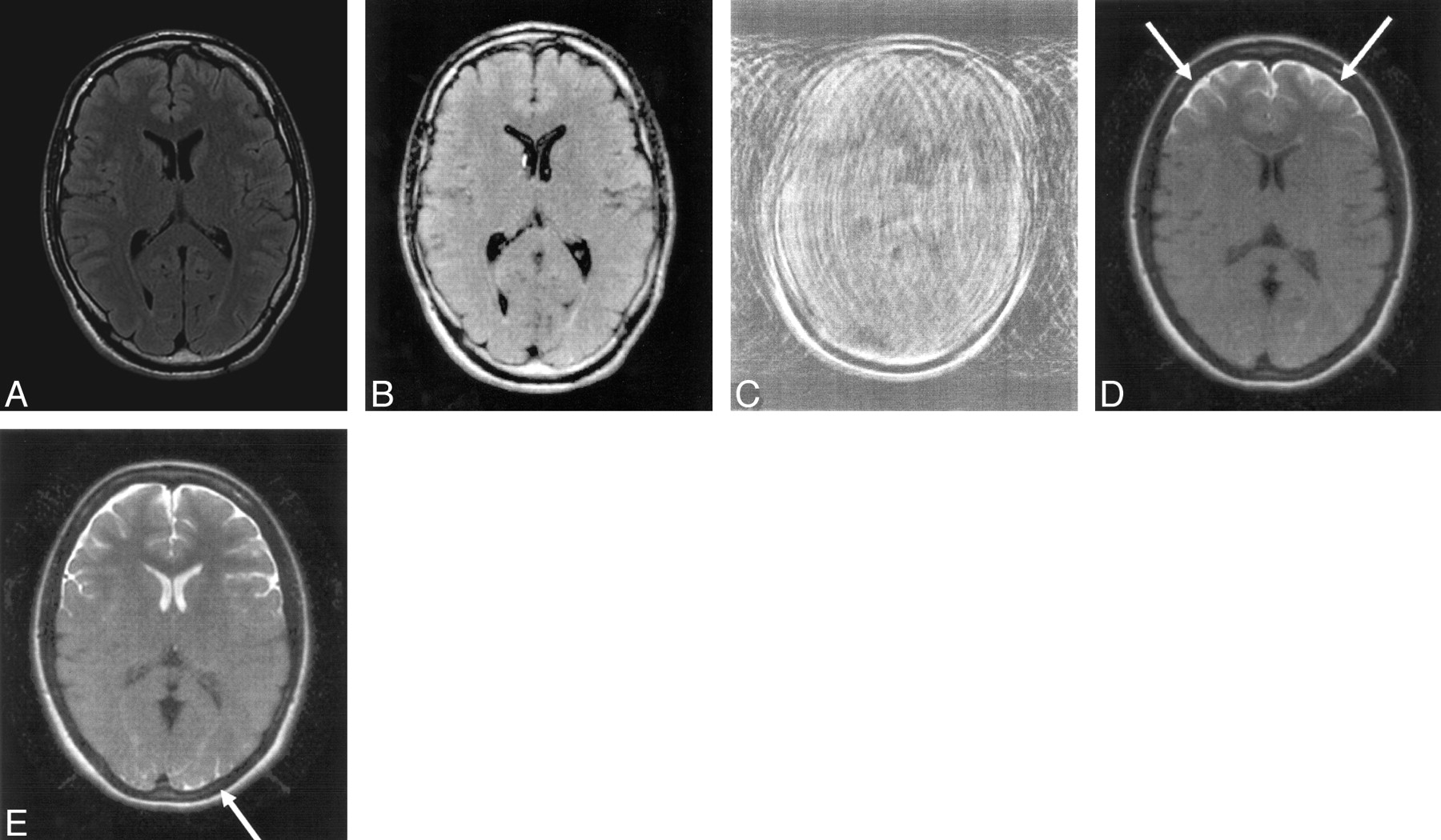
- Fig 1.
Healthy volunteer. Transverse multisection single-shot, fast spin-echo, fluid-attenuated inversion recovery (SS-FSE-FLAIR) (repetition time [TR]/echo time [TE]eff/inversion time [TI] = 8800/124/2200 ms) (A) and standard SS-FSE-FLAIR (B) images at rest, corresponding images with slow nodding (C and D), and a standard SS-FSE-FLAIR image with moderate nodding (E). Pulsatile CSF flow artifacts are seen in the region of the foramen of Monro, but no high-signal-intensity artifacts are seen elsewhere in the CSF (A and B). In C, the multisection image is badly degraded by motion. The corresponding standard SS-FSE-FLAIR image (D) is not degraded by motion artifact, but high signal intensity is seen in the CSF around the frontal lobes (arrows). With moderate nodding additional high-signal-intensity artifacts are seen on the SS-FSE-FLAIR image in the frontal horns of the lateral ventricles and in sulci of the occipital lobes (E, arrow). There is still no overall motion degradation. Slightly higher gray-white matter contrast is seen in the frontal lobes in C and E compared with B.
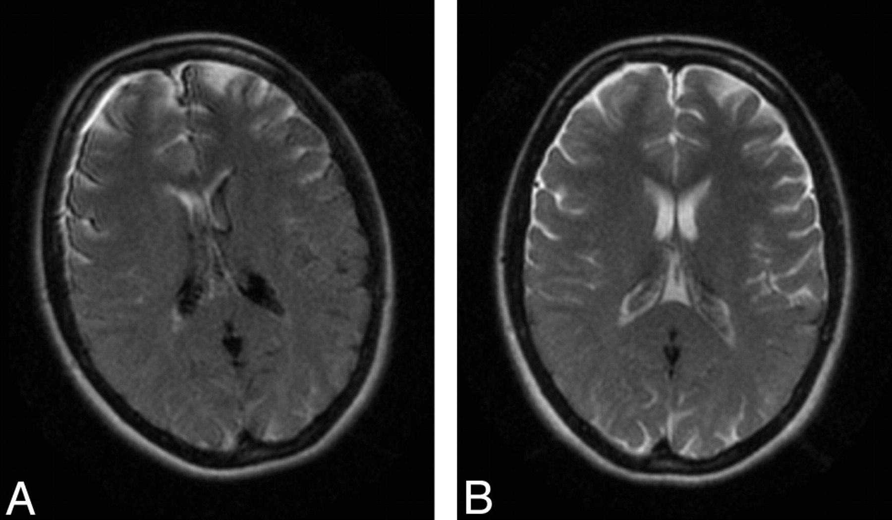
- Fig 2.
Healthy volunteer. Transverse standard single-shot, fast spin-echo, fluid-attenuated inversion recovery (SS-FSE-FLAIR) images with moderate rotation (A) and moderate nodding (B). The high signal intensity in the subarachnoid space and ventricular system is more extensive in panel B.
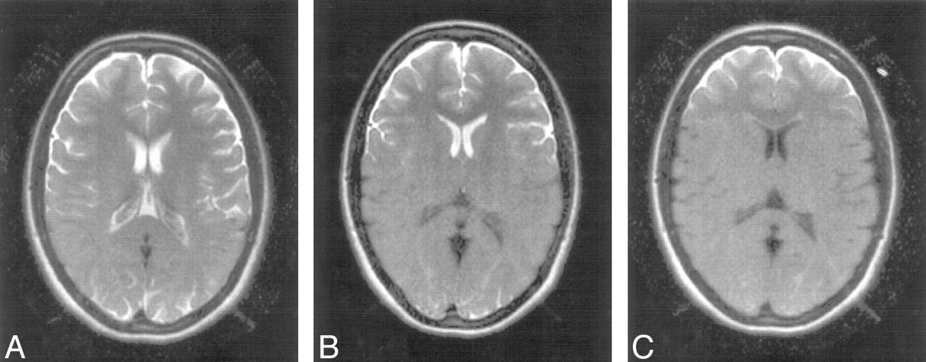
- Fig 3.
Healthy volunteer. Transverse standard single-shot, fast spin-echo, fluid-attenuated inversion recovery (SS-FSE-FLAIR) with 5-mm initial inversion pulse section width (A), a section widened (10 mm) SS-FSE-FLAIR (B), and another section-widened (30 mm) SS-FSE-FLAIR sequence with moderate nodding. With increasing section width there is a progressive reduction in both subarachnoid and intraventricular artifact.
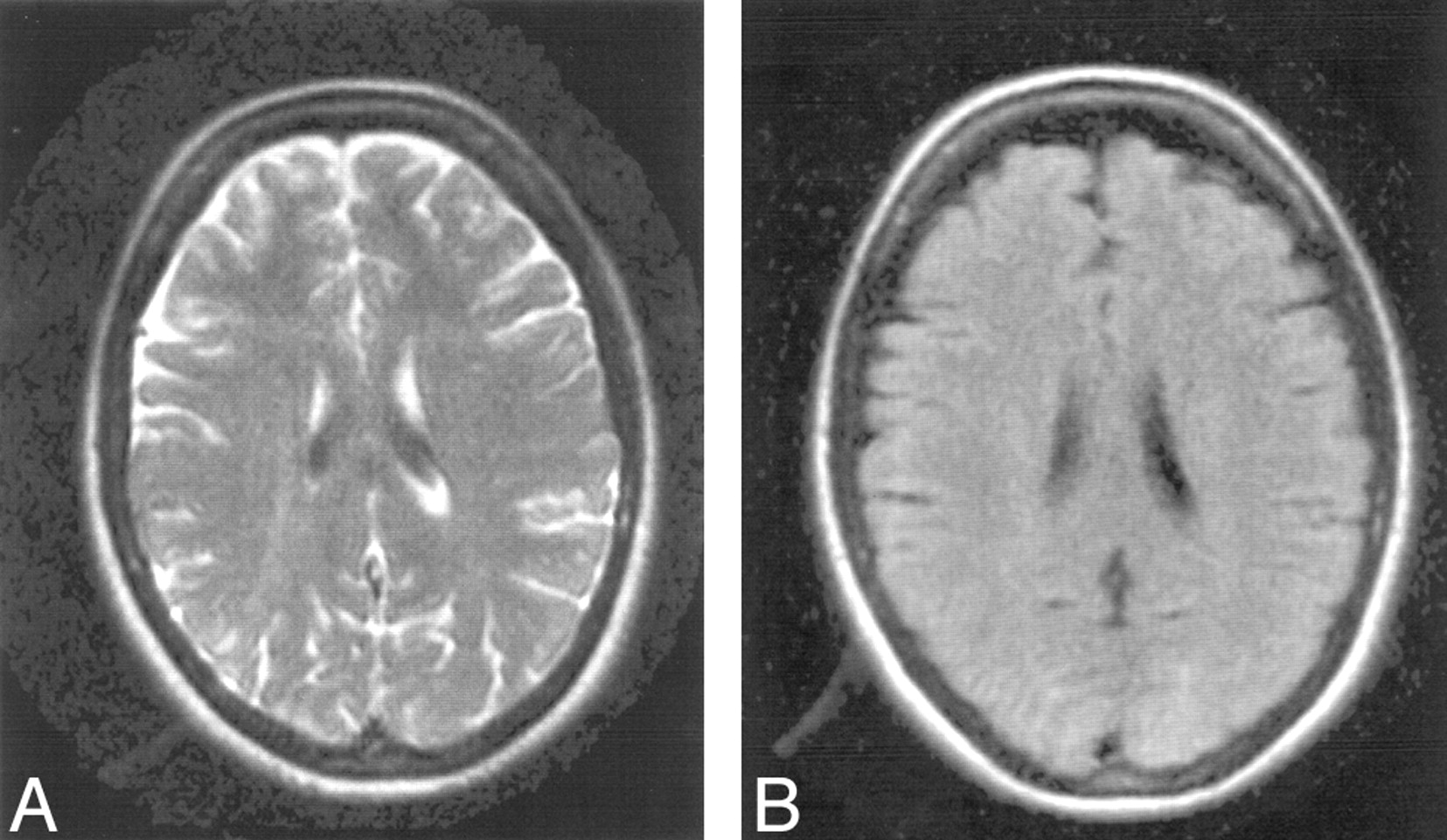
- Fig 4.
Healthy volunteer. Transverse standard single-shot, fast spin-echo, fluid-attenuated inversion recovery (SS-FSE-FLAIR) (A) and image obtained with a non–section-selective inversion pulse (B). Both were acquired with moderate nodding. There is much better control of motion artifact in B.
- Fig 5.
Images from a 53-year-old man with chronic hypertensive encephalopathy and acute mental status change. The patient was agitated and confused. Multisection T2-weighted FSE (repetition time [TR]/echo time [TE]eff = 2800/120 ms) image (A) and standard single-shot, fast spin-echo, fluid-attenuated inversion recovery (SS-FSE-FLAIR) images at different levels (B–D). The T2-weighted FSE image is severely degraded by motion artifact. Images in B to D show no obvious motion artifact, though the patient moved during the acquisition of the images as shown by the change in angulation of the head between sections. In D, obvious high-signal-intensity areas are seen anteriorly in the subarachnoid space. More subtle changes are seen posteriorly. These mimic subarachnoid hemorrhage. A slight increase in gray-white matter contrast is seen in the frontal region in D, consistent with the brain in this region not having experienced the initial inversion pulse. The brain shows extensive white matter change.
- Fig 6.
Sagittal drawing of the head showing the region covered by an initial section selective inversion pulse (shaded area) and that covered by the subsequent 90° pulse and acquisition without head movement (A) and with head movement downward in the superior-inferior direction between the initial inversion pulse and the 90° pulse (B). Possible directions of head motion–induced CSF flow at different times are also shown (curved arrows). With movement (B), the excitation and acquisition includes posterior areas of the subarachnoid space, which have experienced the inversion pulse as well as anterior areas that have not experienced it. These latter areas are likely to display high signal intensity, though the final result may be affected by CSF flow induced by the head motion. Initially, some CSF may move with the head while other portions of the CSF may move in the same direction later. When the head stops moving, there may be overshoot of the CSF moving with the head, followed by a later reversal of its direction of flow.


