
Article Figures & Data
Figures
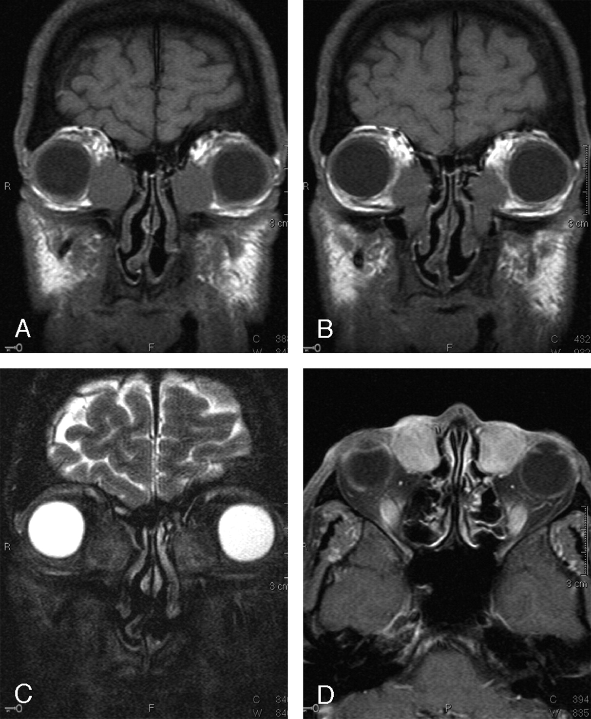
- Fig 1.
Coronal T1 (A and B), coronal T2 fat-saturated (C), and axial T1 fat-saturated postgadolinium (D) images of the orbits. Homogeneous, well-defined T1-hypointense masses are seen in the lacrimal fossae bilaterally (A), extending inferiorly into the nasolacrimal ducts (B). These masses are largely hypointense on T2-weighted imaging with heterogeneous areas of hyperintensity (C). Diffuse enhancement is seen after gadolinium administration (D).
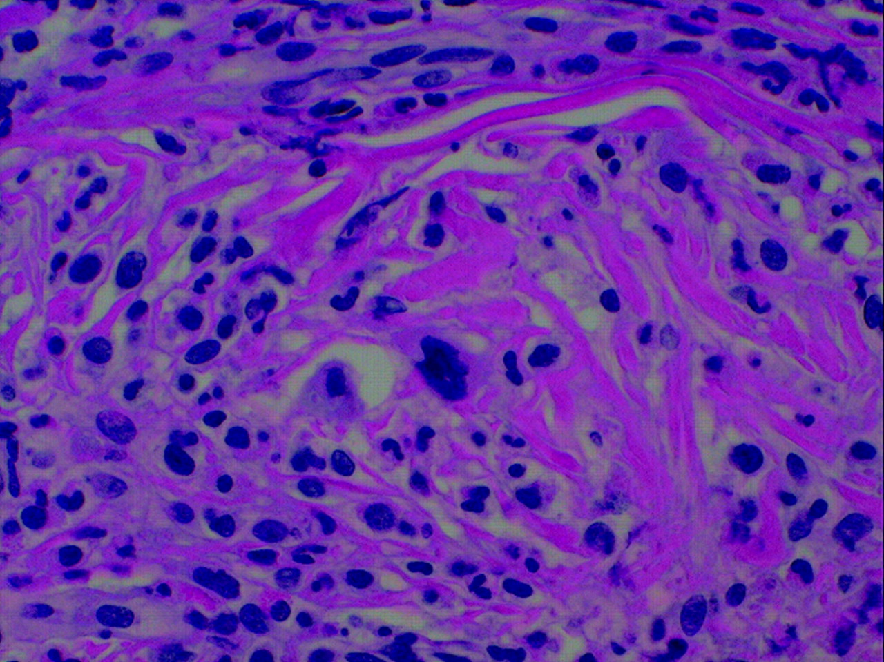
- Fig 2.
Histologic features. These masses were characterized by a proliferation of pleomorphic, large cells, and fibrosis that infiltrated, surrounded, and replaced the normal tissue. Every specimen consisted of attenuated, sclerotic connective tissue within which large, atypical cells (megakaryocyte-like) with lobulated nuclei were found as well as scattered lymphocytes and granulocytes.


In this issue


Jump to section
Related Articles
Cited By...
- No citing articles found.