
Article Figures & Data
Figures
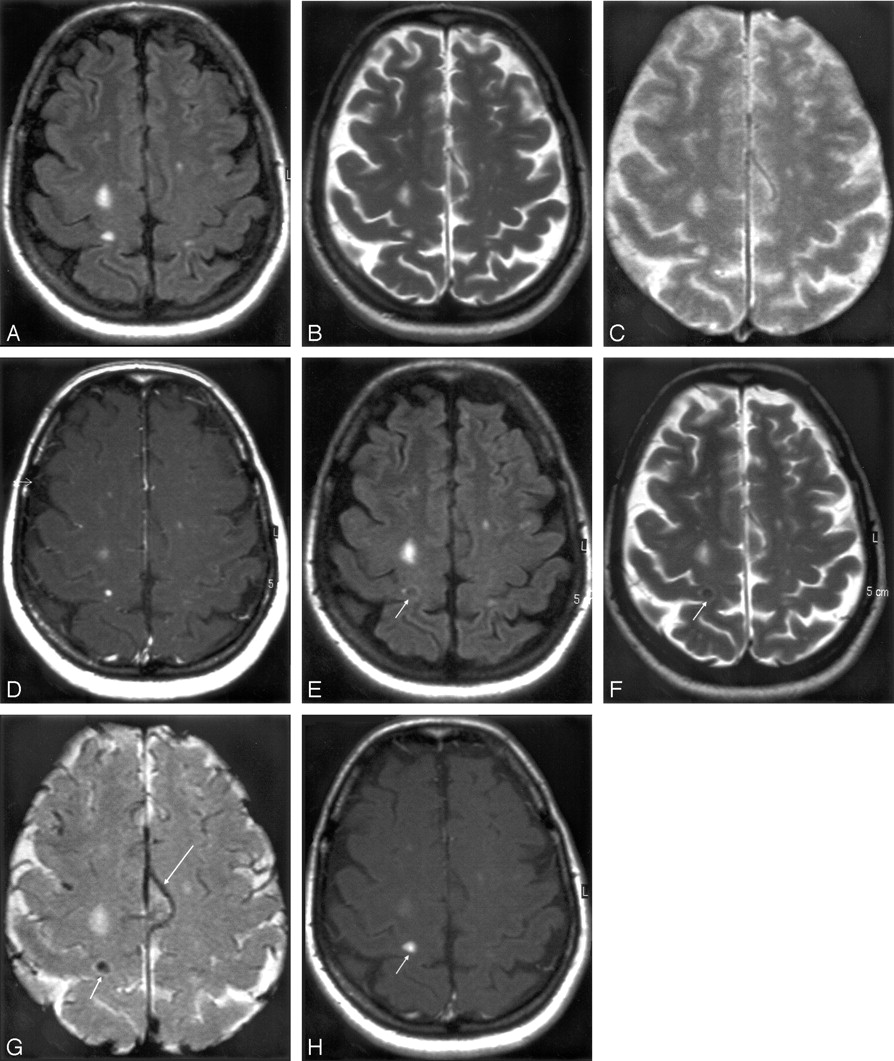
- Fig 1.
Ultra-small-particle iron oxide (USPIO)- and gadolinium-enhanced multiple sclerosis (MS) lesions on several MR imaging sequences. MR imaging 1 performed with fluid-attenuated inversion recovery (FLAIR) (A), T2-weighted (B), T2*-weighted (C), and magnetization transfer T1-weighted postgadolinium images (D). Some MS lesions were enhanced by gadolinium. MR imaging 2, performed 24 hours after USPIO injection, shows an uptake of USPIO in 1 lesion on FLAIR (E), turbo spin echo (TSE) T2-weighted (F), T2*-weighted (G), and T1-weighted (H) images (short arrows). Veins are clearly seen as dark signal intensity on T2*-weighted images (G) (long arrow).
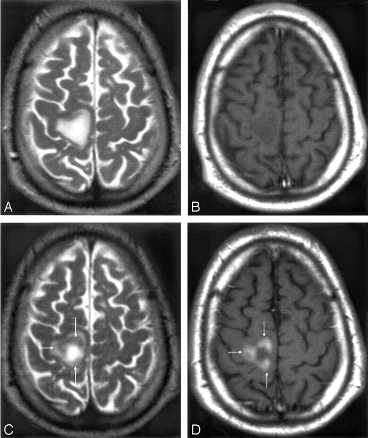
- Fig 2.
Mismatch of contrast agents uptake in an ultra-small-particle iron oxide (USPIO)-enhanced acute multiple sclerosis (MS) plaque. MR imaging 1 T2-weighted (A) and T1-weighted postgadolinium images (B) shows a large MS lesion that was not enhanced by gadolinium. MR imaging 2 shows the USPIO uptake at the periphery of the lesion (arrows), seen as a decreased signal intensity on T2-weighted images (C) and a high signal intensity on T1-weighted images (D). According to histologic observations (Lucchinetti et al, 2000), the number of macrophages at the center of acute MS lesions is usually minor.
- Fig 3.
Mismatch of contrast agent uptake in gadolinium-enhanced acute multiple sclerosis (MS) plaques. MR imaging 1 T1-weighted image (A) revealing the presence of 3 gadolinium-enhanced MS lesions (arrows). MR imaging 2 T1-weighted image (B) reveals USPIO enhancement in only one small lesion (arrow).


Tables
Comparison of ultra-small particle iron oxide (USPIO) versus gadolinium MRI to detect multiple sclerosis brain lesions with macrophage infiltration and/or blood–brain barrier permeability
No. of Patients Out of 10 No. of MRI Brain Lesions (n = 57) USPIO total 9 33 Gadolinium total 7 55 USPIO and gadolinium 7 31 USPIO only 2 2 Gadolinium only 7 24
In this issue


Jump to section
Related Articles
Cited By...
- In vivo nanoparticle imaging of innate immune cells can serve as a marker of disease severity in a model of multiple sclerosis
- Cervical Disk Pathology in Patients With Multiple Sclerosis: Two Case Reports
- Imaging Microglial/Macrophage Activation in Spinal Cords of Experimental Autoimmune Encephalomyelitis Rats by Positron Emission Tomography Using the Mitochondrial 18 kDa Translocator Protein Radioligand [18F]DPA-714
- Radioisotopic Imaging of Neuroinflammation
- Systemic Inflammatory Response Reactivates Immune-Mediated Lesions in Rat Brain
- Can imaging techniques measure neuroprotection and remyelination in multiple sclerosis?
- Imaging Inflammation in Acute Brain Ischemia
- MRI Monitoring of Neuroinflammation in Mouse Focal Ischemia