
Article Figures & Data
Figures
- Fig 1.
An example of a 1HMR spectroscopy study on a nonhuman primate 3 days after left hemispheric stroke (infarct volume, 15% of the hemisphere). A, Selected T2-weighted axial image with 2D MR spectroscopy overlay grid (FOV of the 2D-CSI dataset, 240 mm; 16 phase-encoding steps were acquired per side; section thickness, 15 mm; voxel cubic volume, 3.38 mL) demonstrates an intracerebral hyperintensity localized mainly to the anteromedial area of the left hemisphere, diagnostic for infarction. Remnants of the surgical approach to the circle of Willis are seen in the left orbit. Inside each voxel on the grid, the corresponding 1H spectrum is shown after processing (see Methods). Spectra from the 4 highlighted voxels are expanded in B. Signal intensity assignments are shown on the expanded spectra, correlating with both normal and abnormal signals.
- Fig 2.
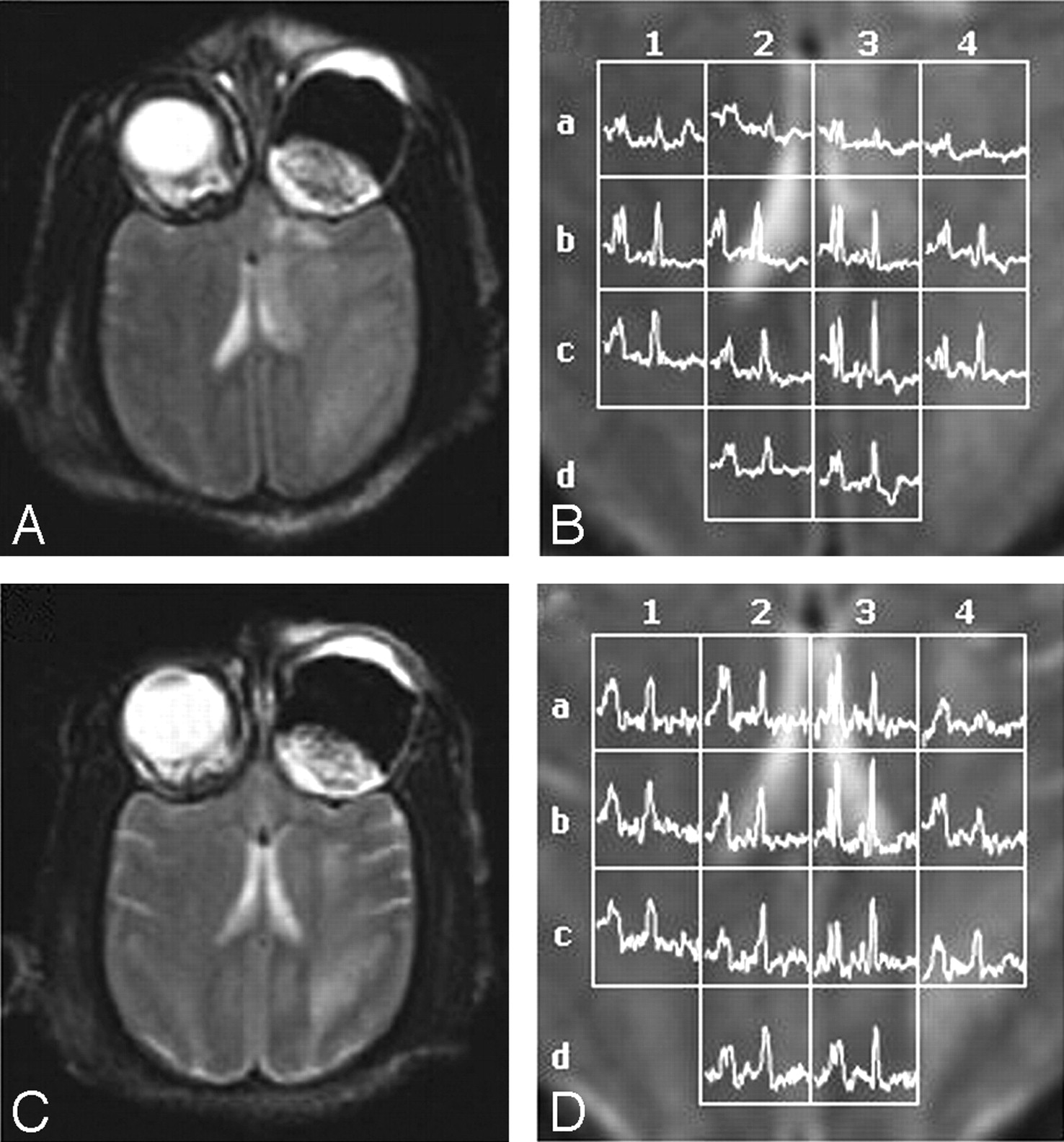
Representative data from a 1H-MR spectroscopy study on a nonhuman primate at both 3 days (A, -B) and 10 days (C, -D) following a small left hemispheric stroke (infarct volume, 28% of hemisphere). A and C, T2-weighted MR images are used to prescribe the spectral sections at (A) day 3 and (C) day 10. B and D, 1H-MR spectroscopy voxel grid and corresponding normalized spectra (FOV, 200 mm; thickness, 15 mm; each voxel was a volume of 15 × 12.5 × 12.5 mm per side with a capacity of 2.34 mL) are overlaid on magnified views of the T2-weighted MR images from (B) day 3 and (D) day 10. The voxel grids are labeled horizontally with numbers (1-4) and vertically with letters (a–d) to allow direct comparison between spectra collected at 3 and 10 days.
- Fig 3.
Representative data from a 1H-MR spectroscopy study on a nonhuman primate at both 3 days (A, -B) and 10 days (C, -D) following a large left hemispheric stroke (infarct volume, 67% of hemisphere). A and C, T2-weighted MR images are used to prescribe the spectral sections at (A) day 3 and (C) day 10. B and D, 1H-MR spectroscopy voxel grid and corresponding normalized spectra (same parameters as in Fig 2) are overlaid on magnified views of the T2-weighted MR images from (B) day 3 and (D) day 10. The voxel grids are labeled horizontally with numbers (1-4) and vertically with letters (a-d) to allow direct comparison between spectra collected at 3 and 10 days.
- Fig 4.
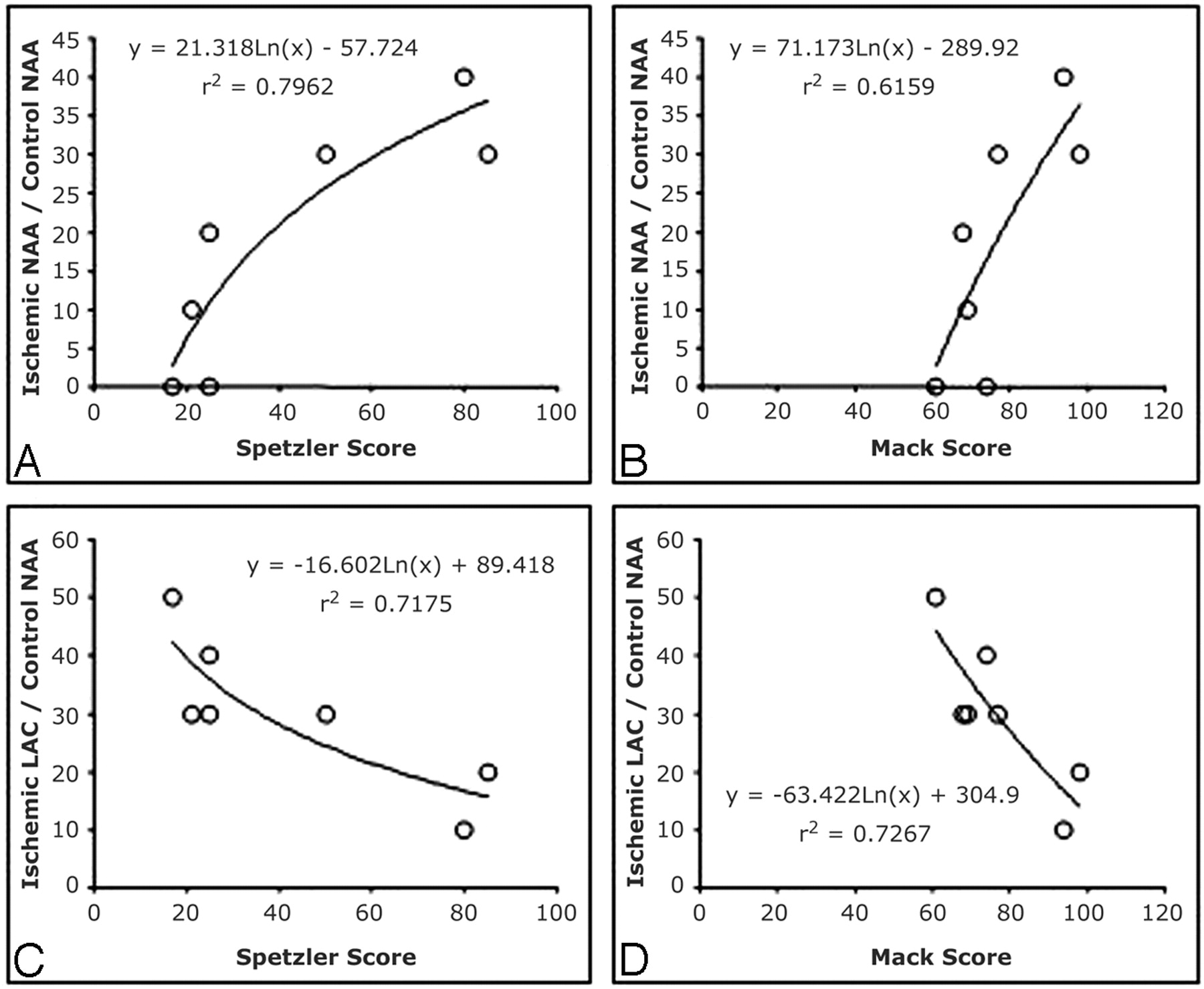
Plots demonstrate the correlation between the ratio of ischemic hemisphere mean integral value (area under the curve) of NAA (A, B) and LAC (C, D) with the control hemisphere mean integral value of NAA and the Spetzler score (A, C) and Mack scale (B, D) functional outcome measurements at 10 days postischemia. There is a positive correlation shown in (A) and (B) for the metabolite NAA on both scoring systems, with higher fractional NAA correlating with better functional outcome (higher scores). In contrast, there is a strong negative correlation observed for LAC (C, -D), with higher fractional LAC ratios being associated with lower (worse) functional outcome scores (Spetzler, r2 = 0.72; Mack, r2 = 0.73).


