Advertisement

Main menu

  • Home
  • Content
    • Current Issue
    • Accepted Manuscripts
    • Article Preview
    • Past Issue Archive
    • Video Articles
    • AJNR Case Collection
    • Case of the Week Archive
    • Case of the Month Archive
    • Classic Case Archive
  • Special Collections
    • AJNR Awards
    • Low-Field MRI
    • Alzheimer Disease
    • ASNR Foundation Special Collection
    • Photon-Counting CT
    • View All
  • Multimedia
    • AJNR Podcasts
    • AJNR SCANtastic
    • Trainee Corner
    • MRI Safety Corner
    • Imaging Protocols
  • For Authors
    • Submit a Manuscript
    • Submit a Video Article
    • Submit an eLetter to the Editor/Response
    • Manuscript Submission Guidelines
    • Statistical Tips
    • Fast Publishing of Accepted Manuscripts
    • Graphical Abstract Preparation
    • Imaging Protocol Submission
    • Author Policies
  • About Us
    • About AJNR
    • Editorial Board
    • Editorial Board Alumni
  • More
    • Become a Reviewer/Academy of Reviewers
    • Subscribers
    • Permissions
    • Alerts
    • Feedback
    • Advertisers
    • ASNR Home

User menu

  • Alerts
  • Log in

Search

  • Advanced search
American Journal of Neuroradiology

American Journal of Neuroradiology

ASHNR American Society of Functional Neuroradiology ASHNR American Society of Pediatric Neuroradiology ASSR
  • Alerts
  • Log in

Advanced Search

  • Home
  • Content
    • Current Issue
    • Accepted Manuscripts
    • Article Preview
    • Past Issue Archive
    • Video Articles
    • AJNR Case Collection
    • Case of the Week Archive
    • Case of the Month Archive
    • Classic Case Archive
  • Special Collections
    • AJNR Awards
    • Low-Field MRI
    • Alzheimer Disease
    • ASNR Foundation Special Collection
    • Photon-Counting CT
    • View All
  • Multimedia
    • AJNR Podcasts
    • AJNR SCANtastic
    • Trainee Corner
    • MRI Safety Corner
    • Imaging Protocols
  • For Authors
    • Submit a Manuscript
    • Submit a Video Article
    • Submit an eLetter to the Editor/Response
    • Manuscript Submission Guidelines
    • Statistical Tips
    • Fast Publishing of Accepted Manuscripts
    • Graphical Abstract Preparation
    • Imaging Protocol Submission
    • Author Policies
  • About Us
    • About AJNR
    • Editorial Board
    • Editorial Board Alumni
  • More
    • Become a Reviewer/Academy of Reviewers
    • Subscribers
    • Permissions
    • Alerts
    • Feedback
    • Advertisers
    • ASNR Home
  • Follow AJNR on Twitter
  • Visit AJNR on Facebook
  • Follow AJNR on Instagram
  • Join AJNR on LinkedIn
  • RSS Feeds

AJNR Awards, New Junior Editors, and more. Read the latest AJNR updates

Research ArticleBrain

Iron Particle–Enhanced Visualization of Inflammatory Central Nervous System Lesions by High Resolution: Preliminary Data in an Animal Model

R.A. Linker, A. Kroner, T. Horn, R. Gold, M. Mäurer and M. Bendszus
American Journal of Neuroradiology June 2006, 27 (6) 1225-1229;
R.A. Linker
  • Find this author on Google Scholar
  • Find this author on PubMed
  • Search for this author on this site
A. Kroner
  • Find this author on Google Scholar
  • Find this author on PubMed
  • Search for this author on this site
T. Horn
  • Find this author on Google Scholar
  • Find this author on PubMed
  • Search for this author on this site
R. Gold
  • Find this author on Google Scholar
  • Find this author on PubMed
  • Search for this author on this site
M. Mäurer
  • Find this author on Google Scholar
  • Find this author on PubMed
  • Search for this author on this site
M. Bendszus
  • Find this author on Google Scholar
  • Find this author on PubMed
  • Search for this author on this site
  • Article
  • Figures & Data
  • Info & Metrics
  • Responses
  • References
  • PDF
Loading

Article Figures & Data

Figures

  • Fig 1.
    • Download figure
    • Open in new tab
    • Download powerpoint
    Fig 1.

    SPIO-based macrophage imaging in EAE lesions of the rat brain by sonography. rhMOG-EAE was induced in congenic 1AV1 Lewis rats. During relapse, whole brains were removed and imaged ex vivo in a water tank by using a 13-MHz phased-array sonographic transducer 24 hours after SPIO injection. On coronal planes, sharply delineated circumscribed areas of focal echogenicity could be identified in different rats, predominantly involving the frontal, periventricular, cerebellar, and brain stem region (upper row, lesions marked by white arrows; representative images from different rats are shown), a pattern that is typical for MOG-EAE. Imaging of rat brains with injection of PBS after induction of rhMOG-EAE (middle row, anatomically equivalent areas from different rats are shown) or SPIO injection without prior induction of EAE (lower row) did not reveal similar areas with focal hyperechogenicity and served as negative controls.
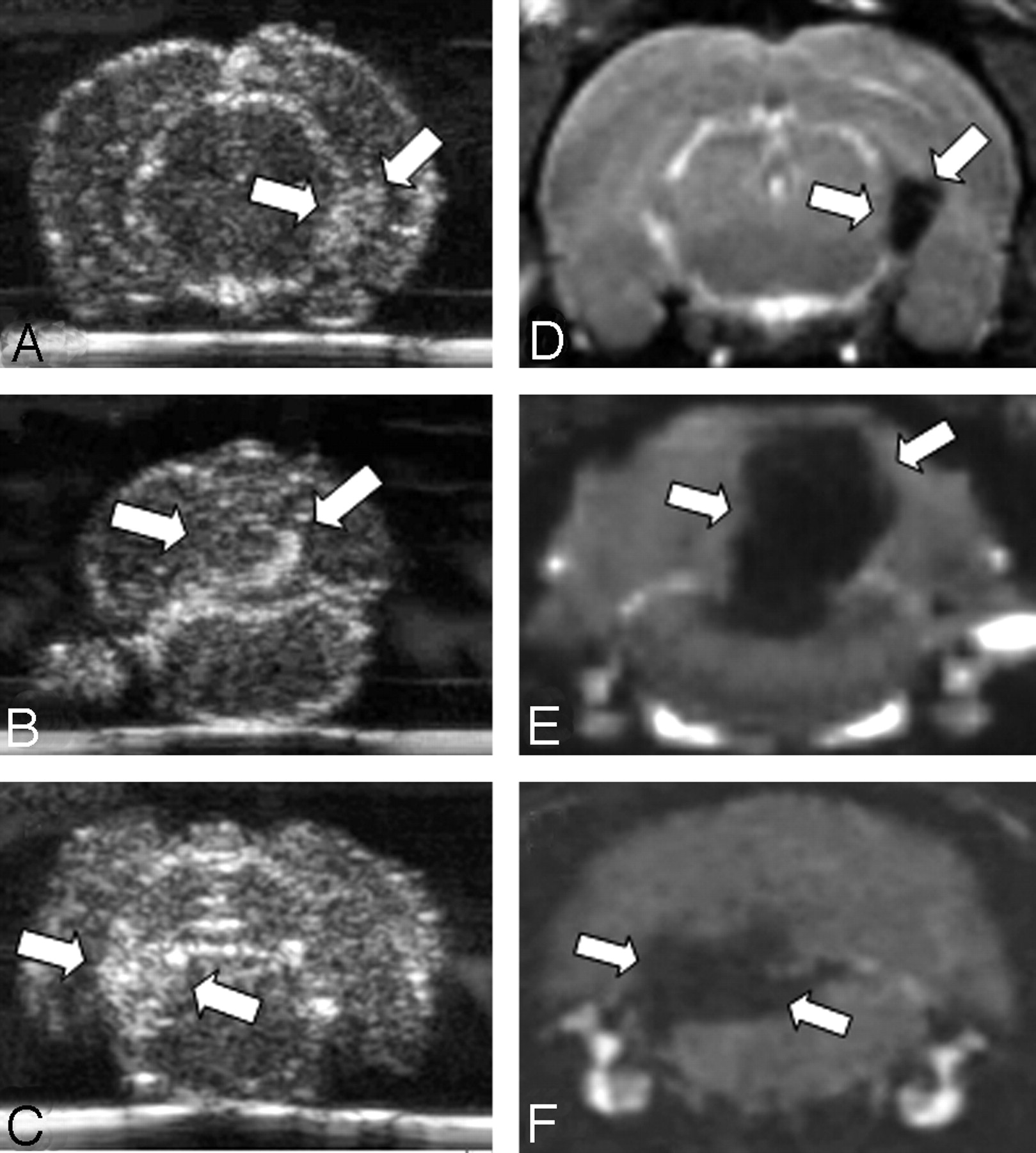
  • Fig 2.
    • Download figure
    • Open in new tab
    • Download powerpoint
    Fig 2.

    Comparison of SPIO-enhanced sonographic imaging of EAE lesions with MR imaging. We performed in vivo MR imaging to correlate the sonographic results with a well-established technique to monitor SPIO-particle accumulation. On coronal CISS images (D–F) corresponding to the brain regions depicted by sonography (A–C), we identified areas of focal hypointensity in the periventricular area (A, -D) and in the cerebellar (B, -E) or brain stem region (C, -F) of rat brains (lesions marked by white arrows). Hypointense areas are indicative of local SPIO-particle accumulation. Note the marked similarity of lesion distribution between sonograms and MR images (compare A–C with D–F).

  • Fig 3.
    • Download figure
    • Open in new tab
    • Download powerpoint
    Fig 3.

    Histologic evaluation. Paraffin sections were cut through the lesions as detected on MR imaging and sonography. A and B, The histology corresponds to the cerebellar lesion depicted in Figs 1A and 2A. A, Staining of macrophages and activated microglia by immunohistochemistry for ED1 (bar = 50 μm). Note the massive infiltrate of brown-stained ED1–positive cells. On a consecutive section (B, bar = 15 μm), Perls blue staining was performed to detect focal iron accumulation. Note the extensive perivascular iron deposition. Perls staining of liver sections with massively iron-laden Kupffer cells served as a histologically positive control for correct intravenous SPIO-particle injection (B, inset). Combination of Perls staining with ED1 immunohistochemistry reveals a clear colocalisation (C, bar = 10 μm), thereby identifying macrophage-like cells as one of the definitively iron-laden cell types in the CNS.

PreviousNext
Back to top

In this issue

American Journal of Neuroradiology: 27 (6)
American Journal of Neuroradiology
Vol. 27, Issue 6
June 2006
  • Table of Contents
  • Index by author
Advertisement
Print
Download PDF
Email Article

Thank you for your interest in spreading the word on American Journal of Neuroradiology.

NOTE: We only request your email address so that the person you are recommending the page to knows that you wanted them to see it, and that it is not junk mail. We do not capture any email address.

Enter multiple addresses on separate lines or separate them with commas.
Iron Particle–Enhanced Visualization of Inflammatory Central Nervous System Lesions by High Resolution: Preliminary Data in an Animal Model
(Your Name) has sent you a message from American Journal of Neuroradiology
(Your Name) thought you would like to see the American Journal of Neuroradiology web site.
CAPTCHA
This question is for testing whether or not you are a human visitor and to prevent automated spam submissions.
Cite this article
R.A. Linker, A. Kroner, T. Horn, R. Gold, M. Mäurer, M. Bendszus
Iron Particle–Enhanced Visualization of Inflammatory Central Nervous System Lesions by High Resolution: Preliminary Data in an Animal Model
American Journal of Neuroradiology Jun 2006, 27 (6) 1225-1229;

Citation Manager Formats

  • BibTeX
  • Bookends
  • EasyBib
  • EndNote (tagged)
  • EndNote 8 (xml)
  • Medlars
  • Mendeley
  • Papers
  • RefWorks Tagged
  • Ref Manager
  • RIS
  • Zotero
0 Responses
Respond to this article
Share
Bookmark this article
Iron Particle–Enhanced Visualization of Inflammatory Central Nervous System Lesions by High Resolution: Preliminary Data in an Animal Model
R.A. Linker, A. Kroner, T. Horn, R. Gold, M. Mäurer, M. Bendszus
American Journal of Neuroradiology Jun 2006, 27 (6) 1225-1229;
del.icio.us logo Twitter logo Facebook logo Mendeley logo
  • Tweet Widget
  • Facebook Like
  • Google Plus One

Jump to section

  • Article
    • Abstract
    • Methods
    • Results
    • Discussion
    • Conclusion
    • Acknowledgments
    • Footnotes
    • References
  • Figures & Data
  • Info & Metrics
  • Responses
  • References
  • PDF

Related Articles

  • No related articles found.
  • PubMed
  • Google Scholar

Cited By...

  • Iron deposition in periaqueductal gray matter as a potential biomarker for chronic migraine
  • In vivo nanoparticle imaging of innate immune cells can serve as a marker of disease severity in a model of multiple sclerosis
  • Crossref
  • Google Scholar

This article has not yet been cited by articles in journals that are participating in Crossref Cited-by Linking.

More in this TOC Section

  • Fast Contrast-Enhanced 4D MRA and 4D Flow MRI Using Constrained Reconstruction (HYPRFlow): Potential Applications for Brain Arteriovenous Malformations
  • Multimodal CT Provides Improved Performance for Lacunar Infarct Detection
  • Optimal MRI Sequence for Identifying Occlusion Location in Acute Stroke: Which Value of Time-Resolved Contrast-Enhanced MRA?
Show more Brain

Similar Articles

Advertisement

Indexed Content

  • Current Issue
  • Accepted Manuscripts
  • Article Preview
  • Past Issues
  • Editorials
  • Editor's Choice
  • Fellows' Journal Club
  • Letters to the Editor
  • Video Articles

Cases

  • Case Collection
  • Archive - Case of the Week
  • Archive - Case of the Month
  • Archive - Classic Case

More from AJNR

  • Trainee Corner
  • Imaging Protocols
  • MRI Safety Corner
  • Book Reviews

Multimedia

  • AJNR Podcasts
  • AJNR Scantastics

Resources

  • Turnaround Time
  • Submit a Manuscript
  • Submit a Video Article
  • Submit an eLetter to the Editor/Response
  • Manuscript Submission Guidelines
  • Statistical Tips
  • Fast Publishing of Accepted Manuscripts
  • Graphical Abstract Preparation
  • Imaging Protocol Submission
  • Evidence-Based Medicine Level Guide
  • Publishing Checklists
  • Author Policies
  • Become a Reviewer/Academy of Reviewers
  • News and Updates

About Us

  • About AJNR
  • Editorial Board
  • Editorial Board Alumni
  • Alerts
  • Permissions
  • Not an AJNR Subscriber? Join Now
  • Advertise with Us
  • Librarian Resources
  • Feedback
  • Terms and Conditions
  • AJNR Editorial Board Alumni

American Society of Neuroradiology

  • Not an ASNR Member? Join Now

© 2025 by the American Society of Neuroradiology All rights, including for text and data mining, AI training, and similar technologies, are reserved.
Print ISSN: 0195-6108 Online ISSN: 1936-959X

Powered by HighWire