
Article Figures & Data
Figures
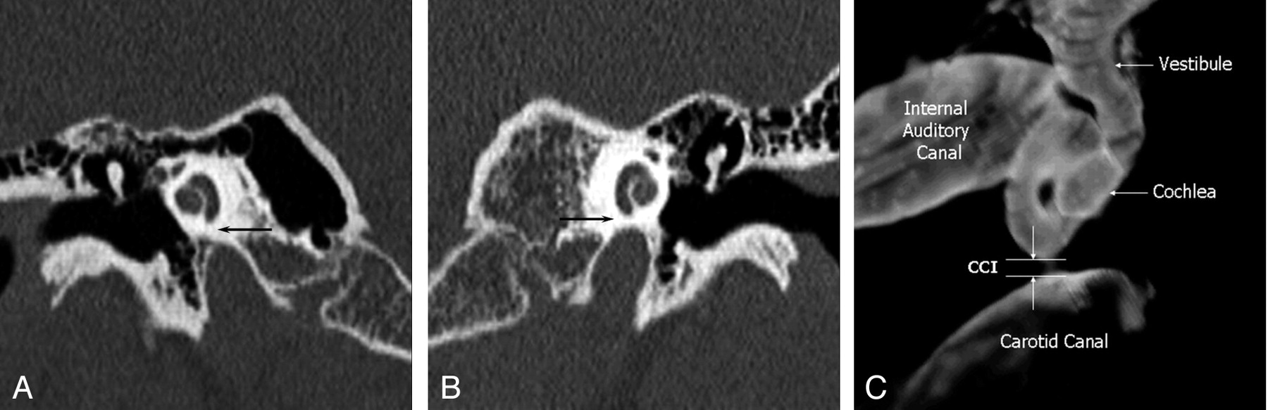
- Fig. 1.
Normal cochlea-carotid interval (CCI), measured on coronal CT images as the minimal distance from the basal turn of the cochlea to the carotid canal. Both sides are from a single patient.
A, Right CCI measures 2.1 mm (arrow).
B, Left CCI measures 1.0 mm (arrow).
C, 3D temporal bone reconstruction of a normal CCI shows the normal separation between the cochlea and carotid canal.
- Fig. 2.
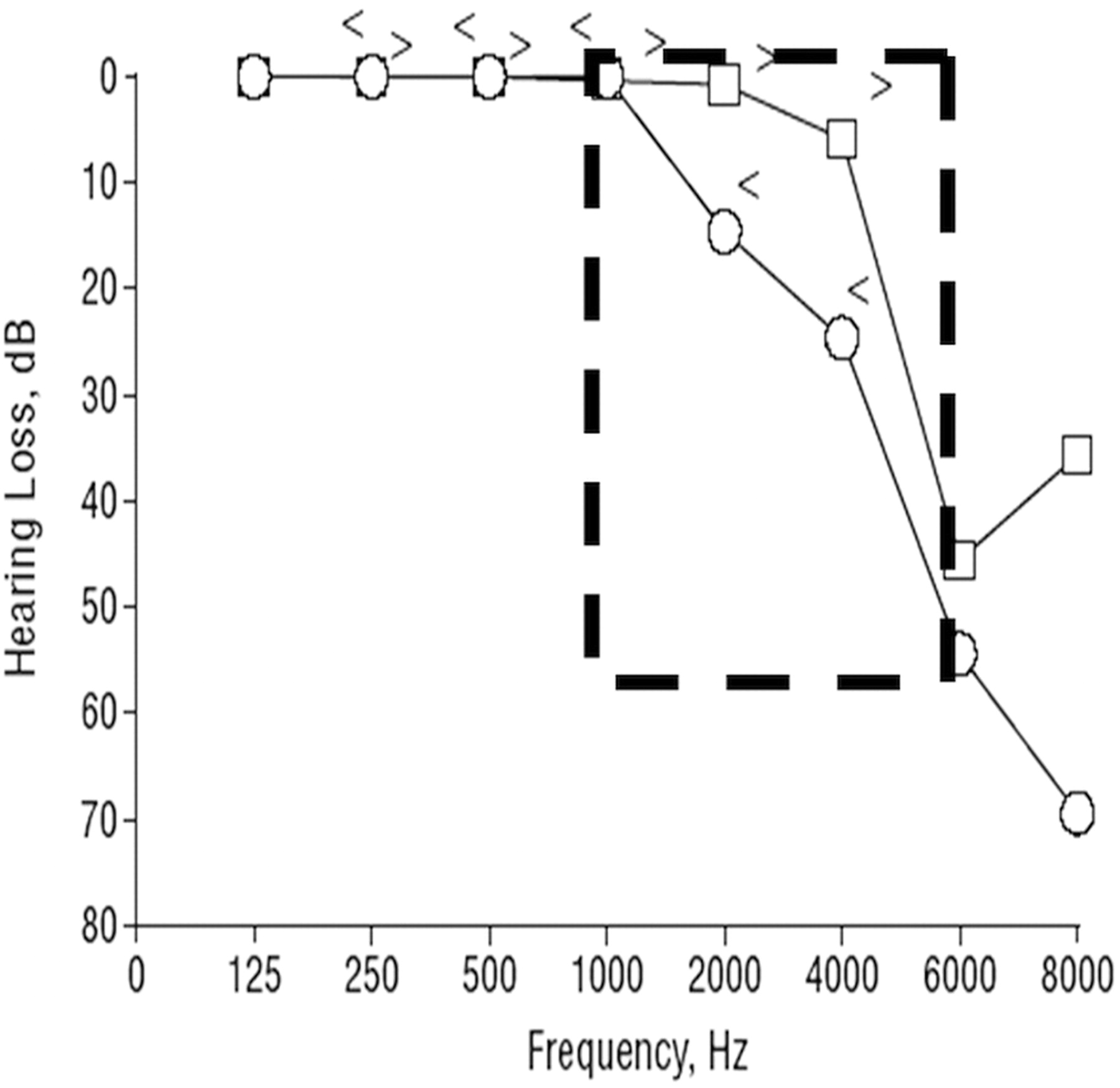
Case report: masked bone conduction thresholds are shown for a series of audiograms performed on September 8 (A), October 19 (B), November 11 (C), and December 2 (D).
A, The initial baseline audiogram demonstrated high-frequency sensorineural hearing loss (SNHL) that is symmetric and bilateral (dashed box).
B, Six weeks later, the patient complained of left-sided hearing loss and the audiogram revealed left-sided mid-tone SNHL (dashed box).
C, Three weeks later, when the patient presented with contralateral SNHL, the audiogram showed resolution of left-sided mid-tone SNHL and new right-sided mid-tone SNHL (dashed box).
D, Three weeks later, when the patient again presented with left-sided symptoms, the audiogram showed resolution of right-sided mid-tone SNHL and new left-sided mid-tone SNHL (dashed box). Subsequent audiogram was similar to the initial evaluation; however, the fluctuation continued.
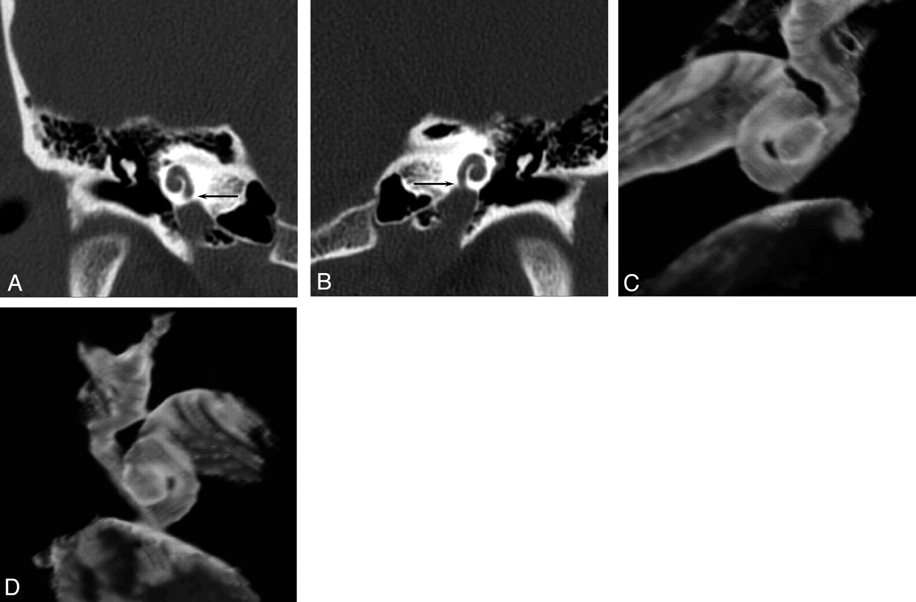
- Fig. 3.
Case report with mid-tone SNHL: coronal temporal bone CT images. Coronal CT (A) and (C) 3D reconstruction show right CCI to measure 0.2 mm (arrow). Coronal CT (B) and (D) 3D reconstruction show left CCI to measure 0.0 mm (arrow).
- Fig. 4.
Audiogram from Modugno et al’s reported patient with bilateral dehiscence of the bony cochlea shows typical mid-tone SNHL that is identical to that seen in Fig 2. (Figure reproduced from Modugno GC, Brandolini C, Cappello I, et al. Bilateral dehiscence of the bony cochlear basal turn. Arch Otolaryngol Head Neck Surg 2004;130:1427–29. Copyright © 2004 American Medical Association, All rights reserved. Used with permission.)


Tables
Carotid-cochlear interval measures for all temporal bones, normal temporal bones, and abnormal temporal bones
All sides (mm)(n = 59) Normal Only (mm)(n =33) Abnormal Only (mm)(n = 26) Right Range 0.4–3.7 0.5–2.2 0.6–2.5 Mean ± SD 1.2 ± 0.8 1.2 ± 0.5 1.0 ± 0.4 Left Range 0.4–4.9 0.4–3.7 0.4–4.9 Mean ± SD 1.1 ± 0.9 1.3 ± 1.0 1.3 ± 1.2 Note:—With regards to abnormal temporal bones, 12 showed otomastoiditis; 4, postoperative for inflammatory disease; 3, cholesteatoma; 2, vestibular aqueduct syndrome; 2, vestibulocochlear dysplasia; 2, otosclerosis; 1 labyrinthitis ossificans.


