
Article Figures & Data
Figures
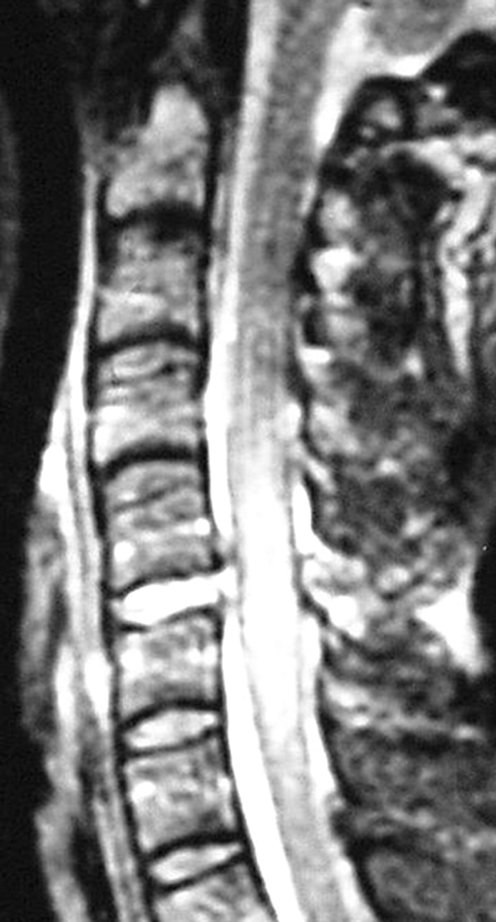
- Fig 1.
True-positive injuries to the ALL, disk, and PLL. Sagittal STIR image demonstrates disruption of the ALL (arrow), intervertebral disk, and PLL (arrowhead) at C6–7. Injuries were confirmed at surgery.
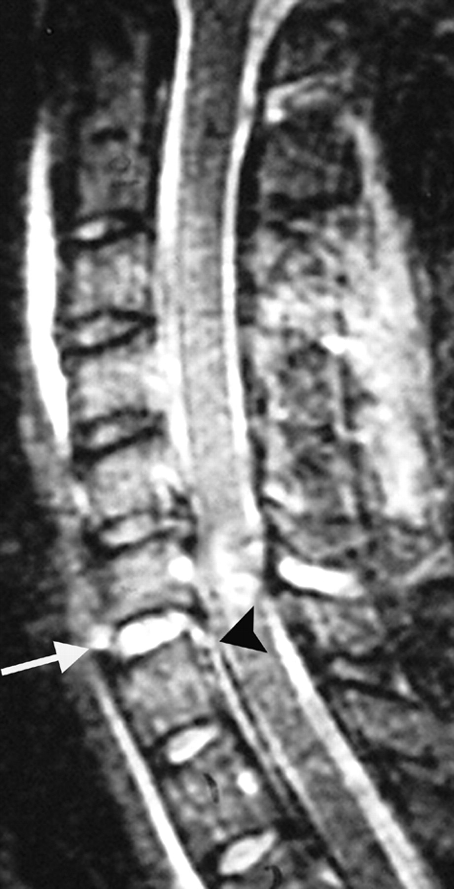
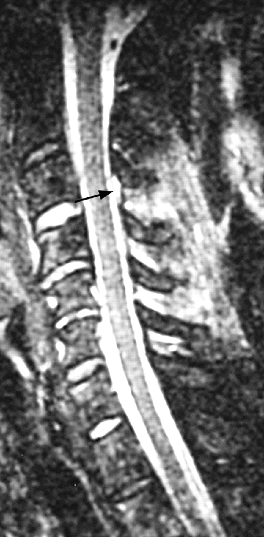
- Fig 2.
True-positive injuries to the ALL, disk, and PLL. Sagittal fast spin-echo T2-weighted image shows elevation of the ALL (white arrow), disruption of the intervertebral disk, and elevation of the PLL at C4–5 (black arrow). Injuries were confirmed at surgery.
- Fig 3.
True-positive ligamentum flavum and interspinous ligament injuries. Sagittal STIR image demonstrates complete disruption of the ligamentum flavum (arrow) and interspinous ligament complex (paired small arrows) at C6–7, which was confirmed at surgery.
- Fig 4.
True-positive facet fracture-dislocation. Parasagittal fast spin-echo T2 image shows a C6 facet fracture (arrowhead) with C6–7 facet dislocation (arrows), which were confirmed at surgery.
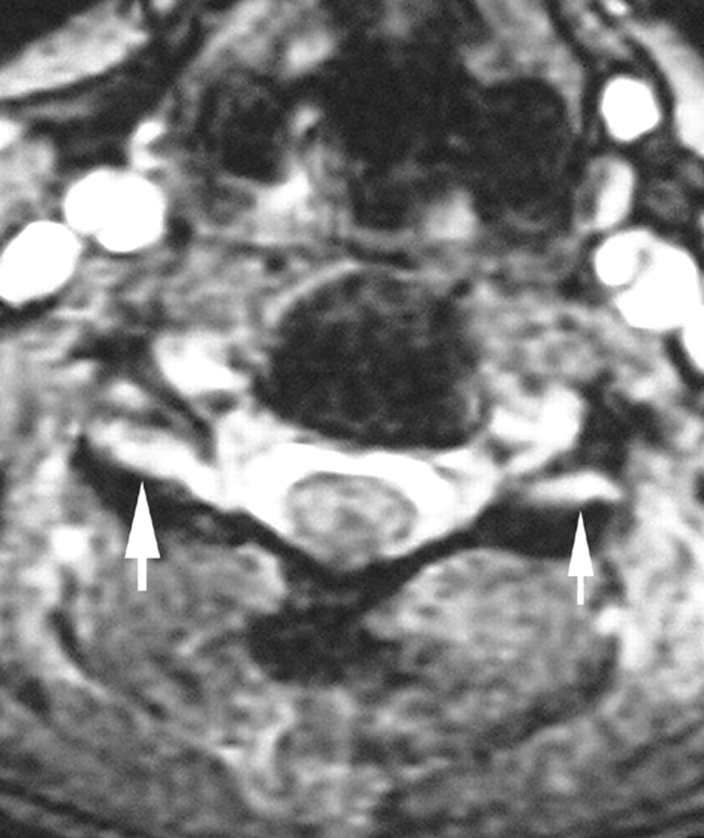
- Fig 5.
Axial image of true-positive facet joint injury. Axial fast spin-echo T2-weighted image demonstrates widening of bilateral facet joints, more so on the right. Both facet capsules were injured at surgery.
- Fig 6.
False-negative ALL. Sagittal fast spin-echo T2-weighted image demonstrates widening of the intervertebral disk and disruption of the PLL at C5–6, which were confirmed at surgery. The ALL, however, appears intact on the MR imaging but was found injured at surgery.
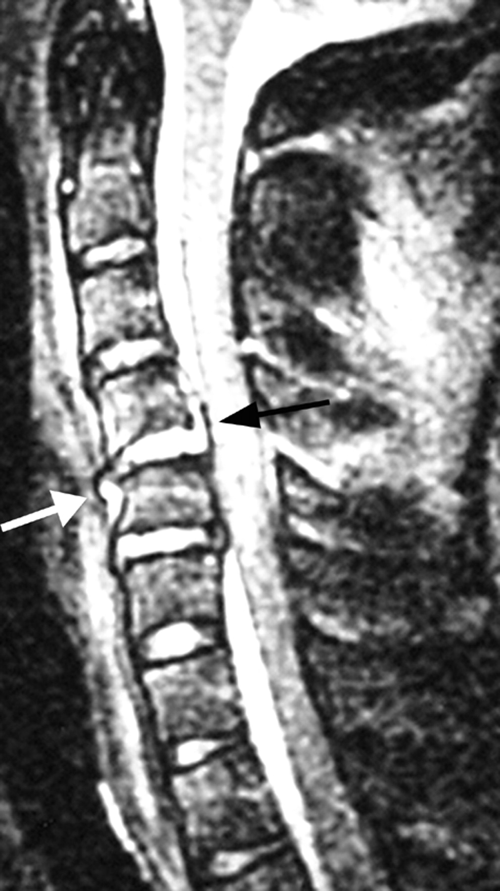
- Fig 7.
False-positive ALL and disk and true-positive PLL injury manifesting as high T2 signal intensity. On this sagittal fast spin-echo T2-weighted image, there is high signal intensity along the PLL (arrow) manifesting as interruption of the normal dark linear PLL at C5–6. At the same level, the ALL appears disrupted and the disk appears widened compared with the level above, especially anteriorly. However, at surgery only the PLL was found injured and the disk and ALL were intact.
- Fig 8.
True-positive ligamentum flavum and false-positive interspinous soft tissues. Sagittal STIR image demonstrates disruption of the ligamentum flavum at multiple levels. Injury at the operative level C3–4 (arrow) was confirmed at surgery. On the image, there is also increased T2 signal intensity with stretching of the interspinous ligamentous complex; however, at surgery, this complex was intact.
- Fig 9.
False-negative ligamentum flavum and true-positive interspinous soft tissue injury. Sagittal T2-weighted image shows spinous process fractures of C7 and T1 (arrows). There is increased T2 signal intensity and stretching of the interspinous ligament complex (injury confirmed at surgery). The ligamentum flavum at the level of injury appears intact on the MR imaging but was found to be injured at surgery.


Tables
- Table 1:
Patterns of abnormality seen on MR imaging for ligamentous structures and disks as described in the literature8–15
Structure Abnormality on MR Images Anterior longitudinal ligament High T2 signal Displacement/elevation Disruption Disk High T2 signal Widening Posterior longitudinal ligament High T2 signal Displacement/elevation Disruption Vertebral body Abnormal marrow signal (bone contusion) Deformity of shape/contour (fracture) Posterior osseous structures Deformity of shape/contour (fracture) Facet capsules High T2 signal Widening Dislocation Ligamentum flavum High T2 signal Disruption Interspinous soft tissues High T2 signal Disruption - Table 2:
Sensitivity of MR imaging relative to intraoperative findings for soft tissue and ligamentous structures in the subdental cervical spine
Structure No. Injured at Surgery N Sensitivity (%) Anterior longitudinal ligament 14 18 71 Disk 15 18 93 Posterior longitudinal ligament 15 18 93 Vertebral body 6 18 100 Posterior osseous structures 22 77 45 Facet capsules 22 26 86 Ligamentum flavum 6 14 67 Interspinous soft tissues 6 14 100 Note:—Sensitivity here is defined as the proportion of injuries at surgery that were abnormal on MR imaging: that is, (True-positive)/(True-positive + False-negative).
- Table 3:
Kappa values for agreement between MR imaging findings and intraoperative findings of injury to osseous and soft tissue/ligamentous structures
Anatomic Structure Surgical Finding(s) MRI Finding(s) κ Anterior longitudinal ligament Partial or complete tear Abnormal signal minus;0.12 Partial or complete tear Ligament elevation 0.069 Partial or complete tear Complete disruption 0.053 Partial or complete tear Any of above abnormalities −0.033 Partial or complete tear Elevation or disruption 0.18 Complete tear Abnormal signal 0.11 Complete tear Ligament elevation −1.0 Complete tear Complete disruption 0.32 Complete tear Any of above abnormalities 0.18 Complete tear Elevation or disruption 0.31 Posterior longitudinal ligament Partial or complete tear Abnormal signal −0.054 Partial or complete tear Ligament elevation 0.18 Partial or complete tear Complete disruption 0.0 Partial or complete tear Any of above abnormalities 0.31 Partial or complete tear Elevation or disruption 0.29 Complete tear Abnormal signal 0.16 Complete tear Ligament elevation 0.14 Complete tear Complete disruption −0.07 Complete tear Any of above abnormalities 0.27 Complete tear Elevation or disruption 0.077 Intervertebral disk Partial or complete disruption Abnormal signal 0.077 Partial or complete disruption Complete disruption −0.24 Partial or complete disruption Any of above abnormalities −0.09 Complete disruption Abnormal signal 0.18 Complete disruption Complete disruption −0.09 Complete disruption Any of above abnormalities 0.21 Right facet capsule Partial or complete disruption Abnormal signal 0.041 Partial or complete disruption Widening of joint 0.26 Partial or complete disruption Complete disruption 0.018 Partial or complete disruption Any of above abnormalities 0.58 Partial or complete disruption Widening or disruption 0.44 Left facet capsule Partial or complete disruption Abnormal signal −0.12 Partial or complete disruption Widening of joint 0.20 Partial or complete disruption Complete disruption 0.20 Partial or complete disruption Any of above abnormalities 0.43 Partial or complete disruption Widening or disruption 0.53 Ligamentum flavum Partial or complete tear Abnormal signal −0.13 Partial or complete tear Complete disruption 0.32 Partial or complete tear Any of above abnormalities 0.21 Complete tear Abnormal signal −0.098 Complete tear Complete disruption 0.0187 Complete tear Any of above abnormalities −0.017 Interspinous ligament Partial or complete tear Abnormal signal −0.29 Partial or complete tear Complete disruption 0.42 Partial or complete tear Any of above abnormalities 0.11 Complete tear Abnormal signal −0.29 Complete tear Complete disruption 0.39 Complete tear Any of above abnormalities 0.060 Vertebral body Fracture Abnormal signal −0.37 Fracture Fracture (change in shape) 0.48 Fracture Any of above abnormalities 0.19 Right lamina Fracture Fracture (change in shape) 0.19 Fracture Left lamina Fracture Fracture (change in shape) 0.074 Fracture Right pedicle Fracture Fracture (change in shape) 0.41 Fracture Left pedicle Fracture Fracture (change in shape) 0.62 Fracture Right facet Fracture Fracture (change in shape) 0.39 Fracture Left facet Fracture Fracture (change in shape) 0.51
In this issue


Jump to section
Related Articles
Cited By...
- Regarding "MR Imaging of the Cervical Spine in Nonaccidental Trauma: A Tertiary Institution Experience"
- Spinal Cord Injury after Blunt Cervical Spine Trauma: Correlation of Soft-Tissue Damage and Extension of Lesion
- Evaluation of the Intervertebral Disk Angle for the Assessment of Anterior Cervical Diskoligamentous Injury
- Assessment of the Posterior Ligamentous Complex Following Acute Cervical Spine Trauma
- Imaging after trauma to the neck
- Imaging after trauma to the neck