
Article Figures & Data
Figures
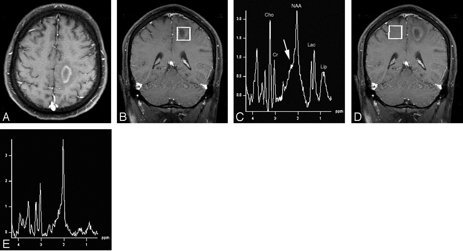
- Fig 1.
A, Axial T1-weighted postcontrast MR image (TR/TE = 437/14) depicts a ring-enhancing lesion in the posterior medial left frontal lobe white matter with surrounding vasogenic edema.
B, Voxel localization for proton MR spectroscopy of the mass.
C, Proton MR spectrum of the mass shows marked elevation of choline, lactate, and lipid, metabolites normally indicative of aggressive neoplastic lesions. Of note, however, is elevation of the β,γ-Glx peaks (arrow) that suggests an inflammatory demyelinating process, which, in conjunction with the anatomic findings, is indicative of tumefactive multiple sclerosis. NAA indicates N-acetylaspartate; Cho, choline; Cr, creatine, Lac, lactate, Lip, lipid.
D, Voxel localization for proton MR spectroscopy of contralateral control.
E, Normal Proton MR spectrum of contralateral control.
- Fig 2.
A, Axial T1-weighted postcontrast MR image (TR/TE = 466/14) shows a small ring-enhancing lesion in the genu of the right internal capsule (arrow) and a barely perceptible lesion in the left globus pallidus (arrowhead). Multiple additional similar small ring-enhancing lesions were identified throughout the brain parenchyma.
B, Voxel localization for proton MR spectroscopy of the right internal capsule lesion.
C, MR spectroscopy of the right internal capsule lesion demonstrates marked elevation of the β,γ-Glx peaks (double arrows) compared with creatine (peak height ratio 1.1 [normal less than 0.5]) compatible with tumefactive multiple sclerosis. There is also mild decrease of N-acetylaspartate and probable mild presence of lactate. NAA indicates N-acetylaspartate; Cho, choline; Cr, creatine; Lac, lactate.
- Fig 3.
A, Axial fluid-attenuated inversion recovery MR image (TR/TE/TI = 9000/109/2500) demonstrates a large region of increased T2-weighted signal intensity in the left frontal and temporal lobes, primarily in the white matter and extending into the corpus callosum. The lesion is exerting mass effect with compression of the left lateral ventricle and left to right subfalcine herniation. There was no significant enhancement of the lesion on postcontrast images.
B, Voxel localization for proton MR spectroscopy of the mass.
C, 1H-MR spectroscopy of the abnormality shows mild choline elevation, presence of lipid, and marked elevation of lactate. There is very significant elevation of the β,γ-Glx peaks (arrow). This large solitary nonenhancing mass lesion is another example of the varied imaging appearance of tumefactive multiple sclerosis. NAA indicates N-acetylaspartate; Cho, choline; Cr, creatine; Lac, lactate; Lip, lipid.
D, Voxel localization for proton MR spectroscopy of contralateral control.
E, Normal proton MR spectrum of contralateral control.
- Fig 4.
A, Axial T1-weighted postcontrast MR image (TR/TE = 481/14) shows a solidly enhancing lesion in the right temporal lobe (arrow). There is also a curvilinear focus of enhancement in the left temporal subcortical white matter (arrowhead) suggestive, but not diagnostic, of the diagnosis of demyelinating disease.
B, Voxel localization for proton MR spectroscopy of the right temporal enhancing mass.
C, MR spectroscopy of the right temporal lobe lesion shows mild elevation of choline and mild decrease of N-acetylaspartate metabolites with probable presence of lipid and lactate possibly leading to the incorrect assumption of neoplastic disease. Again, however, there is elevation of the β,γ-Glx peaks (arrow) consistent with the patient’s correct diagnosis of tumefactive multiple sclerosis.


Tables
Proton MR spectroscopy metabolite ratios of lesions (and controls)
Patient No. NAA/Cr Cho/Cr Glx/Cr Lac Lip 1 2.2 (1.9) 2.0 (0.6) 1.2 (0.5) + + 2 1.6 1.1 1.1 + − 3 2.1 (1.6) 1.2 (0.8) 1.5 (0.4) ++ + 4 1.6 1.4 1.0 + + Note:—NAA indicates N-acetylaspartate; Cr, creatine; Cho, choline; Glx, glutamine and glutamate; Lac, lactate; Lip, lipid; +, mildly elevated; ++, markedly elevated; −, not evident.
In this issue


Jump to section
Related Articles
Cited By...
- Clinicoradiologic features distinguish tumefactive multiple sclerosis from CNS neoplasms
- Utility of Proton MR Spectroscopy for Differentiating Typical and Atypical Primary Central Nervous System Lymphomas from Tumefactive Demyelinating Lesions
- Tumefactive demyelination: an approach to diagnosis and management
- MR Imaging of Neoplastic Central Nervous System Lesions: Review and Recommendations for Current Practice
- Proton MR Spectroscopy Improves Discrimination between Tumor and Pseudotumoral Lesion in Solid Brain Masses