Advertisement

Main menu

  • Home
  • Content
    • Current Issue
    • Accepted Manuscripts
    • Article Preview
    • Past Issue Archive
    • AJNR Case Collection
    • Case of the Week Archive
    • Classic Case Archive
    • Case of the Month Archive
  • Special Collections
    • Spinal CSF Leak Articles (Jan 2020-June 2024)
    • 2024 AJNR Journal Awards
    • Most Impactful AJNR Articles
  • Multimedia
    • AJNR Podcast
    • AJNR Scantastics
    • Video Articles
  • For Authors
    • Submit a Manuscript
    • Author Policies
    • Fast publishing of Accepted Manuscripts
    • Graphical Abstract Preparation
    • Manuscript Submission Guidelines
    • Imaging Protocol Submission
    • Submit a Case for the Case Collection
  • About Us
    • About AJNR
    • Editorial Board
  • More
    • Become a Reviewer/Academy of Reviewers
    • Subscribers
    • Permissions
    • Alerts
    • Feedback
    • Advertisers
    • ASNR Home
  • Other Publications
    • ajnr

User menu

  • Alerts
  • Log in

Search

  • Advanced search
American Journal of Neuroradiology

American Journal of Neuroradiology

ASHNR American Society of Functional Neuroradiology ASHNR American Society of Pediatric Neuroradiology ASSR
  • Alerts
  • Log in

Advanced Search

  • Home
  • Content
    • Current Issue
    • Accepted Manuscripts
    • Article Preview
    • Past Issue Archive
    • AJNR Case Collection
    • Case of the Week Archive
    • Classic Case Archive
    • Case of the Month Archive
  • Special Collections
    • Spinal CSF Leak Articles (Jan 2020-June 2024)
    • 2024 AJNR Journal Awards
    • Most Impactful AJNR Articles
  • Multimedia
    • AJNR Podcast
    • AJNR Scantastics
    • Video Articles
  • For Authors
    • Submit a Manuscript
    • Author Policies
    • Fast publishing of Accepted Manuscripts
    • Graphical Abstract Preparation
    • Manuscript Submission Guidelines
    • Imaging Protocol Submission
    • Submit a Case for the Case Collection
  • About Us
    • About AJNR
    • Editorial Board
  • More
    • Become a Reviewer/Academy of Reviewers
    • Subscribers
    • Permissions
    • Alerts
    • Feedback
    • Advertisers
    • ASNR Home
  • Follow AJNR on Twitter
  • Visit AJNR on Facebook
  • Follow AJNR on Instagram
  • Join AJNR on LinkedIn
  • RSS Feeds

Welcome to the new AJNR, Updated Hall of Fame, and more. Read the full announcements.


AJNR is seeking candidates for the position of Associate Section Editor, AJNR Case Collection. Read the full announcement.


OtherBRAIN

In Vivo Assessment and Visualization of Intracranial Arterial Hemodynamics with Flow-Sensitized 4D MR Imaging at 3T

S. Wetzel, S. Meckel, A. Frydrychowicz, L. Bonati, E.-W. Radue, K. Scheffler, J. Hennig and M. Markl
American Journal of Neuroradiology March 2007, 28 (3) 433-438;
S. Wetzel
  • Find this author on Google Scholar
  • Find this author on PubMed
  • Search for this author on this site
S. Meckel
  • Find this author on Google Scholar
  • Find this author on PubMed
  • Search for this author on this site
A. Frydrychowicz
  • Find this author on Google Scholar
  • Find this author on PubMed
  • Search for this author on this site
L. Bonati
  • Find this author on Google Scholar
  • Find this author on PubMed
  • Search for this author on this site
E.-W. Radue
  • Find this author on Google Scholar
  • Find this author on PubMed
  • Search for this author on this site
K. Scheffler
  • Find this author on Google Scholar
  • Find this author on PubMed
  • Search for this author on this site
J. Hennig
  • Find this author on Google Scholar
  • Find this author on PubMed
  • Search for this author on this site
M. Markl
  • Find this author on Google Scholar
  • Find this author on PubMed
  • Search for this author on this site
  • Article
  • Figures & Data
  • Info & Metrics
  • Responses
  • References
  • PDF
Loading

Article Figures & Data

Figures

  • Tables
  • Fig 1.
    • Download figure
    • Open in new tab
    • Download powerpoint
    Fig 1.

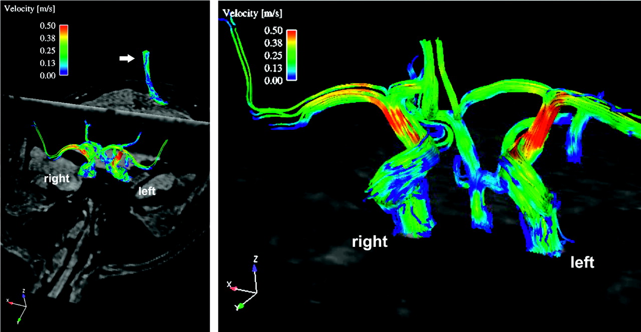
    Normal 3D blood-flow patterns and velocities of the circle of Willis by using 3D streamline visualization reveal complex flow patterns and segmental changes in absolute blood-flow velocities (color-coding indicates local blood flow velocity magnitude). Additional streamline visualization was performed in a short segment of the superior sagittal sinus transecting the axial 3D volume (top left, white arrow).

  • Fig 2.
    • Download figure
    • Open in new tab
    • Download powerpoint
    Fig 2.

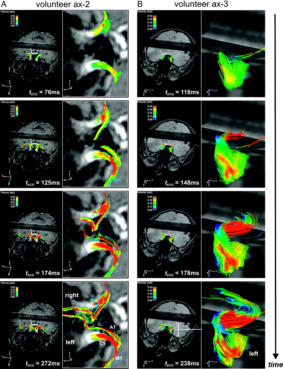
    Time-resolved 3D particle traces for 4 successive systolic timeframes illustrating blood flow in parts of the circle of Willis in 2 healthy volunteers with axial (ax) slab orientations. Color-coding corresponds to the local blood-flow velocity magnitude. A, Overview of the anterior part of the circle of Willis. Simultaneous systolic filling and local blood-flow characteristics of the left and right C1, M1, and A1 segments can clearly be appreciated. B, Targeted view of the left carotid siphon exhibiting complex-flow helical patterns and segmental-flow acceleration.

  • Fig 3.
    • Download figure
    • Open in new tab
    • Download powerpoint
    Fig 3.

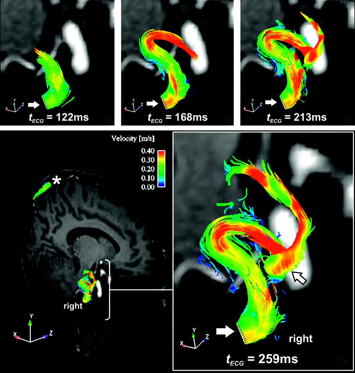
    Time-resolved 3D particle traces in 4 consecutive systolic timeframes illustrate the filling of the right carotid siphon. Note the complex and counter-clockwise helical flow pattern (if viewed along the flow direction) in the C5 segment (open arrow). The emitter plane from which particle traces were released from equidistant grid points is indicated by the solid white arrow. In the superior sagittal sinus (asterisk), particle traces demonstrate lower flow velocities and thus reduced tracer length compared with arterial blood flow.

  • Fig 4.
    • Download figure
    • Open in new tab
    • Download powerpoint
    Fig 4.

    Time-resolved 3D particle trace visualization for a patient with occlusion of the LCA. A, TOF-MRA demonstrating occlusion of the LCA and a small anterior communicating artery (small arrows). B, Arterial filling of the large intracranial vessels for 2 systolic timeframes indicating the absence of flow evolution at the level of the distal left ICA (open white arrows). Blood-flow patterns and systolic filling on the nonoccluded right side are clearly visualized. C and D, Collateral blood flow from right to left via a small anterior communicating artery. C, The emitter plane is localized at the right A1 segment. The crossflow via the small anterior communicating artery to the left A1 segment (arrows) is clearly visible. D, The emitter plane localized in the left A1 segment depicts the retrograde flow direction within that vessel segment.

Tables

  • Figures
  • Bilateral peak systolic flow velocities in cm/s derived from TCD and MR imaging (mean +/− SD)

    Peak Systolic Flow [cm/s]
    Vascular segmentC1M1A1
    MR imaging73 +/− 1171 +/− 1169 +/− 5
    TCD73 +/− 12100 +/− 13116 +/− 53
    • Note:—TCD indicates transcranial Doppler sonography.

PreviousNext
Back to top

In this issue

American Journal of Neuroradiology: 28 (3)
American Journal of Neuroradiology
Vol. 28, Issue 3
March 2007
  • Table of Contents
  • Index by author
Advertisement
Print
Download PDF
Email Article

Thank you for your interest in spreading the word on American Journal of Neuroradiology.

NOTE: We only request your email address so that the person you are recommending the page to knows that you wanted them to see it, and that it is not junk mail. We do not capture any email address.

Enter multiple addresses on separate lines or separate them with commas.
In Vivo Assessment and Visualization of Intracranial Arterial Hemodynamics with Flow-Sensitized 4D MR Imaging at 3T
(Your Name) has sent you a message from American Journal of Neuroradiology
(Your Name) thought you would like to see the American Journal of Neuroradiology web site.
CAPTCHA
This question is for testing whether or not you are a human visitor and to prevent automated spam submissions.
Cite this article
S. Wetzel, S. Meckel, A. Frydrychowicz, L. Bonati, E.-W. Radue, K. Scheffler, J. Hennig, M. Markl
In Vivo Assessment and Visualization of Intracranial Arterial Hemodynamics with Flow-Sensitized 4D MR Imaging at 3T
American Journal of Neuroradiology Mar 2007, 28 (3) 433-438;

Citation Manager Formats

  • BibTeX
  • Bookends
  • EasyBib
  • EndNote (tagged)
  • EndNote 8 (xml)
  • Medlars
  • Mendeley
  • Papers
  • RefWorks Tagged
  • Ref Manager
  • RIS
  • Zotero
0 Responses
Respond to this article
Share
Bookmark this article
In Vivo Assessment and Visualization of Intracranial Arterial Hemodynamics with Flow-Sensitized 4D MR Imaging at 3T
S. Wetzel, S. Meckel, A. Frydrychowicz, L. Bonati, E.-W. Radue, K. Scheffler, J. Hennig, M. Markl
American Journal of Neuroradiology Mar 2007, 28 (3) 433-438;
del.icio.us logo Twitter logo Facebook logo Mendeley logo
  • Tweet Widget
  • Facebook Like
  • Google Plus One

Jump to section

  • Article
    • Abstract
    • Description of Technique
    • Discussion
    • References
  • Figures & Data
  • Info & Metrics
  • Responses
  • References
  • PDF

Related Articles

  • No related articles found.
  • PubMed
  • Google Scholar

Cited By...

  • Intracranial Aneurysm Wall Displacement Predicts Instability
  • Reply:
  • Identification of Vortex Cores in Cerebral Aneurysms on 4D Flow MRI
  • Understanding Angiography-Based Aneurysm Flow Fields through Comparison with Computational Fluid Dynamics
  • Assessment of intra-aneurysmal flow modification after flow diverter stent placement with four-dimensional flow MRI: a feasibility study
  • Early experience in high-resolution MRI for large vessel occlusions
  • Attenuation of Blood Flow Pulsatility along the Atlas Slope: A Physiologic Property of the Distal Vertebral Artery?
  • Cerebral Veins-Why Functional MR Imaging is Worth the Trouble
  • Fast 4D Flow MRI Re-Emerges as a Potential Clinical Tool for Neuroradiology
  • Intracranial 4D Flow MRI: Toward Individualized Assessment of Arteriovenous Malformation Hemodynamics and Treatment-Induced Changes
  • Comparison of Blood Flow Velocity Quantification by 4D Flow MR Imaging with Ultrasound at the Carotid Bifurcation
  • Noninvasive Evaluation of Cerebral Arteriovenous Malformations by 4D-MRA for Preoperative Planning and Postoperative Follow-Up in 56 Patients: Comparison with DSA and Intraoperative Findings
  • Dampening of Blood-Flow Pulsatility along the Carotid Siphon: Does Form Follow Function?
  • Complete Intracranial Arterial and Venous Blood Flow Evaluation with 4D Flow MR Imaging
  • Crossref
  • Google Scholar

This article has not yet been cited by articles in journals that are participating in Crossref Cited-by Linking.

More in this TOC Section

  • Progression of Microstructural Damage in Spinocerebellar Ataxia Type 2: A Longitudinal DTI Study
  • Usefulness of Quantitative Susceptibility Mapping for the Diagnosis of Parkinson Disease
  • Evaluating the Effects of White Matter Multiple Sclerosis Lesions on the Volume Estimation of 6 Brain Tissue Segmentation Methods
Show more Brain

Similar Articles

Advertisement

Indexed Content

  • Current Issue
  • Accepted Manuscripts
  • Article Preview
  • Past Issues
  • Editorials
  • Editors Choice
  • Fellow Journal Club
  • Letters to the Editor

Cases

  • Case Collection
  • Archive - Case of the Week
  • Archive - Case of the Month
  • Archive - Classic Case

Special Collections

  • Special Collections

Resources

  • News and Updates
  • Turn around Times
  • Submit a Manuscript
  • Author Policies
  • Manuscript Submission Guidelines
  • Evidence-Based Medicine Level Guide
  • Publishing Checklists
  • Graphical Abstract Preparation
  • Imaging Protocol Submission
  • Submit a Case
  • Become a Reviewer/Academy of Reviewers
  • Get Peer Review Credit from Publons

Multimedia

  • AJNR Podcast
  • AJNR SCANtastic
  • Video Articles

About Us

  • About AJNR
  • Editorial Board
  • Not an AJNR Subscriber? Join Now
  • Alerts
  • Feedback
  • Advertise with us
  • Librarian Resources
  • Permissions
  • Terms and Conditions

American Society of Neuroradiology

  • Not an ASNR Member? Join Now

© 2025 by the American Society of Neuroradiology All rights, including for text and data mining, AI training, and similar technologies, are reserved.
Print ISSN: 0195-6108 Online ISSN: 1936-959X

Powered by HighWire