
Article Figures & Data
Figures
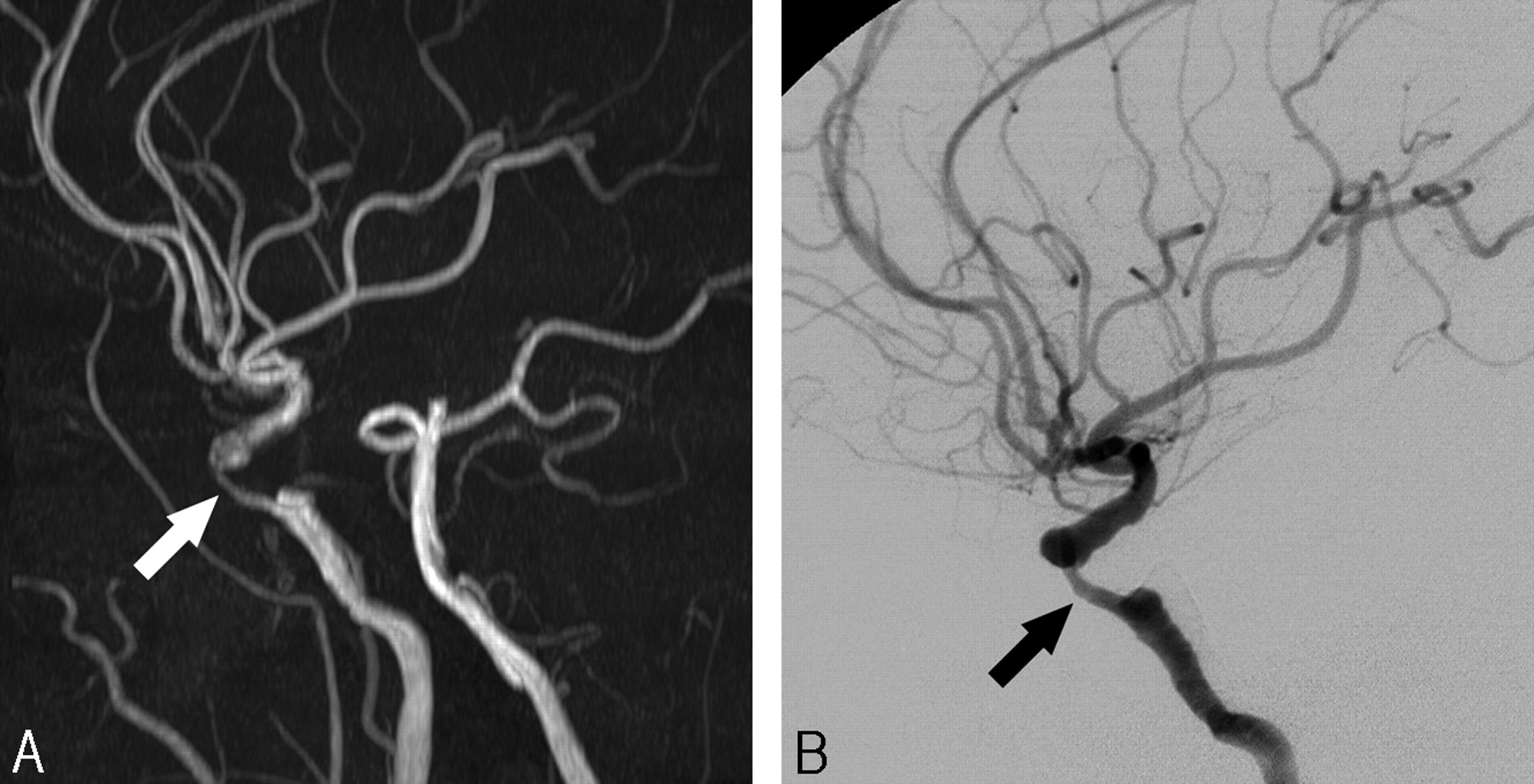
- Fig 1.
A 61-year-old male patient with dysarthria.
A, 3D TOF-MRA shows a segmental high-grade stenosis (white arrow) at the cavernous segment of the distal ICA.
B, DSA lateral view reveals almost identical features of stenosis (black arrow) at the corresponding location.
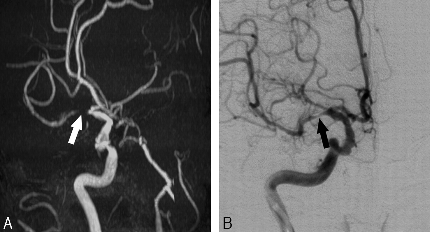
- Fig 2.
A 70-year-old female patient with involuntary movement of the left side.
A, 3D TOF-MRA shows a flow void (white arrow) in the proximal portion of the right middle cerebral artery with visible distal branches.
B, DSA anteroposterior view reveals a focal high-degree stenosis (black arrow) at the corresponding location.
- Fig 3.
A 69-year-old male patient with dizziness.
A, 3D TOF-MRA shows a focal high-grade stenosis (white arrow) at the proximal portion of the basilar trunk.
B, DSA oblique view also shows a focal high-grade stenosis (black arrow) at the corresponding location.
- Fig 4.
A 60-year-old female patient with left side weakness (enrollment number 43 in Table 4).
A, 3D TOF-MRA shows a flow void (white arrow) in the proximal portion of left MCA with visible distal branches.
B, DSA oblique view reveals a focal low-degree stenosis at the corresponding location (black arrow, measured as 40%).
C, Retrospective review of the axial source images also shows a high-grade stenosis at the corresponding location. This type of overestimation may be caused by the turbulent flow and intravoxel phase dispersion within the proximal MCA stenosis close to the distal ICA bifurcation. This patient had a near occlusion of the right MCA that was responsible for her symptoms (not presented).
- Fig 5.
A 61-year-old female patient with left side weakness (enrollment number 10 in Table 4).
A, 3D TOF-MRA shows a faint flow signal intensity along the course of the right ICA (white arrows). Both readers suspected high-grade stenosis in the petrocavernous segment of the ICA.
B, Lateral view, an early phase of DSA, reveals a severe stenosis of the proximal ICA bulb portion (black arrow) with early filling of the external carotid artery.
C, Late-phase DSA shows slow filling and decreased diameter of the petrocavernous segment, compatible with near occlusion of the ICA. Supraclinoidal segment of the distal ICA is filling from the ipsilateral posterior communicating artery (not presented).


Tables
Arteries and Segments 50∼99% Stenosis Complete Occlusion Distal ICA Supraclinoid 5 2 Petrocavernous 6 5* MCA Horizontal (M1) 9 6 Bifurcation or M2 2 0 VBA Basilar trunk 3 0 Distal vertebral 2 0 Total (n = 40) 27 13 Note:—DSA indicates digital subtraction angiography; ICA, internal carotid artery; MCA, middle cerebral artery; M2, vertical segment of MCA; VBA, vertebrobasilar artery.
* Complete occlusion of the petrocavernous portion of internal carotid artery by occlusion of the proximal bulb portion.
3D TOF-MRA DSA Low-Grade High-Grade Occlusion Total Distal internal carotid artery (n = 50) (n = 11) (n = 7) (n = 68) Observer 1 Low-grade 47 1 0 48 High-grade 3 8 0 11 Occlusion 0 2 7 9 Observer 2 Low-grade 47 1 0 48 High-grade 3 9 0 12 Occlusion 0 1 7 8 Middle cerebral artery (n = 51) (n = 11) (n = 6) (n = 68) Observer 1 Low-grade 48 2 0 50 High-grade 3 9 0 12 Occlusion 0 0 6 6 Observer 2 Low-grade 48 1 0 49 High-grade 3 9 0 12 Occlusion 0 1 6 7 Vertebrobasilar artery (n = 19) (n = 5) (n = 0) (n = 24) Observer 1 Low-grade 18 1 0 19 High-grade 1 4 0 5 Occlusion 0 0 0 0 Observer 2 Low-grade 19 0 0 19 High-grade 0 5 0 5 Occlusion 0 0 0 0 All arteries (n = 120) (n = 27) (n = 13) (n = 160) Observer 1 Low-grade 113 4 0 117 High-grade 7 21 0 28 Occlusion 0 2 13 15 Observer 2 Low-grade 114 2 0 116 High-grade 6 23 0 29 Occlusion 0 2 13 15 Note:—Low-grade, normal or <50% diameter stenosis; high-grade, 50%–99% diameter stenosis; DSA, digital subtraction angiography; TOF-MRA, time-of-flight MR angiography.
Sensitivity (%) Specificity (%) PPV (%) NPV (%) Accuracy (%) High-grade stenosis Observer 1 78 (21/27) 95 (126/133) 75 (21/28) 95 (126/132) 92 (147/160) Observer 2 85 (23/27) 95 (127/133) 79 (23/29) 97 (127/131) 94 (150/160) Complete occlusion Observer 1 100 (13/13) 99 (145/147) 87 (13/15) 100 (145/145) 99 (158/160) Observer 2 100 (13/13) 99 (145/147) 87 (13/15) 100 (145/145) 99 (158/160) High-grade stenosis or complete occlusion Observer 1 90 (36/40) 94 (113/120) 84 (36/43) 97 (113/117) 93 (149/160) Observer 2 95 (38/40) 95 (114/120) 86 (38/44) 98 (114/116) 95 (152/160) Note:—TOF-MRA indicates time-of-flight MR angiography; PPV, positive predictive value; NPV, negative predictive value; High-grade stenosis, 50%–99% diameter stenosis.
Enrollment Number Location Stenosis on MRA Observers 1/2 (%) Stenosis on DSA (%) Estimated Causes 3 Right MCA, bifurcation 45/55 0 Venetian blind artifact 3 Left MCA, M1 portion 85/0 0 Venetian blind artifact 41 Right MCA, bifurcation 50/63 34 Intravoxel phase dispersion 43 Left MCA, proximal M1 90/80 40 Intravoxel phase dispersion 43 Right MCA, M1 segment 95/100 99 Near occlusion of M1 5 Right ICA, petrocavernous junction 55/43 29 Observer error 10 Right ICA, petrocavernous segment 66/70 0 Near occlusion of proximal ICA without distal stenosis 13 Right ICA, petrocavernous segment 0/60 0 Near occlusion of proximal ICA without distal stenosis 17 Right ICA, petrocavernous segment 40/51 41 Borderline stenosis 31 Right ICA, petrocavernous segment 100/90 99 Near occlusion of ICA due to multifocal stenosis 36 Right ICA, supraclinoidal segment 100/100 99 Near occlusion of ICA due to multifocal stenosis 36 Left ICA, petrocavernous segment 40/80 38 Intravoxel phase dispersion 10 Left distal VA segment 80/0 0 Left subclavian steal Note:—MCA indicates middle cerebral artery; M1, horizontal segment of MCA; ICA, internal carotid artery; VA, vertebral artery; MRA, 3-dimensional time-of-flight MR angiography; DSA, digital subtraction angiography.
Enrollment Number Location Stenosis on MRA Observers 1/2 (%) Stenosis on DSA (%) Estimated Causes 33 Left MCA, proximal M1 30/57 60 Observer error 36 Left MCA, bifurcation area 40/39 57 Observer error 3 Left ICA, petrocavernous segment 80/0 60 Proximal occlusion with reconstructed distal ICA from ECA collaterals 28 Right ICA, petrocavernous segment 40/57 51 Borderline stenosis 40 Right distal VA segment 45/58 73 Observer error Note:—MCA indicates middle cerebral artery; M1, horizontal segment of MCA; ICA, internal carotid artery; ECA; external carotid artery; VA, vertebral artery; MRA, 3D time-of-flight MR angiography; DSA, digital subtraction angiography.
In this issue


Jump to section
Related Articles
Cited By...
- Automated Detection of Steno-Occlusive Lesion on Time-of-Flight MR Angiography: An Observer Performance Study
- Validation of Zero TE-MRA in the Characterization of Cerebrovascular Diseases: A Feasibility Study
- Concordance of Time-of-Flight MRA and Digital Subtraction Angiography in Adult Primary Central Nervous System Vasculitis
- Quantifying Intracranial Internal Carotid Artery Stenosis on MR Angiography
- In Vivo Assessment of the Impact of Regional Intracranial Atherosclerotic Lesions on Brain Arterial 3D Hemodynamics
- Comparison of High-Resolution MR Imaging and Digital Subtraction Angiography for the Characterization and Diagnosis of Intracranial Artery Disease
- Prevalence of Intracranial Atherosclerotic Stenosis Using High-Resolution Magnetic Resonance Angiography in the General Population: The Atherosclerosis Risk in Communities Study
- One-Year MR Angiographic and Clinical Follow-Up after Intracranial Mechanical Thrombectomy Using a Stent Retriever Device
- Gadolinium Enhancement of Atherosclerotic Plaque in the Middle Cerebral Artery: Relation to Symptoms and Degree of Stenosis
- MR Angiography and Imaging for the Evaluation of Middle Cerebral Artery Atherosclerotic Disease
- Quality-Evaluation Scheme for Cerebral Time-Resolved 3D Contrast-Enhanced MR Angiography Techniques